Ophthalmoscopic Examination
Обзор
Source: Richard Glickman-Simon, MD, Assistant Professor, Department of Public Health and Community Medicine, Tufts University School of Medicine, MA
The simplest ophthalmoscopes consist of an aperture to look through, a diopter indicator, and a disc for selecting lenses. The ophthalmoscope is primarily used to examine the fundus, or the inner wall of the posterior eye, which consists of the choroid, retina, fovea, macula, optic disc, and retinal vessels (Figure 1). The spherical eyeball collects and focuses light on the neurosensory cells of the retina. Light is refracted as it passes sequentially through the cornea, the lens, and the vitreous body.
The first landmark observed during the funduscopic exam is the optic disc, which is where the optic nerve and retinal vessels enter the back of the eye (Figure 2). The disc usually contains a central whitish physiologic cup where the vessels enter; it normally occupies less than half the diameter of the entire disc. Just lateral and slightly inferior is the fovea, a darkened circular area that demarcates the point of central vision. Around this is the macula. A blind spot approximately 15° temporal to the line of gaze results from a lack of photoreceptor cells at the optic disc.
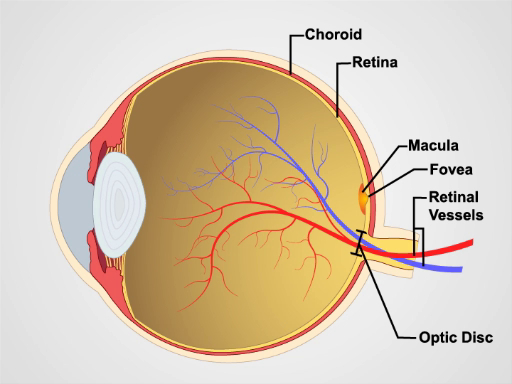
Figure 1. Anatomy of the eye. A diagram showing a sagittal view of the human eye with the structures labeled.

Figure 2: Normal retina. A photograph showing an ophthalmoscopic view on the normal retina.
Процедура
Since mydriatic eye drops are typically not used in general practice, the view of the fundus is limited to only a section of the posterior retina. Be familiar with these features before attempting to examine the patient.
- Unless the patient's refractive errors make it difficult to focus on the retina, it is usually best to remove your own eyeglasses for the exam.
- After darkening the room, turn on the ophthalmoscope and shine the light on your hand or on the wall.
- Turn the lens disc until the largest white
Заявка и Краткое содержание
The ophthalmologic exam is probably the most challenging for students to master. With time, however, it becomes routine. It is also one of the most productive parts of the physical exam, as it not only offers a window into the condition of the eye, but also provides evidence of disease elsewhere in the body. Elevated intracranial pressure from a variety of causes may lead to swelling of the optic nerve, which appears as papilledema on a funduscopic exam. In papilledema, the optic disc is swollen, its margins are blurred,
Перейти к...
Видео из этой коллекции:

Now Playing
Ophthalmoscopic Examination
Physical Examinations II
66.8K Просмотры

Eye Exam
Physical Examinations II
76.0K Просмотры

Ear Exam
Physical Examinations II
53.8K Просмотры

Nose, Sinuses, Oral Cavity and Pharynx Exam
Physical Examinations II
64.7K Просмотры

Thyroid Exam
Physical Examinations II
103.7K Просмотры

Lymph Node Exam
Physical Examinations II
382.1K Просмотры

Abdominal Exam I: Inspection and Auscultation
Physical Examinations II
200.9K Просмотры

Abdominal Exam II: Percussion
Physical Examinations II
245.9K Просмотры

Abdominal Exam III: Palpation
Physical Examinations II
137.6K Просмотры

Abdominal Exam IV: Acute Abdominal Pain Assessment
Physical Examinations II
66.6K Просмотры

Male Rectal Exam
Physical Examinations II
112.9K Просмотры

Comprehensive Breast Exam
Physical Examinations II
85.9K Просмотры

Pelvic Exam I: Assessment of the External Genitalia
Physical Examinations II
300.9K Просмотры

Pelvic Exam II: Speculum Exam
Physical Examinations II
148.5K Просмотры

Pelvic Exam III: Bimanual and Rectovaginal Exam
Physical Examinations II
145.8K Просмотры
Авторские права © 2025 MyJoVE Corporation. Все права защищены