
A subscription to JoVE is required to view this content. Sign in or start your free trial.
Method Article
Laser-Capture Microdissection RNA-Sequencing for Spatial and Temporal Tissue-Specific Gene Expression Analysis in Plants
In This Article
Summary
Presented here is a protocol for laser-capture microdissection (LCM) of plant tissues. LCM is a microscopic technique for isolating areas of tissue in a contamination-free manner. The procedure includes tissue fixation, paraffin embedding, sectioning, LCM and RNA extraction. RNA is used in the downstream tissue-specific, temporally resolved analysis of transcriptomes.
Abstract
The development of a complex multicellular organism is governed by distinct cell types that have different transcriptional profiles. To identify transcriptional regulatory networks that govern developmental processes it is necessary to measure the spatial and temporal gene expression profiles of these individual cell types. Therefore, insight into the spatio-temporal control of gene expression is essential to gain understanding of how biological and developmental processes are regulated. Here, we describe a laser-capture microdissection (LCM) method to isolate small number of cells from three barley embryo organs over a time-course during germination followed by transcript profiling. The method consists of tissue fixation, tissue processing, paraffin embedding, sectioning, LCM and RNA extraction followed by real-time PCR or RNA-seq. This method has enabled us to obtain spatial and temporal profiles of seed organ transcriptomes from varying numbers of cells (tens to hundreds), providing much greater tissue-specificity than typical bulk-tissue analyses. From these data we were able to define and compare transcriptional regulatory networks as well as predict candidate regulatory transcription factors for individual tissues. The method should be applicable to other plant tissues with minimal optimization.
Introduction
Plant development and growth involve the coordinated action of transcriptional regulatory networks within different cells that exist in a complex cellular environment. To understand the activity of these regulatory networks, we require the knowledge of spatial and temporal gene expression within different cell types across developmental stages. However, analyses of gene expression are more commonly conducted in whole organs or bulk tissue samples due to the technical challenge of isolating and analyzing small numbers of cells. The method we describe here has allowed obtaining spatial and temporal tissue-specific transcriptome analysis by coupling LCM with RNA-seq.
LCM was developed two decades ago by Emmert-Buck and colleagues1. The technique enabled researchers to precisely isolate single-cells or clusters of cells from their environment using direct microscopic visualization and manipulation with a narrow beam laser1. Since then the method has been widely used in cancer biology and pathology2,3. Many plant research groups have also adapted LCM for the use with different plant species and different tissue types4,5,6,7,8,9,10,11. Recently, several papers have also used LCM on eudicot and monocot seeds to study embryo, endosperms and other seed structures during seed development and germination10,12,13. Most of the other commonly used single-cell isolation methods such as micro-pipetting, cell sorting, magnetic separation and microfluidic platforms depend on the enzymatic digestion or mechanical homogenization to dissociate cells. This may perturb gene expression, introducing technical artefacts that confound data interpretation14,15. These methods also require previous knowledge of marker genes for each cell type to relate the dissociated cells to their spatial location and true cell-type. A further group of techniques depends on affinity-based isolation of subcellular structures instead of whole cells, for example INTACT (Isolation of Nuclei Tagged in Cell Types) and TRAP (Translating Ribosome Affinity Purification)16,17. However, affinity labeling and purification of nuclei or ribosomes are technically challenging in plant species that do not have well-established transformation protocols. LCM takes advantage of quick tissue fixation to preserve transcript levels and conventional histological identification by direct visualization of cells within their normal tissue/organ context, which allows discrete cells to be isolated in a short period of time18,19.
The protocol presented here is an optimized method for the isolation of specific cells or cell types from the tissue sections of cereal seeds, which can be applied to most of the cells that can be histologically identified. LCM provides a contact-free method of cell isolation, greatly reducing contamination and increasing integrity of recovered RNA. Furthermore, the method illustrates the power of LCM on large-scale genome wide studies starting with small quantities of biological materials. We also describe linear amplification of RNA for generating sufficient input material for downstream transcript/transcriptome analyses.
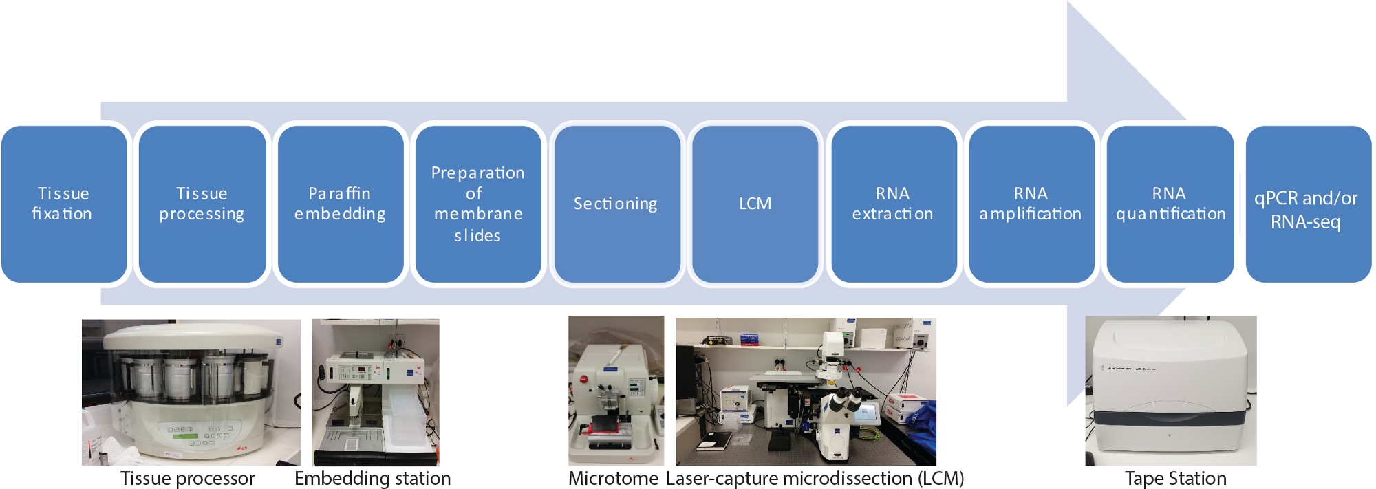
There are ten main steps in this LCM RNA-seq protocol for spatial and temporal tissue-specific transcriptomes, including fixation of tissue samples, dehydration, paraffin infiltration, embedding, sectioning, LCM, RNA extraction, RNA amplification, RNA quantification and qRT-PCR and/or RNA-seq (Figure 1).

Figure 1: Flowchart of LCM followed by RNA-seq or qRT-PCR. LCM is a spatially precise and contact-free technique to collect cells from fixed tissue sections using a laser beam under microscopic visualization. The process starts with fixation of tissue samples, followed by dehydration using a gradient series of ethanol and xylene, and finished with paraffin infiltration. The process can be fully automated by using a tissue processor. Once the tissue is infiltrated with paraffin, it is embedded in a mold with molten paraffin using an embedding station. Sectioning is carried out using microtome set to the desired thickness. Slides are prepared and LCM conducted immediately before RNA is to be extracted from captured cells. RNA extraction is followed directly by two rounds of RNA amplification prior to qRT-PCR and/or RNA-seq. Please click here to view a larger version of this figure.
Protocol
As the final product is RNA, take care to avoid contaminating the work with RNases. Wearing gloves is a must. Use diethyl pyrocarbonate (DEPC) -treated water, buffers, etc. Autoclave buffers and bake glassware before use.
1. Tissue fixation
- Prepare fixative of choice depending on the species and tissue types; for barley seed, use Farmer’s fixative (75% ethanol, 25% glacial acetic acid (v/v)).
- Chill the fixative on ice prior to harvesting tissues.
- Collect the plant material of interest and if necessary, dissect it into pieces of appropriate size to fit into the selected embedding mold. For barley seed, cut the seed into half longitudinally to help penetration of the fixative solution and to fit into the embedding mold.
- Submerge the tissue into at least 10x volume of ice-cold fixative. For barley seed, submerge the seed cut into half into the fixative.
- Use vacuum infiltration to accelerate the penetration of fixative. The tissues should sink after the vacuum infiltration of the fixative. For barley seed, use 30 min of vacuum infiltration.
- Replace the fixative and incubate at 4 °C to allow the fixative to fully penetrate the tissue. For barley seed, incubate the samples overnight (~12-16 h).
NOTE: Thinner or small tissues will require shorter fixation time due to the higher diffusion rate of fixative into the tissue. - Remove the tissue from fixative and transfer the tissue into cassettes and then commence tissue processing.
NOTE: Small or fragile tissue, such as leaf tissue, can be placed into cassettes for fixing to ensure it is not damaged during fixation. Biopsy bags, pads or wraps could be used to hold the tissue securely inside the cassettes during tissue fixation and tissue processing steps.
2. Tissue processing
- Use an automated tissue processor in step 2 with a minimum of 10 solution chambers and 2 heated paraffin chambers (see Table of Materials).
- Check that there are adequate amounts of solution in each chamber; replace solutions after every few uses of the tissue processor.
- Place cassettes with tissue into the metal basket. Attach the metal basket to its holder above chamber 1. The holder will rotate and “dunk and dip” the cassettes into the chambers, following the designated program.
- Set the program by performing button clicks on the control panels of the tissue processor which involve setting length of time for each chamber.
- Press the “Start” button to start the processing program. The following program is designed for barley seeds and runs overnight (~18 h)
- Perform dehydration by dipping the cassette for 1 h 30 min each in the gradient series of ethanol (75%, 85%, 100%, 100%, and 100% (v/v) ethanol).
- Perform clearing using ethanol: xylene gradient for 1 h 30 min each at 75:25, 50:50, 25:75 (ethanol: xylene %, v/v). Then dip the cassette for 1 h 30 min each of 100% xylene then 100% xylene.
- Perform paraffin infiltration at 55-60 °C for 1 h 30 min twice in molten paraffin.
NOTE: Temperature of paraffin heater chambers can be set at the back of the tissue processor.
- The next morning, remove the cassettes from the tissue processor and proceed to paraffin embedding.
NOTE: Program time may vary between different tissue type. Vacuum and/or agitation can be used during tissue processing to accelerate the infiltration of selected solutions by pressing “V” and/or “agitation” buttons on the control panel of the tissue processor.
3. Paraffin embedding
- Use an embedding machine in this step (see Table of Materials).
- Preset the embedding machine to turn on at least a few hours prior to embedding to allow time for the paraffin in the reservoirs to melt completely.
- Turn on the cold plate prior to starting.
- Embed samples in molds by holding the samples in position using fine forceps and dispensing molten paraffin into the mold. Ensure proper orientation of samples for each experimental purpose. For barley seed, orientate the seed longitudinal to the cutting direction to obtain longitudinal sections.
NOTE: Embedding molds come in different sizes. Select an appropriate size to allow the sample to be positioned and embedded properly. Orientation of the samples should be considered depending on experimental needs. If longitudinal sections are required, the sample should be orientated longitudinal to the cutting direction whereas for transverse sections the sample should be orientated parallel to the cutting direction. - Place a clean cassette onto the mold and ensure sufficient paraffin fully cover the whole cassette to hold the sample onto the cassette.
- Place the mold onto the cold plate and allow the paraffin to set fully (10-20 min) before releasing the block from mold.
- Proceed to sectioning or transfer the blocks to 4 °C for storage.
NOTE: The protocol can be paused here. The embedded blocks can be stored at 4 °C for up to three months.
4. Preparation of polyethylene naphthalate (PEN) membrane slides
- Submerge PEN membrane slides in RNase deactivating solution for 3 s followed by two brief washes in DEPC-treated water to remove RNases on the slides. Dry the slides in a 37 °C incubator to remove left over solution.
- UV-treat the slides using a UV lamp in a laminar flow cabinet for 30 min to enhance hydrophilic properties for improved paraffin adhesion.
5. Sectioning
- Use a microtome in the sectioning step (see Table of Materials).
- Place a new blade into the knife holder, and always keep knife guard up when not actively sectioning
CAUTION: Microtome blades are extremely sharp and can cause significant harm when handled inappropriately.
NOTE: There are two locking mechanisms on the microtome, one is at the side of the machine and the other is on the handle of the wheel. Both are to be engaged when not actively sectioning. - Adjust the knife block to be as close to the sample as possible without touching. Ensure that the microtome arm never comes into full contact with the knife block as this will cause catastrophic structural damage to the microtome.
- Turn on the cold plate prior to starting. Keep paraffin blocks on the cold plate prior to sectioning and re-cool blocks when necessary during sectioning to prevent the blocks from softening.
- Fill the water bath with DEPC-treated water and heat to 42 °C prior to starting.
- Trim blocks to the desired depth (where the section you are interested in) and section paraffin blocks at desired thickness (6-10 µm) using the microtome; a well-sectioned block will form a ‘ribbon’ at the edge of the blade. For barley seed, section with 8 µm thickness.
- Gently transfer ribbons from the microtome to the water bath using fine paint brush or fine forceps, ensuring that the ribbon is flat on the surface of the water.
- Hold a slide at a 45° angle, using an upward motion, lift a ribbon out of the water onto the slide and carefully remove excess water with a lint-free tissue.
- Dry slides for 30 min at 37 °C to eliminate any remaining water under the paraffin.
- Proceed to paraffin removal or store at 4 °C in a closed box under dehydrating conditions (to be used within several days).
- Remove paraffin by washing the slides 3x for 20 s each in xylene, followed by 2x washes of 30 s in 100% (v/v) ethanol and 2x washes of 30 s in 70% (v/v) ethanol.
- Proceed immediately to laser-capture microdissection after paraffin is removed.
NOTE: Cryosectioning is an alternative method that has been successfully coupled with LCM. Sample preparation for cryosectioning will differ.
6. Laser-capture microdissection
- Use a laser-capture microdissection microscope (see Table of Materials) to microdissect cells from de-paraffinized and dried tissue sections.
- Load slides on the three available slots.
- Use the special adhesive caps of collection tubes to collect the captured samples. Capturing without liquid (“dry” collection) minimizes RNase activity. Load the collection tubes into the available slots.
- Move the stage to locate the region of the sample that needs to be cut. This can be done using mouse or joystick of the LCM machine, or the arrow keys on the keyboard.
- To optimize the cutting speed, cutting energy and focus, laser pressure catapulting (LPC) energy and focus, first cut on a blank segment free of tissue on the membrane slide. For barley seed, cutting speed = 18, CutEnergy = 52 CutFocus = 63, LPCEnergy = 78, LPC focus = 61 at 10x magnification.
NOTE: Cutting focus and energy have to be adjusted for different slides, different tissues, and captured area but general rules are the catapulting power is higher than the cutting power and the laser has to be defocused for catapulting. The higher the magnification of the objective lens, the smaller the focus of the laser and the higher the energy. - Use the Drawing tools to select cells by outlining the area of interest.
- Select RoboLPC function from the function toolbar to catapult cells into the adhesive caps based on the optimized parameters obtained by cutting on a black segment above.
NOTE: LCM parameters vary between tissue types as well as thickness of section, tissue hardness and objective lenses. Therefore, it is best to optimize each slide on a plain membrane area without tissue specimen before cutting the actual sample. - Use the Flag tools to mark regions of interest to locate them immediately by selecting that flag from the elements list.
- Check by “CapCheck” button to inspect the adhesive cap to confirm samples were captured. Typically, LCM of 10-15 sections (~200 cells) per cap is required for RNA extraction.
- Keep the captured samples on ice. Proceed immediately for RNA extraction to avoid RNA degradation.
NOTE: Some LCM microscopy are equipped with fluorescent light which allows the capture of cells labeled with fluorescent markers.
7. RNA extraction
- Use a low input RNA isolation (see Table of Materials) kit for RNA extraction after LCM. Such kits are designed to recover high-quality total RNA consistently from fewer than ten cells.
- Isolate the total RNA from the captured cell types according to the manufacturer’s instructions including the on-column DNase treatment.
NOTE: The first step of the RNA extraction where the tube is inverted and flicked is crucial to ensure the captured samples on the lid are in contact with the extraction buffer added.
8. RNA amplification
- Use an antisense RNA (aRNA) amplification kit (see Table of Materials) for aRNA amplification from the RNA extracted by in vitro transcription to produce sufficient aRNA for RNA-seq library synthesis.
- Perform two rounds of amplification using the aRNA amplification Kit according to the manufacturer’s instructions.
NOTE: It is important to preheat the thermo-cycler and lid to the temperature instructed by the kit (see Table of Materials) manufacturer. An alternative approach instead of RNA amplification is to use a low input library preparation kit to synthesize the library directly from extracted RNA.
9. RNA quantification
- Quantify and qualify aRNA using an automated electrophoresis system (see Table of Materials).
NOTE: An automated electrophoresis system is preferred as it requires less sample (1-2 µL) and provides a gel-like image and electropherogram for each individual sample.
10. qRT-PCR and/or RNA-seq
- Synthesize cDNA from the aRNA for qRT-PCR or to make RNA-seq libraries using standard RNA-seq library kits.
Results
We generated spatial and temporal tissue-specific transcriptomes from barley seeds during germination using our LCM RNA-seq protocol10. The study was carried out by applying LCM RNA-seq to small number of cells from three embryo organs (plumule, radicle tip, scutellum) every 8 h over a 48 h time course during germination (0-48 h, 7 time points) (Figure 2A,B).
Discussion
Many tissue-specific gene expression studies have been limited by hand dissection of samples, which is time-consuming, labor intensive, has a high risk of contamination and can only utilize samples that a human operative is sufficiently dexterous to harvest. LCM is a precise and contact-free technique to collect cells from fixed tissue sections using a mechanically operated laser beam under microscopic visualization.
Good sample preparation is critical for LCM. The process relies upon proper f...
Disclosures
The authors have nothing to disclose.
Acknowledgements
This work was supported by the Australian Research Council Centre of Excellence in Plant Energy Biology (CE140100008) to JW. M.G.L was supported by a La Trobe University starting grant. We thank the La Trobe Genomics Platform for their support in high-throughput sequencing and data analysis. We thank Associate Professor Matthew Tucker for expert advice on establishing LCM in our lab.
Materials
| Name | Company | Catalog Number | Comments |
| Acetic acid 100 % ACS/R. | AnalaR NORMAPUR (BioStrategies) | VWRC20104.323 | |
| AdhesiveCap 200 opaque | Zeiss | 415190-9181-000 | |
| Clear base moulds 8 X 10 | Leica | 3803015 | |
| Diethyl pyrocarbonate | Sigma-Aldrich | 40718-25ML | |
| High Sensitivity RNA ScreenTape | Agilent | 5067-5579 | |
| Lowprofile disp.blades DB80LS | Leica | 14035843489 | |
| MembraneSlide 1.0 PEN | Zeiss | 415190-9041-000 | |
| MessageAmp II aRNA Amplification Kit | Ambion (ThermoFisher) | AMB17515 | |
| On-Column DNase I Digestion Set | Sigma-Aldrich | DNASE70 | |
| Ovation RNA-Seq System V2 | NuGen (Integrated Science) | 7102-08 | |
| Paraffin (Surgipath Paraplast) | Leica | 39601006 | |
| PicoPure RNA Isolation Kit | ABI (ThermoFisher) | KIT0214 | |
| RNaseZap RNase Decontamination Solution | Ambion (ThermoFisher) | AM9780 | |
| Xylene | AnalaR NORMAPUR (BioStrategies) | VWRC28975.360 | |
| Leica Benchtop Tissue Processor | Leica Biosystems | TP1020 | |
| Leica Heated Paraffin Embedding Module | Leica Biosystems | EG1150H | |
| Leica Cold Plate | Leica Biosystems | EG1150C | |
| Safemate Class 2 Biological Safety Cabinets | LAF Technologies Pty Ltd | Safemate 1.5 | |
| Leica Fully Automated Rotary Microtome | Leica Biosystems | RM2265 | with PALMRobo v 4.6 software |
| Zeiss PALM MicroBeam LCM system | Zeiss miscroscopy | ||
| TapeStation | Agilent | TapeStation 2200 |
References
- Emmert-Buck, M. R., et al. Laser capture microdissection. Science. 274 (5289), 998-1001 (1996).
- Alevizos, I., et al. Oral cancer in vivo gene expression profiling assisted by laser capture microdissection and microarray analysis. Oncogene. 20 (43), 6196-6204 (2001).
- Cong, P., et al. In situ localization of follicular lymphoma: description and analysis by laser capture microdissection. Blood, The Journal of the American Society of Hematology. 99 (9), 3376-3382 (2002).
- Blokhina, O., et al. Laser capture microdissection protocol for xylem tissues of woody plants. Frontiers in Plant Science. 7, 1965 (2017).
- Casson, S., Spencer, M., Walker, K., Lindsey, K. Laser capture microdissection for the analysis of gene expression during embryogenesis of Arabidopsis. The Plant Journal. 42 (1), 111-123 (2005).
- Chen, Z., et al. LCM-seq reveals the crucial role of LsSOC1 in heat-promoted bolting of lettuce (Lactuca sativa L.). The Plant Journal. 95 (3), 516-528 (2018).
- Jiao, Y., et al. A transcriptome atlas of rice cell types uncovers cellular, functional and developmental hierarchies. Nature Genetics. 41 (2), 258-263 (2009).
- Kivivirta, K., et al. A protocol for laser microdissection (LMD) followed by transcriptome analysis of plant reproductive tissue in phylogenetically distant angiosperms. Plant Methods. 15 (1), 1-11 (2019).
- Li, P., et al. The developmental dynamics of the maize leaf transcriptome. Nature Genetics. 42 (12), 1060-1067 (2010).
- Liew, L. C., et al. Temporal tissue-specific regulation of transcriptomes during barley (Hordeum vulgare) seed germination. The Plant Journal. 101 (3), 700-715 (2020).
- Matas, A. J., et al. Tissue-and cell-type specific transcriptome profiling of expanding tomato fruit provides insights into metabolic and regulatory specialization and cuticle formation. The Plant Cell. 23 (11), 3893-3910 (2011).
- Sakai, K., et al. Combining laser-assisted microdissection (LAM) and RNA-seq allows to perform a comprehensive transcriptomic analysis of epidermal cells of Arabidopsis embryo. Plant Methods. 14 (1), 10 (2018).
- Zhan, J., et al. RNA Sequencing of Laser-Capture Microdissected compartments of the maize kernel identifies regulatory modules associated with endosperm cell differentiation. The Plant Cell. 27 (3), 513-531 (2015).
- Hwang, B., Lee, J. H., Bang, D. Single-cell RNA sequencing technologies and bioinformatics pipelines. Experimental & Molecular Medicine. 50 (8), 1-14 (2018).
- Zeb, Q., Wang, C., Shafiq, S., Liu, L. . Single-Cell Omics. , 101-135 (2019).
- Deal, R. B., Henikoff, S. The INTACT method for cell type-specific gene expression and chromatin profiling in Arabidopsis thaliana. Nature Protocols. 6 (1), 56 (2011).
- Heiman, M., Kulicke, R., Fenster, R. J., Greengard, P., Heintz, N. Cell type-specific mRNA purification by translating ribosome affinity purification (TRAP). Nature Protocols. 9 (6), 1282 (2014).
- Bevilacqua, C., Ducos, B. Laser microdissection: A powerful tool for genomics at cell level. Molecular Aspects of Medicine. 59, 5-27 (2018).
- Nelson, T., Tausta, S. L., Gandotra, N., Liu, T. Laser microdissection of plant tissue: what you see is what you get. Annual Reviews in Plant Biology. 57, 181-201 (2006).
- Day, R. C., Grossniklaus, U., Macknight, R. C. Be more specific! Laser-assisted microdissection of plant cells. Trends in Plant Science. 10 (8), 397-406 (2005).
- Takahashi, H., et al. A method for obtaining high quality RNA from paraffin sections of plant tissues by laser microdissection. Journal of Plant Research. 123 (6), 807-813 (2010).
- Schroeder, A., et al. The RIN: an RNA integrity number for assigning integrity values to RNA measurements. BMC Molecular Biology. 7 (1), 3 (2006).
- Ferreira, E. N., et al. Linear mRNA amplification approach for RNAseq from limited amount of RNA. Gene. 564 (2), 220-227 (2015).
- Schneider, J., et al. Systematic analysis of T7 RNA polymerase based in vitro linear RNA amplification for use in microarray experiments. BMC Genomics. 5 (1), 29 (2004).
- Shanker, S., et al. Evaluation of commercially available RNA amplification kits for RNA sequencing using very low input amounts of total RNA. Journal of Biomolecular Techniques. 26 (1), 4 (2015).
- Bhattacherjee, V., et al. Laser capture microdissection of fluorescently labeled embryonic cranial neural crest cells. Genesis. 39 (1), 58-64 (2004).
- Clément-Ziza, M., Munnich, A., Lyonnet, S., Jaubert, F., Besmond, C. Stabilization of RNA during laser capture microdissection by performing experiments under argon atmosphere or using ethanol as a solvent in staining solutions. RNA. 14 (12), 2698-2704 (2008).
- Blokhina, O., et al. Parenchymal Cells Contribute to Lignification of Tracheids in Developing Xylem of Norway Spruce. Plant Physiology. 181 (4), 1552-1572 (2019).
- Schad, M., Lipton, M. S., Giavalisco, P., Smith, R. D., Kehr, J. Evaluation of two-dimensional electrophoresis and liquid chromatography-tandem mass spectrometry for tissue-specific protein profiling of laser-microdissected plant samples. Electrophoresis. 26 (14), 2729-2738 (2005).
- Schad, M., Mungur, R., Fiehn, O., Kehr, J. Metabolic profiling of laser microdissected vascular bundles of Arabidopsis thaliana. Plant Methods. 1 (1), 2 (2005).
- Latrasse, D., et al. The quest for epigenetic regulation underlying unisexual flower development in Cucumis melo. Epigenetics & Chromatin. 10 (1), 22 (2017).
- Turco, G. M., et al. DNA methylation and gene expression regulation associated with vascularization in Sorghum bicolor. The New Phytologist. 214 (3), 1213-1229 (2017).
- Gomez, S. K., Harrison, M. J. Laser microdissection and its application to analyze gene expression in arbuscular mycorrhizal symbiosis. Pest Management Science: Formerly Pesticide Science. 65 (5), 504-511 (2009).
- Roux, B., et al. An integrated analysis of plant and bacterial gene expression in symbiotic root nodules using laser-capture microdissection coupled to RNA sequencing. The Plant Journal. 77 (6), 817-837 (2014).
- Tang, W., Coughlan, S., Crane, E., Beatty, M., Duvick, J. The application of laser microdissection to in planta gene expression profiling of the maize anthracnose stalk rot fungus Colletotrichum graminicola. Molecular Plant-Microbe Interactions. 19 (11), 1240-1250 (2006).
Reprints and Permissions
Request permission to reuse the text or figures of this JoVE article
Request PermissionExplore More Articles
This article has been published
Video Coming Soon
Copyright © 2025 MyJoVE Corporation. All rights reserved