
Live-cell Video Microscopy of Fungal Pathogen Phagocytosis
January 9th, 2013
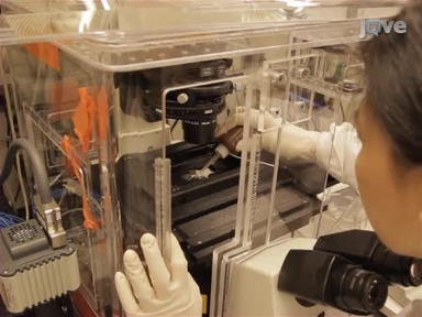
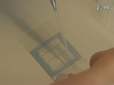
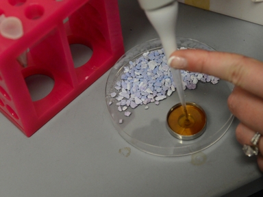
•We describe methods for live-cell video microscopy of Candida albicans phagocytosis by macrophages. These methods enable stage-specific analysis of macrophage migration, recognition, engulfment and phagosome maturation and reveal novel aspects of phagocytosis.
Tags
Related Videos

Direct Observation of Phagocytosis and NET-formation by Neutrophils in Infected Lungs using 2-photon Microscopy
Use of an Optical Trap for Study of Host-Pathogen Interactions for Dynamic Live Cell Imaging
Live Cell Imaging of Bacillus subtilis and Streptococcus pneumoniae using Automated Time-lapse Microscopy

Visualization of Bacterial Toxin Induced Responses Using Live Cell Fluorescence Microscopy

Isolation, Purification and Labeling of Mouse Bone Marrow Neutrophils for Functional Studies and Adoptive Transfer Experiments

Live Cell Imaging of Alphaherpes Virus Anterograde Transport and Spread

Assessing Anti-fungal Activity of Isolated Alveolar Macrophages by Confocal Microscopy

Loop-mediated Isothermal Amplification (LAMP) Assays for the Species-specific Detection of Eimeria that Infect Chickens
Biolistic Transformation of a Fluorescent Tagged Gene into the Opportunistic Fungal Pathogen Cryptococcus neoformans

A New Method for Qualitative Multi-scale Analysis of Bacterial Biofilms on Filamentous Fungal Colonies Using Confocal and Electron Microscopy
ABOUT JoVE
Copyright © 2024 MyJoVE Corporation. All rights reserved