
Preparation of CD4+ T Cells for Analysis of GD3 and GD2 Ganglioside Membrane Expression by Microscopy
November 8th, 2016
•We describe a standard antibody staining protocol for use in microscopy to determine the membrane expression and localization of gangliosides in resting and activated human naïve CD4+ T cells. Also described are real-time PCR experiments using <40,000 cells that do not require additional low input RNA kits.
Tags
Related Videos
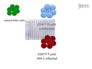
Preparation and Use of HIV-1 Infected Primary CD4+ T-Cells as Target Cells in Natural Killer Cell Cytotoxic Assays
Accelerated Type 1 Diabetes Induction in Mice by Adoptive Transfer of Diabetogenic CD4+ T Cells

Adenoviral Transduction of Naive CD4 T Cells to Study Treg Differentiation
Isolation and Th17 Differentiation of Naïve CD4 T Lymphocytes
Assessing the Innate Sensing of HIV-1 Infected CD4+ T Cells by Plasmacytoid Dendritic Cells Using an Ex vivo Co-culture System.
In Situ Detection of Autoreactive CD4 T Cells in Brain and Heart Using Major Histocompatibility Complex Class II Dextramers
Induction of Murine Intestinal Inflammation by Adoptive Transfer of Effector CD4+CD45RBhigh T Cells into Immunodeficient Mice
Analysis of Yersinia enterocolitica Effector Translocation into Host Cells Using Beta-lactamase Effector Fusions
Imaging CD4 T Cell Interstitial Migration in the Inflamed Dermis

Cortical Actin Flow in T Cells Quantified by Spatio-temporal Image Correlation Spectroscopy of Structured Illumination Microscopy Data
ABOUT JoVE
Copyright © 2024 MyJoVE Corporation. All rights reserved