
Reconstruction of Single-Cell Innate Fluorescence Signatures by Confocal Microscopy
May 27th, 2020
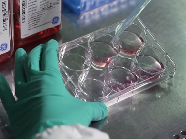
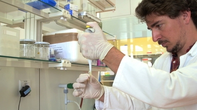
•Here, a protocol is presented for optically extracting and cataloging innate cellular fluorescence signatures (i.e., cellular autofluorescence) from every individual live cell distributed in a three-dimensional space. This method is suitable for studying the innate fluorescence signature of diverse biological systems at a single-cell resolution, including cells from bacteria, fungi, yeasts, plants, and animals.
Tags
Related Videos

Live Imaging of the Zebrafish Embryonic Brain by Confocal Microscopy

Time-lapse Imaging of Mitosis After siRNA Transfection

Fluorescence-based Measurement of Store-operated Calcium Entry in Live Cells: from Cultured Cancer Cell to Skeletal Muscle Fiber

In vivo Clonal Tracking of Hematopoietic Stem and Progenitor Cells Marked by Five Fluorescent Proteins using Confocal and Multiphoton Microscopy

In Vivo 4-Dimensional Tracking of Hematopoietic Stem and Progenitor Cells in Adult Mouse Calvarial Bone Marrow
Detection of Ligand-activated G Protein-coupled Receptor Internalization by Confocal Microscopy
Isolation and Fluorescence Imaging for Single-particle Reconstruction of Chlamydomonas Centrioles

Quantification of Interbacterial Competition using Single-Cell Fluorescence Imaging

Isolation and Transcriptome Analysis of Plant Cell Types

Imaging of mtHyPer7, a Ratiometric Biosensor for Mitochondrial Peroxide, in Living Yeast Cells
ABOUT JoVE
Copyright © 2024 MyJoVE Corporation. All rights reserved