Method Article
Obtenir un A avec les 3C: Capture de conformation chromosomique pour les étudiants de premier cycle
Dans cet article
Résumé
Ici, nous présentons une adaptation de la technique de capture de conformation chromosomique (3C) en détail en mettant l’accent sur la participation et l’apprentissage au premier cycle.
Résumé
La capture de conformation chromosomique (3C) est un outil puissant qui a engendré une famille de techniques similaires (par exemple, Hi-C, 4C et 5C, appelées ici techniques 3C) qui fournissent des informations détaillées sur l’organisation tridimensionnelle de la chromatine. Les techniques 3C ont été utilisées dans un large éventail d’études, allant de la surveillance des changements dans l’organisation de la chromatine dans les cellules cancéreuses à l’identification des contacts d’amplificateur établis avec les promoteurs de gènes. Alors que de nombreuses études utilisant ces techniques posent de grandes questions à l’échelle du génome avec des types d’échantillons complexes (c.-à-d. analyse de cellules uniques), ce qui est souvent perdu, c’est que les techniques 3C sont fondées sur des méthodes de biologie moléculaire de base applicables à un large éventail d’études. En abordant des questions étroitement ciblées sur l’organisation de la chromatine, cette technique de pointe peut être utilisée pour améliorer l’expérience de laboratoire de recherche et d’enseignement de premier cycle. Cet article présente un protocole 3C et fournit des adaptations et des points d’accent pour la mise en œuvre dans les établissements principalement de premier cycle dans les expériences de recherche et d’enseignement de premier cycle.
Introduction
Le génome d’un organisme contient non seulement tous les gènes nécessaires à leur fonctionnement, mais aussi toutes les instructions sur la façon et le moment de les utiliser. Cela fait de la régulation de l’accès au génome l’une des fonctions les plus importantes de la cellule. Il existe de nombreux mécanismes pour contrôler la fonction des gènes; Cependant, à son niveau de base, la régulation des gènes se résume à la capacité des facteurs de transcription régulateurs (trans-facteurs) à se lier à leurs séquences d’ADN spécifiques (séquences cis-régulatrices). Ce n’est pas une capacité innée; Au lieu de cela, il est régi par l’organisation / structure du génome dans le noyau, qui contrôle la disponibilité / exposition des séquences cis-régulatrices aux transfacteurs 1,2,3. Si les transfacteurs ne peuvent pas trouver leurs séquences cis-régulatrices, alors les transfacteurs ne peuvent pas effectuer leurs tâches de régulation. Cela a fait de la compréhension de la façon dont les génomes sont organisés dans le noyau une source importante de recherche.
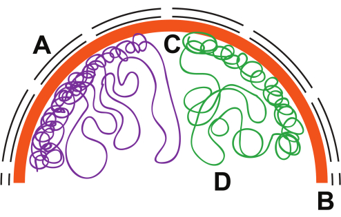
Il est largement admis que pendant l’interphase, les chromosomes eucaryotes du noyau occupent leur propre domaine ancré à la lame nucléaire et à la matrice nucléaire (Figure 1), faisant ainsi du chromosome plus une tranche de pizza, plutôt qu’une nouille sur une assiette de spaghettis. Les chromosomes sont partiellement condensés par des interactions protéine-ADN (chromatine) qui tordent et bouclent des parties du chromosome. Grâce à la microscopie électronique, à l’hybridation in situ par fluorescence tridimensionnelle de l’ADN (FISH) et aux techniques de marquage de l’ADN (c.-à-d. méthylation fluorescente et artificielle de l’ADN), on a constaté que les domaines inactifs de la chromatine étaient étroitement emballés le long de la périphérie nucléaire 4,5,6, tandis que des portions de chromatine active, moins condensée, se trouvent à l’intérieur du noyau 7,8,9, 10. Ces expériences fournissent une vue d’ensemble de la dynamique chromosomique, mais ne font pas grand-chose pour capturer les changements qui se produisent localement autour des promoteurs de gènes observés dans les études DNase11,12 et nucléosome13,14,15.
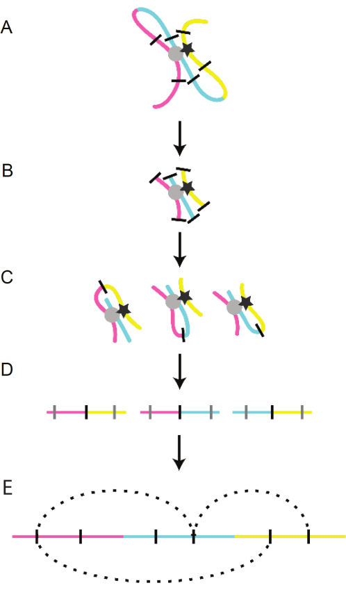
La clé pour déverrouiller la dynamique de la chromatine à plus haute résolution a été la formulation de la technique de cartographie chromosomique 3D, 3C. La technique 3C elle-même comprend quatre étapes principales: la réticulation de la chromatine, la digestion de la chromatine par des enzymes de restriction, la ligature de la chromatine et la purification de l’ADN (Figure 2). Les nouveaux fragments d’ADN artificiels générés par ce processus peuvent ensuite être caractérisés pour révéler l’association physique étroite entre des morceaux d’ADNlinéairement éloignés 16. La technique 3C est devenue la base de la création de multiples techniques dérivées qui utilisent les étapes initiales de 3C pour poser des questions plus larges à l’échelle du génome (par exemple, Hi-C, 4C, ChIP-C). Cette famille de techniques 3C a identifié que les chromosomes sont organisés en plusieurs unités discrètes appelées domaines topologiquement associés (TAD). Les TAD sont codés dans le génome et sont définis par des boucles de chromatine flanquées de frontières non bouclées16,17,18,19. Les limites du TAD sont maintenues par deux facteurs évolutifs conservés et omniprésents, y compris le facteur de liaison CCCT (CTCF) et la cohésion, qui empêchent les boucles au sein de TADs distincts d’interagir16,20. Les boucles sont médiées par l’interaction des transfacteurs avec leurs séquences régulatrices, ainsi que par le CTCF et la cohésion21.
Bien que de nombreuses études utilisant les technologies 3C posent des questions générales à l’échelle du génome et utilisent des techniques complexes de collecte d’échantillons, la formulation de la technique 3C est basée sur des techniques de biologie moléculaire de base. Cela rend 3C intrigant pour le déploiement dans les laboratoires de recherche et d’enseignement de premier cycle. La technique 3C peut être utilisée pour des questions ciblées plus petites et est intrinsèquement flexible pour la mise à l’échelle vers le haut ou vers le bas (gènes uniques22, chromosomes16 et / ou génomes18) en fonction de l’objectif et de la direction des questions posées. Cette technique a également été appliquée à une large gamme de systèmes modèles 7,16,19,23 et s’est avérée polyvalente dans son utilisation. Cela fait de 3C une excellente technique pour les étudiants de premier cycle en ce sens que les étudiants peuvent acquérir de l’expérience dans les techniques courantes de biologie moléculaire tout en acquérant une expérience précieuse dans la réponse aux questions dirigées.
Présenté ici est un protocole adapté pour la préparation de la bibliothèque 3C basé sur les protocolesprécédemment publiés 24,25,26,27. Ce protocole a été optimisé pour environ 1 × 10⁷ cellules, bien qu’il ait généré des bibliothèques 3C avec aussi peu que 1 × 10⁵ cellules. Ce protocole s’est avéré polyvalent et a été utilisé pour générer des bibliothèques 3C à partir d’embryons de poisson zèbre, de lignées cellulaires de poisson zèbre et de Caenorhabditis elegans (vers ronds) pour jeunes adultes (YA). Le protocole devrait également être approprié pour les lignées cellulaires de mammifères et, avec une adaptation supplémentaire, la levure.
L’objectif de ces adaptations est de rendre le 3C plus accessible aux étudiants de premier cycle. On a pris soin d’utiliser des techniques semblables à celles qui peuvent être accomplies dans un laboratoire d’enseignement de premier cycle. La technique 3C offre de nombreuses possibilités d’apprentissage aux étudiants de premier cycle pour apprendre les techniques de base de la biologie moléculaire qui bénéficieront à leur développement sur le banc, en classe et dans leurs efforts après l’obtention du diplôme.
Protocole
1. Conception de l’apprêt
REMARQUE: Les outils de conception des amorces 3C sont disponibles en ligne28. Alternativement, des amorces personnalisées peuvent être conçues par les étudiants (voir ci-dessous).
- Identification des emplacements des amorces
- Ouvrez le navigateur de génome UCSC (http://genome.ucsc.edu/), sélectionnez l’organisme à étudier et recherchez la région du génome à évaluer à l’aide de 3C.
- Dans la fenêtre suivante, activez la piste enzymatique dans l’onglet Mappage et séquençage sous le navigateur en cliquant sur Enzymes Retr.
- Entrez la ou les enzymes de restriction à utiliser et réglez le mode d’affichage sur pack. Cliquez sur Soumettre.
- Dans Variation et répétitions, assurez-vous que RepeatMasker est défini sur dense.
- En utilisant les sites de restriction comme guides, identifiez les sites d’intérêt qui ne se trouvent pas dans les régions de répétition masquées (barres noires). Mettez en surbrillance 300 paires de bases (pb) flanquant la séquence amont et aval entourant le site de restriction en cliquant sur la position (piste la plus haute) et en faisant glisser jusqu’à la longueur souhaitée (~600 bp).
- Relâchez la souris et une fenêtre contextuelle apparaîtra. Prenez note de l’emplacement génomique, cliquez sur Zoom avant et laissez le navigateur se réajuster à la sélection.
- Passez la souris sur l’onglet Affichage du ruban supérieur du navigateur, puis dans le menu déroulant, sélectionnez DNA.
- Laissez l’option par défaut, notez la désignation des séquences masquées (il existe une option pour en faire des N). Cliquez sur obtenir l’ADN.
- La séquence en surbrillance sera alors affichée dans la fenêtre. Copiez et collez cette séquence dans Primer3 (https://primer3.ut.ee/).
- En utilisant les paramètres par défaut d’Primer3, générez des amorces en cliquant sur Choisir les amorces.
- Dans les résultats, prenez note de l’emplacement de l’enzyme de restriction et sélectionnez des amorces qui créent un produit PCR de 200 à 500 pb avec le site de restriction situé au milieu.
- Après ces étapes, les amorces d’essai de conception sont situées à 1 kilobase (kb), 2 kb, 5 kb, 10 kb et 20 kb du ou des sites d’intérêt.
- Pour concevoir les amorces de contrôle d’entrée, répétez ces étapes pour un site situé à 1-2 Ko du site d’intérêt qui n’a pas de site de restriction, ce qui signifie qu’il n’est jamais coupé.
- Validation de l’amorce fonctionnelle
- Pour valider la fonctionnalité de l’amorce, configurez des réactions PCR en utilisant des concentrations d’amorce titrées et de l’ADN génomique purifié. Que ce soit par des moyens quantitatifs ou semi-quantitatifs, déterminez si les amorces créent le produit attendu.
- Reconcevez les amorces dont la validation échoue.
2. Jour 1
NOTE: Le protocole peut être mis en pause (congelé à -20 ° C) après la réticulation de la chromatine et après la collecte des noyaux. Les étapes prennent, en moyenne, 5-6 heures avec les étudiants de premier cycle.
- Collection de noyaux de C. elegans pour jeunes adultes (YA) (adapté de Han et al.29)
- Réticulation de la chromatine
- Prélever 5 000 vers YA dans 30 mL de milieu M9 dans un tube conique de 50 mL et faire tourner à 400 × g pendant 2 minutes à température ambiante (RT).
- Lavez la pastille de ver 3x plus avec M9 pour éliminer les bactéries.
- Retirer le surnageant, remettre en suspension la pastille de ver dans 47,3 mL de M9 contenant 2,7 mL de formaldéhyde à 37 % (2 % final) et incuber en agitant (berçage ou nutation) pendant 30 min à TA.
ATTENTION : Manipulez le formaldéhyde avec précaution et travaillez sous une hotte avec un équipement de protection individuelle approprié (blouse de laboratoire EPI, gants appropriés et protection oculaire). Le formaldéhyde est un irritant qui affecte les yeux, le nez, la gorge et les poumons. - Faire tourner les vers à 400 × g pendant 2 min à TA.
- Retirer le surnageant et remettre en suspension la pastille de ver dans 50 mL de glycine 1 M. Faire tourner les vers à 400 × g pendant 2 min à TA.
- Collection de noyaux
- Resuspendre la pastille de ver dans 6 mL de tampon NP réfrigéré (50 mM HEPES à pH 7,5, 40 mM NaCl, 90 mM KCl, 2 mM EDTA, 0,5mM EGTA, 0,1% Tween 20, 0,2 mM DTT, 0,5 mM de spermidine, 0,25 mM de spermine, 1x inhibiteur complet de la protéase).
- Transférer la suspension à vis sans fin dans un rebond lâche de 7 ml sur glace. Faire rebondir l’échantillon 15x et maintenir sur la glace pendant 5 min.
- Transférer la suspension à vis sans fin dans une rebondance hermétique de 7 ml sur glace. Faire rebondir l’échantillon 20x et maintenir sur la glace pendant 5 min.
- Transférer la suspension de vis sans fin dans un tube conique propre de 15 ml et ajouter le tampon NP à 10 ml au total (environ 4 ml).
- Vortex la suspension à vis sans fin sur un réglage élevé pendant 30 s. Incuber l’échantillon sur glace pendant 5 min.
- Répétez l’étape précédente.
- Faire tourner la suspension de vis sans fin à 100 × g pendant 5 min à 4 °C.
- Transférer le surnageant dans un tube conique frais de 15 mL.
- Vérifiez la présence de débris de vers dans l’échantillon. Visualisez 10 μL de l’échantillon avec un microscope optique. En présence de débris de vers, faire tourner l’échantillon à 2 000 × g pendant 5 min à 4 °C, remettre la pastille en suspension dans un tampon NP frais de 10 ml et faire tourner l’échantillon à nouveau à 100 × g pendant 5 min à 4 °C. Jeter la pastille, vérifier la présence de débris de vers dans le surnageant et répéter jusqu’à ce que l’échantillon soit exempt de débris de vers.
REMARQUE: Alternativement, les débris du ver peuvent également être éliminés en filtrant l’échantillon à travers une crépine cellulaire de 40 μm (6x) suivie d’une crépine cellulaire de 20 μm (6x). - Si l’échantillon est exempt de débris de vers, prélever 5 μL de l’échantillon, ajouter 5 μL de pyronine au vert de méthyle (les noyaux seront bleus) et compter les noyaux avec un hémocytomètre.
- Faire tourner l’échantillon à 2 000 × g pendant 5 min à 4 °C
- Retirer le surnageant, placer les échantillons sur de la glace et continuer; Sinon, congelez les noyaux et stockez-les à −80 °C.
- Réticulation de la chromatine
- Digestion de la chromatine
- Remettez les noyaux en suspension dans 450 μL d’eau propre. Transférer les noyaux dans un tube microcentrifuge propre de 1,5 mL.
- À l’échantillon réticulé, ajouter 60 μL de tampon enzymatique de restriction DpnII 10x et bien mélanger.
REMARQUE: D’autres enzymes de restriction qui coupent encore l’ADN réticulé peuvent être utilisées. - Ajouter 15 μL de dodécylsulfate de sodium (SDS) à 10 % pour perméabiliser les noyaux et incuber en agitant (berçage ou nutation) à 37 °C pendant 1 h.
ATTENTION : Les FDS sont irritantes et toxiques si elles sont ingérées ou absorbées par la peau. Manipulez avec précaution et utilisez l’EPI approprié (blouse de laboratoire, gants appropriés et protection oculaire). - Éteindre le SDS en ajoutant 75 μL de Triton X-100 à 20% et en incubant avec agitation à 37 °C pendant 1 h.
- Prélever 10 μL de l’échantillon comme témoin non digéré. Conserver à 4 °C.
- Ajouter 400 U de DpnII et incuber à 37 °C pendant une nuit en agitant.
3. Jour 2
REMARQUE: En moyenne, il faut 5 heures aux étudiants de premier cycle pour compléter ces étapes.
- Digestion de la chromatine
- Ajouter 200 U supplémentaires de DpnII et incuber les échantillons pendant 4 h à 37 °C en agitant pour assurer la digestion complète de l’échantillon réticulé.
- Prélever une partie aliquote de 10 μL des échantillons comme contrôle de la digestion.
- Inactiver l’enzyme de restriction par la chaleur en incubant l’échantillon pendant 20 min à 65 °C (ou selon les instructions du fabricant). Passez à l’étape 2.1.3.
- Si l’enzyme ne peut pas être inactivée par la chaleur, ajouter 80 μL de SDS à 10 % et incuber l’échantillon pendant 30 min à 65 °C. Ensuite, ajoutez 375 μL de Triton X-100 à 20% et mélangez en tourbillonnant. Incuber l’échantillon pendant 1 h à 37 °C et passer à l’étape 2.2.
- Ligature de la chromatine
- Transférer l’échantillon dans un tube conique propre de 50 mL, ajuster le volume de l’échantillon à 5,7 mL avec du H2O de qualité moléculaire et mélanger en agitant en agitant.
- Ajouter 700 μL de 10x tampon T4 Ligase et mélanger en tourbillonnant.
- Ajouter 60 U de T4 DNA Ligase et mélanger en tourbillonnant.
- Incuber pendant une nuit à 16 °C.
4. Jour 3
REMARQUE: En moyenne, il faut aux étudiants de premier cycle 15-30 minutes pour compléter ces étapes. Après la nuit d’incubation, les échantillons peuvent être congelés.
- Digestion des protéines et réticulation inverse
- Ajouter 30 μL de protéinase K (10 mg/mL) à l’échantillon 3C et incuber à 65 °C pendant une nuit en agitant. Ajouter 5 μL de protéinase K (10 mg/mL) au contrôle non digéré et digestif, et incuber à 65 °C pendant une nuit en agitant.
5. Jour 4
REMARQUE: En moyenne, il faut 4-5 heures aux étudiants de premier cycle pour compléter ces étapes.
- Purification de la bibliothèque 3C
- Ajouter 30 μL de RNase A (10 mg/mL) à l’échantillon 3C et agiter pour mélanger. Incuber l’échantillon pendant 45 min à 37 °C.
- Ajouter 7 mL de phénol-chloroforme à l’échantillon et mélanger en agitant.
MISE EN GARDE : Le phénol-chloroforme est irritant pour la peau et les yeux et peut causer des brûlures si le contact n’est pas traité. Le phénol-chloroforme doit être utilisé dans une hotte avec une protection oculaire appropriée et des gants (nitrile). - Centrifuger l’échantillon pendant 15 min à 3 270 × g à TA. Prélever la phase aqueuse et transférer dans un tube conique propre de 50 mL. Ajouter des volumes égaux de chloroforme et mélanger l’échantillon en agitant.
- Centrifuger l’échantillon pendant 15 min à 3 270 × g à TA. Prélever la phase aqueuse et transférer dans un tube conique propre de 50 mL. Ajouter 7,5 mL de qualité moléculaireH2O, 35 mL d’éthanol à 100 % et (facultatif) 7 μL de glycogène (1 mg/mL). Mélanger en agitant et incuber à −80 °C jusqu’à congélation de l’échantillon.
REMARQUE: Cela peut prendre 1 heure ou plus pour que l’échantillon gèle. Le protocole peut être mis en pause ici.- Pendant que l’échantillon 3C gèle, purifiez les échantillons témoins. Pour les échantillons témoins, ajuster le volume à 500 μL avec de l’eau de qualité moléculaire et ajouter 2 μL de mélange de RNase A (10 mg/mL) en agitant le tube.
- Tourner brièvement pour recueillir l’échantillon au fond du tube. Incuber les échantillons pendant 45 min à 37 °C.
- Aux échantillons témoins, ajouter 1 mL de phénol-chloroforme et mélanger en agitant les tubes. Centrifuger l’échantillon pendant 15 min à 3 270 × g à TA. Prélever la phase aqueuse des témoins et placer dans un tube microcentrifuge propre de 1,5 mL.
- Ajouter des volumes égaux de chloroforme et mélanger l’échantillon en agitant. Centrifuger l’échantillon pendant 15 min à 3 270 × g à TA. Prélever la phase aqueuse des témoins et placer dans un tube microcentrifuge propre de 1,5 mL.
- Ajouter 1 mL d’éthanol à 100 % et 2 μL de glycogène (1 mg/mL). Mélanger en agitant et incuber à −80 °C pendant 30 min.
- Centrifuger l’échantillon pendant 15 min à 3 270 × g à 4 °C. Retirez le surnageant et ajoutez 750 μL d’éthanol réfrigéré à 70 %.
- Centrifuger l’échantillon pendant 10 min à 3 270 × g à 4 °C. Retirer le surnageant et sécher les échantillons à l’air. Remettez la pastille en suspension dans 50 μL de qualité moléculaire H2O. Congelez ou passez à l’étape 6.
- Centrifuger l’échantillon pendant 60 min à 3 270 × g à 4 °C. Retirer le surnageant et ajouter 10 ml d’éthanol réfrigéré à 70 %. Perturber et briser la pastille d’ADN en secouant l’échantillon pour mélanger.
- Centrifuger l’échantillon pendant 30 min à 3 270 × g à 4 °C. Retirer le surnageant et laisser sécher partiellement l’échantillon à l’air libre à TA. Resuspendre la pastille dans 150 μL de Tris-HCl 10 mM (pH 7,5) en pipetant de haut en bas. C’est la « bibliothèque 3C ».
REMARQUE : Congelez l’échantillon ou passez à l’analyse.
6. Jour 5
REMARQUE: En moyenne, il faut aux étudiants de premier cycle 1-2 h pour compléter ces étapes.
- Détermination de la qualité de l’échantillon à l’aide de la courbe standard de l’amorce de contrôle
- Quantifier la concentration d’ADN de l’échantillon 3C ainsi que de tous les témoins, et ajuster les échantillons à 30 μg/μL (si les concentrations le permettent). En série, les échantillons dilués sont multipliés par deux fois quatre fois, ce qui donne cinq dilutions pour chaque échantillon (1x, 0,5x, 0,25x, 0,125x, 0,0625x).
REMARQUE : Si la quantification ne peut pas être facilement effectuée, diluer les échantillons en série comme indiqué ci-dessus et continuer. - Mettre en place des réactions PCR pour le 3C, le contrôle génomique et le contrôle digéré pour chaque échantillon dilué avec les amorces de contrôle : 1 μL d’ADN, 10 μL de tampon de réaction 5x, 1 μL de 10 mM de dNTP, 1 μL d’amorce de contrôle 10 mM (mélange avant et arrière), 1 μL de Taq polymérase et 36 μL d’eau.
- En suivant les instructions du logiciel spécifique à la machine de PCR, configurez le programme de PCR à courbe standard en utilisant les conditions de cyclage suivantes: 30 s à 98 °C; 30 cycles de 5 s à 98 °C, 5 s à 60 °C et 10 s à 72 °C; 1 min à 72 °C; Maintien à 4 °C.
- À l’aide du logiciel, générez les courbes standard pour les échantillons. Une fois que le logiciel a généré la courbe, prenez note de l’efficacité de la PCR et de la valeur R2 .
- Quantifier la concentration d’ADN de l’échantillon 3C ainsi que de tous les témoins, et ajuster les échantillons à 30 μg/μL (si les concentrations le permettent). En série, les échantillons dilués sont multipliés par deux fois quatre fois, ce qui donne cinq dilutions pour chaque échantillon (1x, 0,5x, 0,25x, 0,125x, 0,0625x).
- Détermination de la concentration d’ADN
- Pour déterminer la concentration d’ADN des échantillons 3C, comparez les échantillons à un échantillon d’ADN génomique de concentration connue. Diluer les échantillons à 30 ng/μL et diluer en série le témoin génomique de 10 ng/μL à 0,01 ng/μL en deux étapes pour créer une courbe standard.
REMARQUE : Si la quantification ne peut pas être facilement effectuée, diluer l’échantillon 1:10 et continuer. - Mettre en place les réactions PCR pour le contrôle 3C, le contrôle génomique et le contrôle digéré pour chaque échantillon dilué avec les amorces de contrôle : 1 μL d’ADN, 10 μL de tampon de réaction 5x, 1 μL de 10 mM de dNTP, 1 μL d’amorce de contrôle 10 mM (mélange avant et arrière), 1 μL de Taq polymérase et 36 μL d’eau.
- En suivant les instructions du logiciel spécifique à la machine de PCR, configurez le programme de PCR à courbe standard en utilisant les conditions de cyclage comme suit: 30 s à 98 °C; 30 cycles de 5 s à 98 °C, 5 s à 60 °C et 10 s à 72 °C; 1 min à 72 °C; Maintien à 4 °C.
- À l’aide du logiciel, générez les courbes standard pour les échantillons, en traçant les échantillons 3C sur la courbe. Comparer la position de l’abondance de l’échantillon 3C à la courbe standard créée par les réactions de l’ADN génomique de concentration connue, et ajuster les échantillons 3C à 30 ng / μL.
- Pour déterminer la concentration d’ADN des échantillons 3C, comparez les échantillons à un échantillon d’ADN génomique de concentration connue. Diluer les échantillons à 30 ng/μL et diluer en série le témoin génomique de 10 ng/μL à 0,01 ng/μL en deux étapes pour créer une courbe standard.
- Déterminer la présence ou l’absence d’interaction chromatine
- Mettre en place des réactions qPCR avec 30 ng de l’échantillon 3C, de l’échantillon de contrôle génomique et de l’échantillon témoin digéré : 1 μL d’ADN, 10 μL de tampon de réaction 5x, 1 μL de 10 mM de dNTP, 1 μL d’amorce 10 mM (mixte avant et arrière), 1 μL de Taq polymérase et 36 μL d’eau.
REMARQUE : Les réactions doivent être configurées à l’aide de l’amorce témoin, des amorces d’essai pour les loci génomiques individuels et des combinaisons souhaitées des amorces d’essai (site « a » avant et site « b » inverse) utilisées pour déterminer l’interaction chromatinique (figure 3). - En suivant les instructions du logiciel de la machine PCR, configurez le programme PCR comme suit: 30 s à 98 °C; 40 cycles de 5 s à 98 °C, 5 s à 60 °C et 10 s à 72 °C; 1 min à 72 °C; Maintien à 4 °C.
- Une fois la PCR exécutée, inspectez le graphique d’amplification. Assurez-vous que les réactions de PCR présentent une amplification exponentielle, doublant chaque cycle.
REMARQUE : Les réactions qui n’affichent pas d’amplification exponentielle ne peuvent pas être analysées plus avant. - En suivant les instructions pour le logiciel de la machine PCR, définissez le seuil de l’expérience qPCR.
- Exportez les valeurs Ct pour tous les échantillons.
- Déterminer l’efficacité de la digestion à l’aide des valeurs Ct de l’amorce d’essai (TP) et de l’amorce de contrôle (CP) à partir des échantillons témoins non digérés (UC) et des échantillons témoins digérés (DC) à l’aide de l’équation (1).
Pourcentage digéré = 100 - (1)
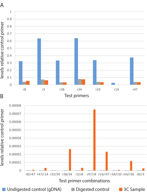
(1) - Ces valeurs devraient être comprises entre 80% et 90% de digestion. Notez ces valeurs (Figure 4A).
- Déterminer l’interaction relative de la chromatine à l’aide des valeurs Ct de la combinaison d’amorces d’essai (TPC) et de l’amorce témoin (CP) provenant des échantillons témoins non digérés (UC) et 3C (3C) à l’aide de l’équation (2).
Interaction chromatine = 100 - (2)
(2) - Tracez les valeurs de chaque échantillon sur un graphique à barres pour chaque combinaison d’amorces de test.
- Comparer le signal des échantillons 3C aux échantillons témoins pour déterminer si l’échantillon 3C a enrichi une interaction chromatine particulière sur les échantillons témoins. Les échantillons 3C qui montrent un enrichissement par rapport au contrôle peuvent être considérés comme conditionnellement positifs et nécessitent une validation à l’aide du séquençage de Sanger (Figure 4B).
REMARQUE : Lors de l’analyse des données du graphique, il est important de se rappeler qu’à moins d’utiliser un « modèle de contrôle » (voir la discussion), l’abondance entre les réactions (c.-à-d. d’un ensemble d’amorces à l’autre) ne peut être comparée.
- Mettre en place des réactions qPCR avec 30 ng de l’échantillon 3C, de l’échantillon de contrôle génomique et de l’échantillon témoin digéré : 1 μL d’ADN, 10 μL de tampon de réaction 5x, 1 μL de 10 mM de dNTP, 1 μL d’amorce 10 mM (mixte avant et arrière), 1 μL de Taq polymérase et 36 μL d’eau.
- Identification des produits 3C
- Exécutez les produits PCR sur un gel d’agarose à 1,5%.
- Visualisez le gel pour la taille correcte des produits PCR à l’aide d’une boîte de lumière UV ou d’un système de documentation de gel.
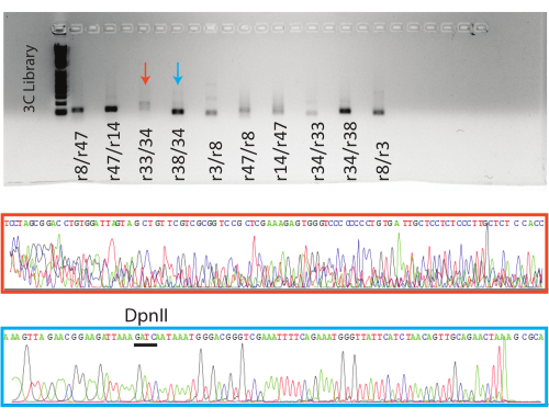
REMARQUE : Des bibliothèques 3C bien construites contiendront une gamme variée de fragments d’ADN, et malgré la validation préalable des amorces pour la spécificité, les réactions de PCR des bibliothèques 3C ont le potentiel de contenir de nombreuses bandes (Figure 5A). Cela n’empêche pas l’analyse des bibliothèques 3C. - Urinez les bandes de produits correspondant à la taille de fragment prévue et effectuez l’extraction du gel des produits PCR en suivant les instructions d’une trousse commerciale ou le protocole maison décrit ci-dessous.
ATTENTION: La lumière UV est un cancérogène connu, et une grande prudence doit être prise pour limiter le temps d’exposition à la lumière UV. Une protection oculaire résistante aux UV, un écran de spécimen, des gants et une blouse de laboratoire doivent être portés.- Pour construire une cartouche de purification maison, percez un trou dans le fond d’un tube de 0,5 ml avec une aiguille.
- Emballez une petite quantité de coton d’une boule de coton dans le fond du tube de 0,5 mL, en ne remplissant pas plus de la moitié du tube.
- Placer le tube de 0,5 mL dans un tube de 1,5 mL; Assurez-vous que le plus petit tube repose sur la lèvre du plus grand tube, et non dans le tube.
- Coupez soigneusement un fragment de gel en petits morceaux et placez-le dans le tube de 0,5 ml de la cartouche.
- Placer l’ensemble dans un congélateur à −20 °C pendant 5 min. Faire tourner l’ensemble pendant 3 min à 13 000 × g à TA.
- Conservez le tube de 1,5 mL contenant l’ADN extrait dans un tampon et jetez le tube de 0,5 mL contenant les débris d’agarose.
- L’ADN purifié peut être envoyé pour le séquençage de Sanger en utilisant des amorces avant et arrière pour le fragment 3C.
- Identification des contacts de chromatine à l’aide de Blat
- Une fois le séquençage terminé, déterminer si les échantillons répondent aux normes de contrôle de la qualité indiquées dans le rapport de séquençage.
- Pour les séquences passant le contrôle qualité, ouvrez les fichiers .seq et .ab1 (trace) dans un programme d’édition de séquençage tel qu’Another Plasmid Editor (ApE), inspectez le fichier .ab1 pour détecter les pics d’appel de base clairs et modifiez les pics mal appelés dans le fichier .seq.
- À l’aide du fichier .seq modifié, recherchez le site DpnII. Selon le résultat de la séquence, cela devrait être à mi-chemin dans la séquence rapportée.
- Pour déterminer si la séquence est la séquence cible attendue, mettez en évidence le site DpnII et 30-50 pb supplémentaires de la séquence en amont correspondant à l’amorce avant des loci génomiques testés pour 3C. Effectuez une recherche Blat de cette séquence par rapport à l’espèce cible. Assurez-vous que les loci génomiques correspondent à ceux de l’amorce.
- Répétez l’étape ci-dessus pour la séquence d’amorce inverse, en vous assurant que le locus génomique renvoyé correspond à celui de l’amorce inverse.
Résultats
Cette procédure produira un échantillon expérimental de 3C et deux échantillons témoins (non digérés et digérés). À l’aide de ces trois échantillons, la qPCR a été réalisée. À partir de ces résultats, l’efficacité de la digestion a été calculée (équation 1) et enregistrée (tableau 1). À partir de ces calculs, il a été déterminé que l’échantillon 3C avait une efficacité de digestion d’environ 88% (moyenne du tableau 1) sur les sept loci génomiques testés.
Ensuite, les échantillons ont été testés pour la présence de contacts chromatiniques à longue distance entre les différents loci génomiques en utilisant des combinaisons d’amorces spécifiques aux loci (tableau 2) et qPCR. À l’aide de ces résultats, les abondances de produits par rapport à un ensemble d’amorces témoins ont été calculées (équation 2) et représentées graphiquement à des fins de comparaison (figure 4). Ces données indiquaient que 8 des 10 réactions étaient conditionnelles positives pour les interactions à longue distance.
Les réactions PCR ont ensuite été exécutées sur un gel d’agarose. Le produit PCR attendu pour les huit positifs conditionnels et une réaction négative ont été purifiés sur gel et envoyés pour le séquençage de Sanger. Les résultats pour les réactions positives représentatives (flèche bleue, case bleue) et négatives (flèche rouge, case rouge) sont présentés (figure 5).

Figure 1 : Structure chromosomique dans le noyau. Organisation chromosomique hypothétique à l’intérieur du noyau. A) Enveloppe nucléaire, lignes noires; B) lame nucléaire, orange; (C) hétérochromatine, lignes compactées; (D) euchromatine, boucles lâches. Veuillez cliquer ici pour voir une version agrandie de cette figure.

Figure 2 : Schéma du protocole 3C. Des portions éloignées de chromosomes linéaires (bleu, jaune et rose) sont rapprochées dans le noyau par des boucles de régulation. (A) Les structures de la boucle sont médiées par des facteurs de transcription (cercle gris et étoile noire); Ces interactions sont préservées grâce à la réticulation chimique. (B) Les boucles sont brisées par digestion enzymatique (lignes noires). (C) Les morceaux de chromatine distants sont ligaturés ensemble par les extrémités collantes créées par la digestion. (D) L’ADN est purifié à partir de protéines. (E) La séquence dans les fragments est identifiée et cartographiée jusqu’au génome. Veuillez cliquer ici pour voir une version agrandie de cette figure.

Figure 3 : Le schéma d’amorce 3C. Les amorces 3C sont conçues autour de sites de restriction à différentes distances de l’emplacement génomique d’intérêt. Les flèches roses représentent les ensembles d’amorces expérimentales entourant un site DpnII. Les amorces expérimentales peuvent être mélangées et appariées pour évaluer la boucle de chromatine dans la région. Les amorces orange représentent un contrôle négatif sans site DpnII. Veuillez cliquer ici pour voir une version agrandie de cette figure.

Figure 4 : Données représentatives de qPCR pour une expérience 3C. Abondance relative des ensembles d’amorces d’essai 3C. (A) Graphique témoin montrant l’abondance relative du produit dans l’échantillon témoin non digéré (bleu), témoin digéré (gris) et 3C (orange). B) L’expérience 3C; l’abondance relative du produit provenant de combinaisons d’amorces d’essai dans l’échantillon témoin non digéré (bleu), témoin digéré (gris) et 3C (orange). Veuillez cliquer ici pour voir une version agrandie de cette figure.

Figure 5 : Visualisation des produits 3C qPCR. En haut, gel avec les produits de point final qPCR avec les échantillons indiqués. En bas, des fichiers de trace de séquence de Sanger représentatifs pour les réactions indiquées. L’échantillon orange est un exemple de faux positif (voir Figure 4, r38/34), et l’échantillon bleu est un exemple de vrai positif avec le site DpnII indiqué au-dessus de la trace. Veuillez cliquer ici pour voir une version agrandie de cette figure.
| Site | % de digestion |
| R8 | 86.98 |
| R3 | 88.44 |
| R38 | 89.64 |
| R34 | 87.55 |
| R33 | 87.85 |
| R14 | 86.97 |
| R47 | 89.45 |
Tableau 1 : Efficacité de digestion calculée.
| Nom | Séquence | Loci génomiques | ||
| r3 FWD | ACGCAAGTAAAATTCTGGTTTTTGACC | chrX:11361475 | ||
| r3 RVS | TTTCCTGAGCTCTAACCATGTTTGC | chrX:11361561 | ||
| r38 FWD | TTACTTCTGAAGTAATCTTTTCTTATCCCC | chrX:5859700 | ||
| r38 RVS | AGACGAGCTGATTAAAAGTAGTTGAGAG | chrX:5859775 | ||
| r34 FWD | ATTTGTGGATTGCGTGGAGACG | chrX:5429702 | ||
| r34 RVS | AATAATCCTCTTAACAAACGTGGCC | chrX:5429777 | ||
| r33 FWD | AAGAGTTGTCCAAAATAAATTGAGCTAAC | chrX:6296704 | ||
| r33 RVS | TTCAGAAAAGTAAACTTTGACTTGGAACG | chrX:6296807 | ||
| r14 FWD | AATTATCGATTTTTCCATCGCAG | chrX:8036367 | ||
| r14 RVS | ATTTCAATGAAAATGTAAAAATGTTCCTTC | chrX:8036427 | ||
| r47 FWD | ATCTAGACTTGATAATATTTGTGTGTCCTC | chrX:9464939 | ||
| r47 RVS | AAGTTCTGCAACTGTTAGATGAATAACAC | chrX:9465064 | ||
| r8 FWD | GAGAATGTTGTTCTGTAACTGAAAACTTG | chrX:11094257 | ||
| r8 RVS | TTACGAAATTTGGTAGTTTTGGACC | chrX:11094362 | ||
| Amorce de contrôle FWD | CAATCGTCTCGCTCACTTGTC | chrX:7608049 | ||
| Amorce de contrôle RVS | GATGTGAGCAACAAGGCACC | chrX:7608166 | ||
Tableau 2 : Amorces pour l’expérience représentative 3C.
Discussion
Le 3C est une technique puissante qui est enracinée dans les techniques moléculaires de base. C’est cette base d’outils fondamentaux qui fait de 3C une technique si intrigante à utiliser avec les étudiants de premier cycle. Avec autant d’études récentes observant la dynamique de la chromatine à une si grande échelle, l’utilisation de ces résultats pour concevoir une expérience étroitement ciblée sur un seul gène ou une seule région génomique a le potentiel de créer une expérience unique et percutante dans la recherche de premier cycle. Souvent, de telles expériences sont considérées comme trop avancées pour les étudiants de premier cycle, mais avec une planification minutieuse, elles sont facilement réalisables. Il est important de noter que les tests conçus pour sonder les connexions chromatines capturées par la bibliothèque 3C peuvent varier de la PCR semi-quantitative au séquençage du génome entier. En fait, les données du premier article3C 16 ont été générées à partir de qPCR. Cette large gamme de tests peut tous être utilisés parce que toutes les technologies 3C produisent le même produit - une bibliothèque de fragments d’ADN représentant des connexions 3D dans le noyau.
Présenté ici est une adaptation d’un protocole plus flexible et accommodant qui convient mieux aux chercheurs de premier cycle. Les périodes de pause énumérées ci-dessus impliquent des retards de nuit; Cependant, ces pauses peuvent s’étendre sur les week-ends et, dans le cas des cellules et des noyaux, pendant des semaines. La considération la plus cruciale est le moment où le travail sera terminé. Souvent, dans les protocoles, il y a des étapes urgentes où la pause n’est pas une option. En dehors de quelques points (jour 1 et jour 2), il existe de nombreux endroits pour s’arrêter et congeler l’échantillon. Ceux-ci sont essentiels lorsque vous travaillez avec des étudiants de premier cycle où les horaires et les horaires des travaux de laboratoire doivent être flexibles. En plus d’intégrer ces arrêts dans le protocole, les étudiants de premier cycle sont encouragés à travailler en paires ou même en petits groupes de trois ou quatre. Les groupes fonctionnent bien pour ce protocole, car les étudiants peuvent se soutenir mutuellement et créer un système de jumelage pour que tout le monde travaille en toute sécurité. Le travail de laboratoire est également plus amusant avec les autres personnes impliquées. Avec les groupes, les étudiants peuvent également travailler sur une variété de questions axées sur l’organisation de la chromatine tout en exécutant le même protocole. Ainsi, même si les étudiants travaillent sur des projets distincts, le protocole relie leurs efforts et, de ce fait, ils peuvent se soutenir mutuellement.
D’autres adaptations visent à contourner le fait que certains outils et équipements spécialisés ne se trouvent pas nécessairement dans tous les établissements de premier cycle. Ces pièces d’équipement comprennent, sans toutefois s’y limiter, les thermocycleurs qPCR, les systèmes de documentation sur gel et les spectromètres de nanovolume. En effet, ces équipements sont pratiques mais ne sont pas une exigence. Ici, la méthode classique de 3C est également décrite dans la partie de conception de l’apprêt; Cela implique d’identifier un locus génomique d’intérêt et, à partir de là, d’évaluer d’autres loci génomiques pour les points de contact de la chromatine plus éloignés. Cette technique fonctionne également bien si un ensemble de données publié est utilisé, tel qu’un ensemble de données utilisant Hi-C, où des loci positifs connus (connectants) et négatifs (non connectés) sont identifiés. La conception d’expériences utilisant ces ensembles de données publiés est une autre excellente adaptation pour les laboratoires d’enseignement, car les chances d’identification réussie des connexions chromatiniques sont généralement plus grandes. De plus, l’article de recherche peut être discuté en classe et servir de référence.
Ce protocole utilise une approche qPCR modifiée pour visualiser la formation du produit 3C. Les contrôles sont essentiels au succès de la technique 3C. Chaque expérience utilise à la fois des contrôles d’échantillon et des contrôles d’amorce pour déterminer l’achèvement de la procédure 3C. Les contrôles de l’échantillon comprennent un contrôle non digéré (ADN génomique) et un contrôle digéré. Le contrôle non digéré détermine le signal de base pour les ensembles d’amorces et est utilisé avec le contrôle de digestion réticulé pour déterminer l’efficacité de la digestion. On s’attend à ce qu’il y ait une baisse de produit pour tous les apprêts dirigés à travers un site de restriction. La comparaison de cette valeur avec le témoin non digéré donne une indication de la façon dont l’échantillon a été digéré.
Les amorces pour la PCR comprennent une amorce de contrôle et des amorces de test. L’amorce de contrôle est un ensemble d’amorces qui se trouve près de la région génomique à tester et qui ne contient pas de site de restriction. Cela fournit la base de référence pour déterminer l’abondance des produits PCR d’amorce d’essai. Les amorces d’essai sont des amorces avant et arrière qui flanquent un site de restriction pour un locus génomique d’intérêt particulier (Figure 3). Les réactions utilisant ces jeux d’amorces sont comparées pour déterminer l’efficacité de la digestion, car l’abondance du produit devrait diminuer si le site de restriction a été coupé. Pour déterminer l’organisation de la chromatine, une amorce de test d’un locus est associée à une autre amorce de test d’un locus génomique différent pour déterminer si ces deux loci sont proches l’un de l’autre dans l’espace 3D. Dans ce cas, on s’attend à ce qu’un produit de PCR ne soit trouvé qu’en utilisant l’échantillon 3C comme modèle.
Il est important de noter que même les amorces validées ont tendance à échouer (Figure 4 : jeu d’amorces r14). En outre, les produits de PCR sont fréquemment identifiés dans les réactions de contrôle et dans les réactions dans lesquelles une connexion de chromatine n’est pas prévue (comme le témoin digéré, puisqu’il n’est pas ligaturé). Ces instances sont séquencées et échouent à Sanger QC ou reviennent sans séquence définie (Figure 5). De plus, les expériences 3C traditionnelles génèrent un « modèle de contrôle », un échantillon d’ADN non réticulé, digéré et ligaturé, qui représente tous les fragments ligaturés possibles pouvant être produits avec une quantité donnée d’ADN. Le « modèle de contrôle » joue un rôle important dans la comparaison des intensités des signaux qPCR entre deux loci génomiques afin de déterminer si le signal représente une véritable interaction ou simplement une association aléatoire. La création d’un « modèle de contrôle » peut être problématique, car une grande partie de la chromatine analysée doit être capturée sous la forme d’un chromosome artificiel et traitée avec les échantillons 3C. Sécuriser une telle construction peut ne pas être faisable, et en créer une pourrait sortir de la portée d’un projet semestriel. En raison de ces difficultés, nous suggérons d’utiliser un abécédaire de contrôle. L’introduction au contrôle ne remplace pas toutes les fonctionnalités du « modèle de contrôle », mais offre tout de même la possibilité d’analyser les données pour déterminer la « présence » ou l'« absence ».
Lors de la qPCR, il est important d’utiliser des quantités égales de l’échantillon. Cela devrait être déterminé, même en utilisant un nano-spectrophotomètre tel qu’une nanogoutte, en générant une courbe standard à partir d’ADN génomique d’une concentration connue et en ajustant les échantillons 3C à cette ligne. Ces montants doivent être enregistrés et utilisés dans les PCR ultérieures. La qualité de la réaction PCR est également importante. Comme la PCR s’exécute en qPCR, l’abondance du produit est mesurée par fluorescence et enregistrée. Cet enregistrement est accessible dans le tracé d’amplification. Une fois le programme terminé, il est important de vérifier le graphique d’amplification et de s’assurer que les réactions (à l’exception des contrôles sans modèle) ont trois phases: une ligne de base, une exponentielle et une phase de plateau/saturation. Il est important de vérifier que les réactions ont une phase exponentielle, en particulier pour fixer le seuil (voir ci-dessous). De plus, pour les échantillons dilués en série, il devrait y avoir un changement dans les valeurs Ct correspondant à la dilution de l’échantillon (la concentration la plus élevée aura les valeurs Ct les plus faibles, tandis que la concentration la plus faible aura les valeurs Ct les plus élevées). Les échantillons qui ne reflètent pas ce changement dans la place d’amplification nécessitent une nouvelle dilution ou indiquent un problème plus important avec la formation de l’échantillon 3C. Enfin, tout en générant la courbe standard, le logiciel de PCR calculera l’efficacité de la PCR et la valeurR2 . L’efficacité de la PCR doit être supérieure à 90 % et la valeur de R2 doit être supérieure à 0,99. Si l’une ou l’autre de ces conditions n’est pas remplie, il est probable que quelque chose ne va pas avec l’échantillon ou les amorces PCR.
Après qPCR, le pourcentage de digestion et la présence d’interactions 3C peuvent être calculés en utilisant la qPCR Ct pour chaque réaction. Pour les déterminer, il faut d’abord définir le seuil de la réaction PCR. Cela se fait normalement à l’aide du logiciel fourni avec la machine qPCR. L’établissement du seuil définira la concentration du produit PCR qui sera utilisé pour comparer les valeurs Ct de l’échantillon. Le seuil doit couper en deux les courbes d’amplification des réactions de PCR dans la phase exponentielle d’amplification. Seules les réactions PCR avec amplification exponentielle peuvent être comparées (dans ce cas, les amorces de contrôle et les réactions d’amorce de test), car c’est le seul moyen de s’assurer que les réactions amplifient l’ADN au même rythme et peuvent être comparées fidèlement. Lors de l’analyse des graphiques 3C, les réactions conditionnellement positives sont identifiées comme celles avec plus de produit que les échantillons témoins, le contrôle génomique et le contrôle de la digestion (Figure 4B). Cependant, ces échantillons doivent être validés davantage à l’aide du séquençage de Sanger après la purification du gel du produit PCR.
Après le séquençage de Sanger, les échantillons qui passent le QC peuvent être analysés à l’aide de Blat. Le but de cette analyse est de déterminer si l’échantillon a la séquence des deux loci génomiques cibles flanquant le site de restriction (DpnII dans le cas de ce protocole). Si les deux séquences sont identifiées, le fragment 3C peut être considéré comme validé. Si les résultats du Blat ne renvoient pas la séquence attendue, cela peut indiquer que l’une ou les deux amorces ne sont pas optimales, ce qui entraîne un résultat qPCR faussement positif. Les fichiers de trace des échantillons faussement positifs auront des pics de base non définis et les rapports Seq contiendront principalement « n » appels de base.
La validation de Sanger est essentielle, car les faux positifs de la formation artificielle de produits PCR sont possibles. Ces faux positifs peuvent être identifiés lorsque les produits de séquençage n’ont pas la séquence cible attendue ou un site DpnII caractéristique d’un fragment 3C approprié (Figure 5). Le séquençage des fragments de PCR fournit également un autre point de données pour l’expérience et fait comprendre aux étudiants que la technique 3C identifie des loci génomiques distants qui se rassemblent dans l’espace 3D dans les noyaux.
La technique 3C fournit une multitude de techniques moléculaires fondamentales pour les étudiants de premier cycle dans une procédure flexible et simple. Cette technique 3C est également un point de départ pour les autres techniques 3C qui intègrent le séquençage de nouvelle génération (NGS). Ces types d’expériences peuvent exposer les étudiants de premier cycle à des aspects importants de la bioinformatique et sont enracinés dans les principes de base décrits ici. L’expérience et la participation au premier cycle sont essentielles à leur succès et à leur développement en tant que jeunes scientifiques. En offrant ces opportunités, les étudiants de premier cycle peuvent renforcer leur compréhension des principes de base tout en renforçant leur confiance pour aborder des techniques et des questions de pointe.
Déclarations de divulgation
Les auteurs ne déclarent aucun conflit d’intérêts.
Remerciements
Ce travail a été soutenu en partie par le Rhode Island Institutional Development Award (IDeA) Network of Biomedical Research Excellence du National Institute of General Medical Sciences des National Institutes of Health sous le numéro de subvention P20GM103430 et le Bryant Center of Health and Behavioral Sciences.
matériels
| Name | Company | Catalog Number | Comments |
| 37% Formaldehyde | Millapore-Sigma | F8775 | |
| 100% Ethanol | Millapore-Sigma | E7023 | |
| CaCl2 | MP Biomedical | 215350280 | |
| chloroform | Millapore-Sigma | C0549 | |
| cOmplete, EDTA-free Protease Inhibitor Cocktail | Millapore-Sigma | COEDTAF-RO | mixed to 50x in water. Diluted to 1x in Sucrose buffer and GB buffer fresh |
| Dithiothreitol (DTT) | Millapore-Sigma | D0632 | 1 M stock diluted to 500 µM in Sucrose buffer and GB buffer fresh |
| DpnII | NEB | R0543M | |
| Glycerol | Millapore-Sigma | G9012 | |
| glycine | Millapore-Sigma | G8898 | |
| glycogen | Millapore-Sigma | 10901393001 | |
| HEPES | Millapore-Sigma | H3375 | |
| KCl | Millapore-Sigma | P3911 | |
| KH2PO4 | Millapore-Sigma | P5655 | |
| methyl green pyronin | Millapore-Sigma | HT70116 | |
| MgAc2 | Thermoscientific | 1222530 | |
| Na2HPO4 | Millapore-Sigma | S5136 | |
| NaCl | Millapore-Sigma | S9888 | |
| phenol-chloroform | Millapore-Sigma | P3803 | |
| Pronase | Millapore-Sigma | 11459643001 | |
| Proteinase K | IBI Scientific | IB05406 | |
| qPCR Ready mix (Phire Taq etc) | Millapore-Sigma | KCQS07 | |
| RNase A | Millapore-Sigma | R6148 | |
| Sodium Acetate | Millapore-Sigma | S2889 | |
| sodium dodecyl sulfate (SDS) | Millapore-Sigma | L3771 | |
| Sucrose | Millapore-Sigma | S0389 | |
| T4 DNA Ligase | Promega | M1804 | |
| Tris-HCl | Millapore-Sigma | 108319 | |
| Triton X-100 | Millapore-Sigma | T9284 | |
| Trypsin-EDTA | Millapore-Sigma | T4049 |
Références
- McBryant, S. J., Adams, V. H., Hansen, J. C. Chromatin architectural proteins. Chromosome Research. 14 (1), 39-51 (2006).
- Nalabothula, N., et al. The chromatin architectural proteins HMGD1 and H1 bind reciprocally and have opposite effects on chromatin structure and gene regulation. BMC Genomics. 15, 92(2014).
- John, S., et al. Chromatin accessibility pre-determines glucocorticoid receptor binding patterns. Nature Genetics. 43 (3), 264-268 (2011).
- Fawcett, D. W. On the occurrence of a fibrous lamina on the inner aspect of the nuclear envelope in certain cells of vertebrates. The American Journal of Anatomy. 119 (1), 129-145 (1966).
- Reddy, K. L., Zullo, J. M., Bertolino, E., Singh, H. Transcriptional repression mediated by repositioning of genes to the nuclear lamina. Nature. 452 (7184), 243-247 (2008).
- Finlan, L. E., et al. Recruitment to the nuclear periphery can alter expression of genes in human cells. PLoS Genetics. 4 (3), e1000039(2008).
- Chambeyron, S., Da Silva, N. R., Lawson, K. A., Bickmore, W. A. Nuclear re-organisation of the Hoxb complex during mouse embryonic development. Development. 132 (9), 2215-2223 (2005).
- Chambeyron, S., Bickmore, W. A. Chromatin decondensation and nuclear reorganization of the HoxB locus upon induction of transcription. Genes and Development. 18 (10), 1119-1130 (2004).
- Mahy, N. L., Perry, P. E., Gilchrist, S., Baldock, R. A., Bickmore, W. A. Spatial organization of active and inactive genes and noncoding DNA within chromosome territories. The Journal of Cell Biology. 157 (4), 579-589 (2002).
- Mahy, N. L., Perry, P. E., Bickmore, W. A. Gene density and transcription influence the localization of chromatin outside of chromosome territories detectable by FISH. The Journal of Cell Biology. 159 (5), 753-763 (2002).
- Li, B., Carey, M., Workman, J. L. The role of chromatin during transcription. Cell. 128 (4), 707-719 (2007).
- Chen, A., Chen, D., Chen, Y. Advances of DNase-seq for mapping active gene regulatory elements across the genome in animals. Gene. 667, 83-94 (2018).
- Hughes, A. L., Rando, O. J. Mechanisms underlying nucleosome positioning in vivo. Annual Review of Biophysics. 43, 41-63 (2014).
- Weiner, A., Hughes, A., Yassour, M., Rando, O. J., Friedman, N. High-resolution nucleosome mapping reveals transcription-dependent promoter packaging. Genome Research. 20 (1), 90-100 (2010).
- Hughes, A. L., Jin, Y., Rando, O. J., Struhl, K. A functional evolutionary approach to identify determinants of nucleosome positioning: A unifying model for establishing the genome-wide pattern. Molecular Cell. 48 (1), 5-15 (2012).
- Dekker, J., Rippe, K., Dekker, M., Kleckner, N. Capturing chromosome conformation. Science. 295 (5558), 1306-1311 (2002).
- van Berkum, N. L., Dekker, J. Determining spatial chromatin organization of large genomic regions using 5C technology. Methods in Molecular Biology. 567, 189-213 (2009).
- Smith, E. M., Lajoie, B. R., Jain, G., Dekker, J. Invariant TAD boundaries constrain cell-type-specific looping interactions between promoters and distal elements around the CFTR locus. American Journal of Human Genetics. 98 (1), 185-201 (2016).
- Crane, E. E. Two inputs into C. elegans dosage compensation: Chromosome conformation and the miRNA-specific argonaute ALG-2. University of California, Berkeley. , PhD thesis (2012).
- Bell, A. C., West, A. G., Felsenfeld, G. The protein CTCF is required for the enhancer blocking activity of vertebrate insulators. Cell. 98 (3), 387-396 (1999).
- Cuadrado, A., et al. Specific contributions of cohesin-SA1 and cohesin-SA2 to TADs and polycomb domains in embryonic stem cells. Cell Reports. 27 (12), 3500-3510 (2019).
- Sarro, R., et al. Disrupting the three-dimensional regulatory topology of the Pitx1 locus results in overtly normal development. Development. 145 (7), (2018).
- Tolhuis, B., et al. Interactions among polycomb domains are guided by chromosome architecture. PLoS Genetics. 7 (3), e1001343(2011).
- Splinter, E., de Wit, E., van de Werken, H. J. G., Klous, P., de Laat, W. Determining long-range chromatin interactions for selected genomic sites using 4C-seq technology: From fixation to computation. Methods. 58 (3), 221-230 (2012).
- Fernández-Miñán, A., Bessa, J., Tena, J. J., Gómez-Skarmeta, J. L. Chapter 21 - Assay for transposase-accessible chromatin and circularized chromosome conformation capture, two methods to explore the regulatory landscapes of genes in zebrafish. Methods in Cell Biology. 135, 413-430 (2016).
- Dekker, J. The three "C" s of chromosome conformation capture: controls, controls, controls. Nature Methods. 3 (1), 17-21 (2006).
- Hagège, H., et al. Quantitative analysis of chromosome conformation capture assays (3C-qPCR). Nature Protocols. 2 (7), 1722-1733 (2007).
- Lajoie, B. R., van Berkum, N. L., Sanyal, A., Dekker, J. My5C: Webtools for chromosome conformation capture studies. Nature Methods. 6 (10), 690-691 (2009).
- Han, M., Wei, G., McManus, C. E., Hillier, L. W., Reinke, V. Isolated C. elegans germ nuclei exhibit distinct genomic profiles of histone modification and gene expression. BMC Genomics. 20 (1), 500(2019).
Réimpressions et Autorisations
Demande d’autorisation pour utiliser le texte ou les figures de cet article JoVE
Demande d’autorisationExplorer plus d’articles
This article has been published
Video Coming Soon