Esame oftalmoscopico
Fonte: Richard Glickman-Simon, MD, Assistant Professor, Dipartimento di Sanità Pubblica e Medicina di Comunità, Tufts University School of Medicine, MA
Gli oftalmoscopi più semplici consistono in un'apertura per guardare attraverso, un indicatore diottro e un disco per la selezione delle lenti. L'oftalmoscopio viene utilizzato principalmente per esaminare il fondo oculare, o la parete interna dell'occhio posteriore, che consiste nella coroide, retina, fovea, macula, disco ottico e vasi retinici (Figura 1). Il bulbo oculare sferico raccoglie e focalizza la luce sulle cellule neurosensoriali della retina. La luce viene rifratta mentre passa sequenzialmente attraverso la cornea, la lente e il corpo vitreo.
Il primo punto di riferimento osservato durante l'esame funduscopico è il disco ottico, che è dove il nervo ottico e i vasi retinici entrano nella parte posteriore dell'occhio (Figura 2). Il disco di solito contiene una coppa fisiologica biancastra centrale in cui entrano i vasi; normalmente occupa meno della metà del diametro dell'intero disco. Appena laterale e leggermente inferiore è la fovea, un'area circolare oscurata che delimita il punto di visione centrale. Intorno a questo c'è la macula. Un punto cieco di circa 15° temporale rispetto alla linea di sguardo deriva dalla mancanza di cellule fotorecettrici sul disco ottico.
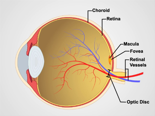
Figura 1. Anatomia dell'occhio. Un diagramma che mostra una vista sagittale dell'occhio umano con le strutture etichettate.

Figura 2: Retina normale. Una fotografia che mostra una vista oftalmoscopica sulla retina normale.
Poiché i colliri midriatici non sono in genere utilizzati nella pratica generale, la vista del fondo è limitata solo a una sezione della retina posteriore. Avere familiarità con queste caratteristiche prima di tentare di esaminare il paziente.
- A meno che gli errori di rifrazione del paziente non rendano difficile concentrarsi sulla retina, di solito è meglio rimuovere gli occhiali per l'esame.
- Dopo aver oscurato la stanza, accendi l'oftalmoscopio e fai brillare la luce sulla mano o sul muro.
- Ruota il disco del
L'esame oftalmologico è probabilmente il più impegnativo da padroneggiare per gli studenti. Con il tempo, tuttavia, diventa routine. È anche una delle parti più produttive dell'esame fisico, in quanto non solo offre una finestra sulla condizione dell'occhio, ma fornisce anche prove di malattia in altre parti del corpo. Un'elevata pressione intracranica da una varietà di cause può portare a gonfiore del nervo ottico, che appare come papilledema in un esame funduscopico. Nel papilledema, il disco ottico è gonfio, i ...
Vai a...
Video da questa raccolta:

Now Playing
Esame oftalmoscopico
Physical Examinations II
66.7K Visualizzazioni

Esame obiettivo dell'occhio
Physical Examinations II
75.9K Visualizzazioni

Esame obiettivo dell'orecchio
Physical Examinations II
53.7K Visualizzazioni

Esame di naso, seni, cavità orale e faringe
Physical Examinations II
64.6K Visualizzazioni

Esame obiettivo della tiroide
Physical Examinations II
103.5K Visualizzazioni

Esame obiettivo dei linfonodi
Physical Examinations II
381.4K Visualizzazioni

Esame obiettivo dell'addome I: ispezione e auscultazione
Physical Examinations II
200.7K Visualizzazioni

Esame obiettivo dell'addome II: percussione
Physical Examinations II
245.6K Visualizzazioni

Esame obiettivo dell'addome III: palpazione
Physical Examinations II
137.5K Visualizzazioni

Esame obiettivo dell'addome IV: valutazione del dolore addominale acuto
Physical Examinations II
66.5K Visualizzazioni

Esame rettale maschile
Physical Examinations II
112.6K Visualizzazioni

Esame completo del seno
Physical Examinations II
85.7K Visualizzazioni

Esame obiettivo ginecologico I: valutazione dei genitali esterni
Physical Examinations II
300.1K Visualizzazioni

Esame obiettivo ginecologico II: esame con lo speculum
Physical Examinations II
148.3K Visualizzazioni

Esame obiettivo ginecologico III: palpazione rettovaginale e bimanuale
Physical Examinations II
145.5K Visualizzazioni
Personale delle biblioteche
Copyright © 2025 MyJoVE Corporation. Tutti i diritti riservati