Method Article
凍結肩甲ラットモデルの推拿:効率的で再現性のあるプロトコル
要約
この研究では、ラットモデルで確立された五十肩を治療するための効率的で再現性のある推拿プロトコルを開発します。このアプローチは、五十肩の推拿療法の治療法を研究するのに役立ちます。
要約
五十肩(FS)は一般的な症状であり、最適な治療法は定義されていません。中国の病院でFS患者の治療に使用される伝統的な漢方薬(TCM)技術である推拿療法は、優れた結果を示していますが、そのメカニズムは完全には理解されていません。この研究は、以前の研究に基づいて、FSラットモデル用のTuinaプロトコルを開発することを目的としていました。20匹のSDラットを対照群(C;n = 5)、FSモデル群(M;n = 5)、FSモデル推拿治療群(MT;n = 5)、およびFSモデル経口治療群(MO;n = 5)にランダムに分割した。本研究では、キャスト固定化法を用いてFSラットモデルを確立した。関節上腕骨可動域(ROM)に対する推拿および経口デキサメタゾンの効果を評価し、組織学的所見を評価した。私たちの研究は、推拿と経口デキサメタゾンが肩の活性ROMを改善し、カプセルの構造を維持できることを示し、推拿療法は経口デキサメタゾンよりも効果的であることが証明されました。結論として、この研究で確立された推拿プロトコルはFSに対して非常に効果的でした。
概要
五十肩(FS)は、肩の癒着性関節包炎とも呼ばれ、肩の痛みと可動性の障害を特徴とする自己限定的な疾患です。通常、30歳から70歳までの平均年齢50歳で、中国の人口では約5%の有病率があります1。女性は男性に比べてFSの発生率が1.6倍高いと報告されています2。FSの有病率は、糖尿病、グルコースおよび脂質代謝障害、またはその他の関連疾患を持つ人で高く、10%から36%の範囲です2,3。FSの現在の臨床治療には、理学療法、ステロイド薬、外科的治療などがあります4。
伝統的な漢方薬(TCM)療法である推拿は、FS患者の肩の痛みを効果的に和らげ、生活の質を向上させることが示されています5,6。しかし、この治療の根本的なメカニズムはよく理解されていません。したがって、動物モデルを使用して、FSの治療における推拿の効果とメカニズムを研究することが重要です。
ラットの肩関節は、ヒトの肩関節に似た複雑な構造をしており、FS7の機構研究でよく用いられます。FSラットモデルは、肩甲上腕骨ROMの低下と被膜線維症を特徴としています8。さらに、このモデルでは肩関節包の観察が可能となり、損傷を修復しながら病理学的研究が可能となる9。さらに、経口コルチコステロイドは、FS治療研究の対照群としてよく使用されます10。この研究は、FSラットモデルのトゥイナプロトコルを開発することを目的としており、トゥイナ療法と経口デキサメタゾンの有効性を比較することにより、トゥイナ研究で動物実験を実施することの実現可能性を実証しています。
プロトコル
この研究は、山東中医薬大学付属病院の倫理委員会によって承認されました(番号:AWE-2022-023)。
1. 実験動物
- 20匹の雄のSprague-Dawley(SD)ラット(7週齢、250-280g)を標準条件(室温[RT] 20-24°C、湿度40%-60%、および12時間/12時間の明暗サイクル)で飼育した。
2. グルーピング方法
- SDラットを対照群(C)、FSモデル対照群(M)、FSモデル推拿治療群(MT)、FSモデル経口治療群(MO)にグループ化し、それぞれ5匹のラットからなる。ケージごとに5匹のラットを飼育します(同じグループ)。
- 7日間の順応後、M、MT、およびMOグループのラットの片方の肩を石膏模型固定化を使用して3週間固定し、次のセクションで説明するようにFSを模倣します。
- セクション4(図1)に記載されているように、MT群のラットに2週間推拿療法を投与します。
- 成人の投与量(0.75 mg /日)とラットの人体表面積の比率(0.018)に基づいて、ラットのキログラムあたりのデキサメタゾンの必要投与量(0.0675 mg /日)を計算します。.
- MO群のラットに0.067 mg/kg/dayの胃内デキサメタゾン溶液を午前7:00に2週間毎日投与します。
注:この研究におけるTuinaプロトコルの効果を確認するために、このグループ化方法を使用してください。さまざまな研究で実験目的に従ってグループ化方法を実行します。
3. FSモデルの開発
- トリブロモエタノール(250 mg / kg、腹腔内注射による)を使用してラットを麻酔します11。
注:施設の倫理委員会の要件に従って、トリボモエタノール(10 g)とtert-アミルアルコール(10 mL)からなるストック溶液を4°Cで保存しました。 使用前に、蒸留水で2%に希釈した。 - ラットの右肩と胸に絆創膏を染み込ませ、右前肢を肩関節の内旋90°に3週間維持します(図2)12。
注意: ラットを監視して、歩行、食事、飲酒などの正常な生理学的活動を実行できることを確認します。ラットが正常な生理学的活動を実行できない場合は、石膏包帯を固定します。 - ラット13において、右肩関節のこわばり、右上肢の収縮、筋萎縮、足を引きずるなどの症状の発現を観察し、FSモデルの確立に成功したことを確認する。
4.推拿法
注:手順全体を通して、調査員は個人用保護具を着用する必要があります。すべての操作を行う必要があるのは、1人の専門の推拿医師だけです(図3、図 4、 および図5)。
- 機械受容器とコンピュータを含むインテリジェントマッサージ技術パラメータ決定システムによってトレーニングします(図3A)。
- 機械受容器と力のパラメータをソフトウェアで表示した3方向の操作を行います(図3B)。
- 親指を使用して、0.5 kgの強度と100〜120回/分の頻度で回転運動で回転混練法を実行します(図3C)。
- 親指の指先を使用して、0.5kgの強さでポイントプレス法を実行します(図3D)。
- ステップ4.1.2および4.1.3で述べた機械的表示を1分間維持することにより、ラットにトゥイナを実行します。
- ネズミが落ち着くまで(~2分)保持します。次に、操作を実行します。ラットを横臥位にしますが、操作方法によって位置が変わる場合があります。
- 右手の人差し指と中指でラットの右前肢を挟み、何度か曲げて伸ばし、ラットの肩関節、肘関節、上腕骨の位置を決めます。
- ラットの右肩、前肢、背中を親指の果肉で時計回りに0.5kgの強さと100〜120回/分の頻度で3分間こねます(図4A-C)。
- 横臥位で前肢の筋肉を操作します。
- うつ伏せの姿勢で肩と背中の筋肉を操作します。
- ツボLI15(Jianyu)、SI11(Tianzong)、HT01(Jiquan)、LI11(Quchi)を親指先でツボごとに30回、0.5kgの強さで垂直に押します(図4D-G)。
- ラット経穴アトラスを使用して、各経穴の位置を定義します(図5)14,15。
- 肩峰端の前方下方のくぼみにあるLI15を腹臥位で押します。
- 肩甲骨脊椎の中点にある棘下窩のくぼみにあるSI11を腹臥位で押します。
- 腋窩の中心にあるHT01を仰臥位で押します。
- 横臥位で肘骨折り目の外側端にある橈骨手根伸筋の内側にあるくぼみLI11を押します。
- 左手の親指と中指で肩関節を持ち、前肢を内転、外転、前方伸展、後伸展の姿勢で10秒間伸ばします(図4H-K)。
注:この延伸方法は、ラットでは抵抗なく実行する必要があります。 - ラットが興奮した場合は、推拿手順を一時停止します。.ラットを10秒間撫でて落ち着かせてから、試験を進めます。
- 手順を2週間毎日実行します。
5.肩甲上腕骨ROMの測定
注意: 関節包組織の変性を防ぐために、測定プロセスをできるだけ早く完了することが重要です。
- 肩甲骨の下端を露出させ、トリブロモエタノールの過剰投与量(腹腔内注射による初期用量の3倍)でラットを犠牲にした後、肩甲骨と上腕骨の近位3分の2を一括して除去します。.
- 注射針(1.2cm×0.45mm)を上腕骨軸に沿って上腕骨頭に挿入します。
- 滅菌手術用シートで包まれたプラスチックフォームの肩甲骨の上下の角に2本の注射針を垂直に挿入します。
- 上腕骨軸の注射針に細い糸を取り付け、上腕骨軸と平行になるように5gの力でもう一方の端を引っ張ります。肩甲骨の下端と上腕骨軸の間の角度を測定します(図6)。
注:信頼性の高い結果を確保するために、別の調査員に測定を行ってもらいます。 - 統計分析ソフトウェアアプリケーションを使用して、データを平均±標準偏差(SD)として報告します。
注: ここでは、SPSS ソフトウェア (SPSS、バージョン 25.0) を使用しました。 - 一元配置分散分析 (ANOVA) を使用してグループ間の差を分析します。
- 適切なソフトウェアを使用してバーグラフィックを取得します。
注:ここではGraphPad Prism 8を使用しました。 - 測定後にH&EおよびMasson染色を使用してカプセルの病理を評価します。
6. 切片の準備
- 肩甲上腕骨ROMを評価した後、全サンプルを4%PFAで3日間固定し、続いてEDTA(pH 7.2)溶液でさらに2か月間脱灰します。
- 脱水後、サンプルを含む包埋組織ブロックを5μmスライス16にスライスする。
- スライスを65°Cで60分間乾燥させます。
- スライスを脱ワックスします。
- スライスをキシレンI、キシレンII、キシレンIIIに7分間浸し、続いて下降エタノール系列(無水エタノール、5分、95%エタノール、2分、80%エタノール、2分、70%エタノール、2分)、最後に超純水に2分間浸します。
7. H&E染色
- ヘマトキシリンで切片を5分間染色し、1%塩酸エタノールで3秒間すすぎ、流水で5分間洗浄します。
- 切片をエオシンで3分間染色し、水道水で洗います。
- 切片をエタノール系列(95%エタノールI、3秒、95%エタノールII、3秒、無水エタノールI、3秒、無水エタノールII、1分)に浸漬し、次にキシレン系列(キシレンI、1分、キシレンII、1分)に浸漬します。
- 各サンプルに中性ガムシーラントを一滴垂らします。各サンプルをカバーガラスで密封します。
- 倒立蛍光顕微鏡(スケールバー=100μm)を用いて画像を収集します。
8.マッソン染色
- 免疫組織化化学ペンを使用して、切片の周囲に円を描き、Bouin溶液中で切片を37°Cで2時間インキュベートして媒染剤にします。続いて、黄色が消えるまで切片を水で洗います。
- サンプルをラピスラズリブルー染料で3分間処理した後、蒸留水で洗浄します。
- 切片をヘマトキシリン(Mayer)で2分間染色した後、切片を酸性エタノール分化溶液で3秒間処理します。次に、流水で10分間セクションを洗浄します。
- 切片をポンソーマゼンタ染料溶液で10分間染色し、続いて水で洗浄します。
- 切片をリンモリブデン酸溶液に10分間浸します。
- 切片にアニリンブルー染色液を5分間添加し、弱酸性作動液で2分間洗浄します。
- 脱水し、手順7.3の説明に従って切片を透明にします。
- 各セクションに中性ガムシーリング剤を一滴垂らし、カバーガラスで覆います。セクションをドラフトに入れたままにして乾かします。
- 手順 7.5 の説明に従って画像を収集します。
結果
ラットの身体活動を観察し、FSモデルの成功または失敗を評価しました。以前の研究では、ギプス固定は正常なラットと比較して移動距離と歩行速度を有意に低下させることが示されました17。別の研究では、FSは移動距離に影響を与えず、足を引きずることが最も一般的な症状であることが示唆されました13。この研究では、モデリング後のラットで右肩関節のこわばり、右上肢の収縮、筋萎縮、足を引きずることが示されました。MT群およびMO群におけるこれらの病変は、2週間の介入によって完全に解消された。しかし、M群には大きな変化は見られなかった。
FS における Tuina の有効性を評価するための主要な基準は、肩関節上腕骨ROM 18 の測定です。肩関節ROMの平均値は、C群で149.3°±5.9°、M群で111.1°±3.9°、MT群で128.5°±2.8°、MO群で119.56°±2.9°であった。 図7に示すように、M群のラットの肩甲上腕骨ROMはC群よりも有意に低かった(P < 0.0001)。また、MT群およびMO群のROMはM群よりも有意に高かった(P < 0.05、P < 0.0001)。しかし、MO群のROMはMT群よりも有意に低かった(P < 0.0001)。この知見は、TuinaがFSラットの肩関節機能を有意に改善できることを示唆しています。
さらに、H&E染色およびマッソン染色は、構造の維持およびカプセル内の線維化の低減におけるTuinaの効果をさらに実証することができます。観察を容易にするために、肩甲上腕骨関節のカプセルを組織学的所見に使用しました。肩関節包は、滑膜層および線維層19を含む。H&E染色により、FSの典型的な特徴であるM群の滑膜細胞増殖、滑膜襞の平坦化、赤血球うっ滞、血管増殖が明らかになりました(図8A、B)。これらの特徴は、推拿とデキサメタゾンの経口療法後にある程度減少しました(図8C、D)。また、MO群はMT群と比較して、滑膜細胞を多くみました。マッソン染色では、各グループの繊維束の配列が示されました(黄色の矢印)。カプセルは、網状繊維の緩いネットワークで構成されており、繊維束がきれいな方向に配列されています(図8E)。M群では、繊維束が無秩序に配列しており、被膜線維化を示しています(図8F)。MT群のラットのカプセルは、繊維束がきれいに明瞭に層状化していることを示しましたが、MO群ではわずかに乱れたままでした(図8G、H)。

図1:FSモデルと推拿介入を確立するためのプロトコル。 ラットは適応摂食を7日間、FSモデル確立を21日間、推拿療法を14日間毎日実施した。36日目に、すべてのラットを犠牲にしました。 この図の拡大版をご覧になるには、ここをクリックしてください。
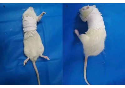
図2:FSのラットモデルを確立するためのキャスト固定化。 この図の拡大版をご覧になるには、ここをクリックしてください。
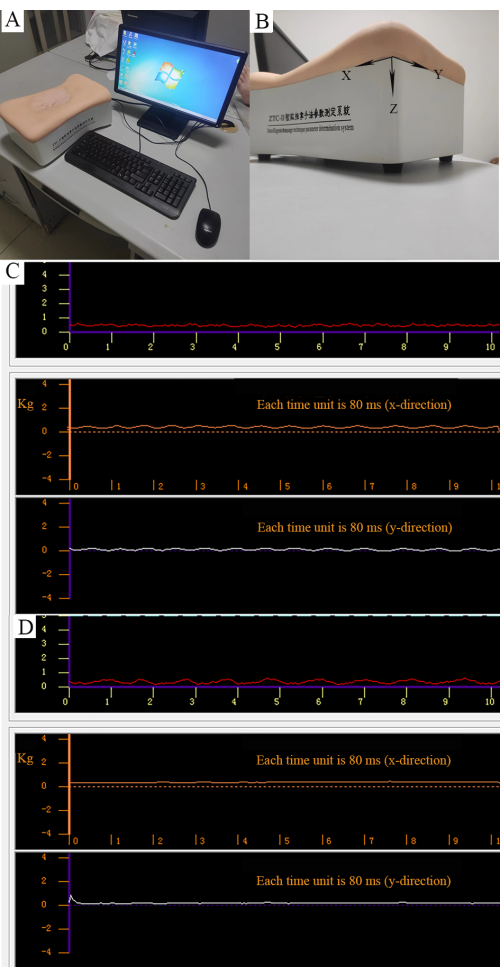
図3:マニピュレーションの定量的制御 。 (A)インテリジェントなマッサージ技術パラメータ決定システム。(B)X方向の平行力、Y方向の縦方向の力、Z方向の垂直方向の力の3つの力を測定できます。(C)回転混練法の強度。赤い曲線は、安定化された垂直方向の力(0.5 kg)を表します。オレンジ色の曲線は、通常の平行な力を表します。白い曲線は、規則的な縦方向の力を表します。(D)ポイントプレス法の強度。赤い曲線は垂直方向の力(0.5 kg)を表します。オレンジ色と白色の曲線は、平行でない縦方向の力を表します。 この図の拡大版をご覧になるには、ここをクリックしてください。
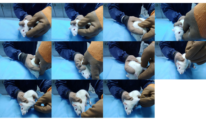
図4:推拿療法で用いられるマニピュレーション。 (A-C)右肩、前肢、背中の筋肉をこねます。(D-G)LI15、SI11、HT01、およびLI11をポイントプレスします。(H-Kさん)内転、外転、前方伸展、後伸展の位置で前肢を伸ばします。 この図の拡大版をご覧になるには、ここをクリックしてください。
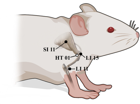
図5:ラットにおけるLI15、SI11、HT01、LI11の解剖学的位置。 ●側面、○内側表面。 この図の拡大版をご覧になるには、ここをクリックしてください。
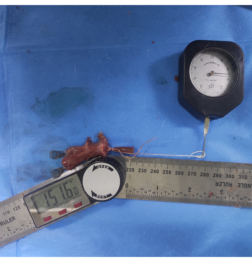
図6:肩関節ROMの測定。 上腕骨軸に挿入した注射針に細い糸を取り付け、上腕骨軸と平行になるように5gの力でもう一方の端を引っ張ります。肩甲骨の下端と上腕骨軸の間の角度は、肩甲上腕骨ROMとして測定されます。 この図の拡大版を表示するには、ここをクリックしてください。
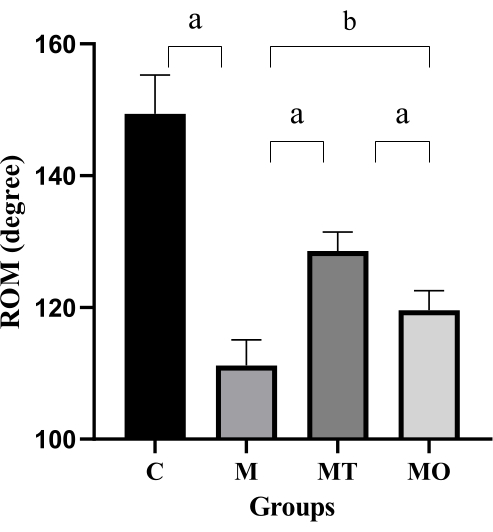
図7:ラットの3つのグループにわたる肩甲骨ROM。値は平均± S.D.、n = 5 です。有意差は一元配置分散分析(a P < 0.001およびbP < 0.0001)によって示されます。 この図の拡大版をご覧になるには、ここをクリックしてください。
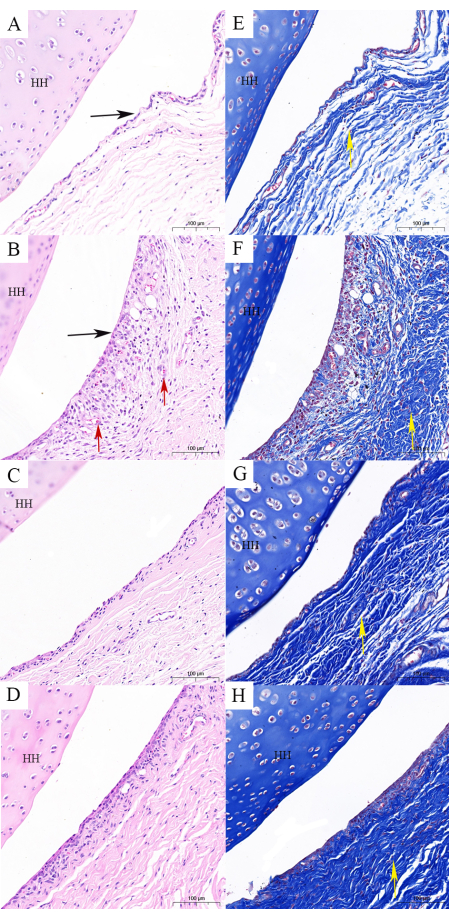
図8:肩関節包の組織学的所見。 (A,E)対照群には、正常なカプセル構造(H&EおよびMasson染色)が含まれています。(B、F)FSモデル群は、滑膜の襞の平坦化、被膜線維症、線維化の乱れ(H&EおよびMasson染色)など、被膜の構造の変化を示しています。(C,G)です。FSモデルとTuinaグループの組み合わせは、カプセルの構造が正常に近く、線維化は明らかではないことを示しています(H&EおよびMasson染色)。(D,H)経口デキサメタゾンと組み合わせたFSモデルは、カプセルの構造が正常に近く、線維化が明らかであることを示しています(H&EおよびMasson染色)。スケールバー = 100 μm。 HH:上腕骨の頭。黒い矢印:滑膜のひだ。赤い矢印:赤血球のうっ滞と血管増殖;黄色の矢印:繊維束。この図の拡大版をご覧になるには、ここをクリックしてください。
ディスカッション
最初の重要なステップは、モデルの選択です。一次FSモデルの実装が困難なため、FSラットモデルを確立するために、ギプス固定と外科的内部固定がしばしば使用されます9,12。肩の可動性と被膜の線維化の最も深刻な制限は、3週間のギプス固定によって確立されたFSモデルで観察されました12,20。この研究では、FSモデルの成功率は優れており、100%の成功でした。
2番目の重要なステップは、このプロトコルで使用される操作です。この研究では、3つの操作(混練、プレス、ストレッチ)を使用しました。肩、肩甲骨、上腕に軟部組織混練マニピュレーションを適用し、筋肉を弛緩させました。FS5,21の臨床診療で最も一般的に使用されるLI15、SI11、HT01、LI11などのツボに圧力をかけることにより、プレス操作を行いました。LI15、SI11、およびHT01は、肩嚢の周囲の位置に位置し、ROMおよび肩機能の改善に有効であり得る22。LI11は上肢の運動障害によく使用され、LI15と同じ子午線にあります。このツボマッチング法は、LI1523の有効性を向上させるのに役立ちます。完全なリラクゼーションの後、機能的な活動を回復させるためにストレッチ技術が使用されました。
このプロトコルで考えられる問題は、ラットが推拗中に強い抵抗を示すことであり、これはラットの耐性を超えるのではなく、恐怖によって引き起こされる可能性があります。この時点で、ラットが落ち着くまで操作を停止する必要があります(10秒間撫でるとラットが落ち着きます)。また、ラットの症状に応じてストレッチの程度を調整する必要があります。当初は肩関節の限界は明らかで、伸展振幅は小さかった。介入に伴い、ラットの肩関節機能は徐々に回復し、伸展の振幅は徐々に増加しました。ラットが抵抗なく延伸法を受け入れることができるのが標準です。最後に、ネズミにはある程度の攻撃性があり、推拿はネズミとの長時間の接触を必要とするため、個人用保護具を着用することが重要です。
操作の定量的制御は、推拿実験で最も困難です。マッサージマニピュレーションシミュレーターは、単一のマニピュレーションの強さと頻度を制御するために使用できますが、複数のマニピュレーションと治療部位が関与する場合、この方法は制限されます24,25。臨床現場では、通常、トゥイナは開業医によって直接行われますが、この研究では、医療機器への介入が困難でした。刺激を制御するために、インテリジェントなマッサージ技術パラメータ決定システムを使用して、推拿のトレーニングを標準化することができます。訓練後、調査員は各ラットにある程度同じ力を加えることができます。このプロトコルの主な制限は、操作を完全に制御できないことです。
TCM推拿療法は中国全土で使用されており、病院のさまざまな医師がさまざまな操作と治療部位の組み合わせを使用しています。したがって、動物実験と臨床研究の両方について、再現可能で効果的なプロトコルを確立することが重要です。この研究では、使用された操作と経穴は、私たちのチームによる以前の研究に基づいており、私たちの臨床経験とFS動物モデル21の特徴を組み合わせています。この研究は、FSラットの肩関節機能の改善とカプセル線維症の減少における開発されたTuinaプロトコルの有効性を実証しました。これらの知見は、推拿治療の根底にあるメカニズムのさらなる研究の基礎となる。さらに、このプロトコルは、FSの代替医療の有効性を調査することに関心のある研究者にとって有用です。
以前の研究では、線維症に対する推拿介入のメカニズムは、MMP-1/TIMP-1のバランスを調節しながらTGF-βとCTGFのダウンレギュレーションに関連している可能性があり、それによって細胞外マトリックス(ECM)の産生が緩和されることがわかった26。肩嚢の線維症に対するTuinaの効果は、さまざまなメカニズムの調節によって達成され得る。しかし、この改善に関与するメカニズムを完全に理解するには、さらなる研究が必要です。
開示事項
著者は何も開示していません。
謝辞
この研究は、済南市2020年科学技術発展計画(助成金番号202019059)、山東省伝統漢方科学技術プロジェクト(助成金番号2021Q080)、およびQilu School of Traditional Chinese Medicine Inherit Project(助成金番号[2022]93)の支援を受けました。
資料
| Name | Company | Catalog Number | Comments |
| 4% paraformaldehyde | Solarbio | P1110 | |
| Embedding machine | Changzhou Paisijie Medical Equipment Co., Ltd | BM450A | |
| Ethylene Diamine Tetraacetic Acid (EDTA) | Solarbio | E1171 | |
| Hematoxylin eosin (HE) staining kit | Sparkjade | EE0012 | |
| Intelligent-massage technique parameter determination system | Shanghai Dukang Intrument Equipment Co. Ltd | ZTC- | |
| Microtome | Leica | 531CM-Y43 | |
Modified Masson Trichrome Staining Solution | Shanghai yuanye Bio-Technology Co., Ltd | R20381-8 | Bouin 50 mL; lapis lazuli blue dye 50 mL; Hematoxylin (Mayer) 50 mL; acidic ethanol differentiation solution 50 mL; ponceau magenta dye solution 50 mL; phosphomolybdic acid solution 50 mL; aniline blue staining solution 50 mL; weak acid 50 mL |
| Tribromoethanol | Macklin | T903147-5 |
参考文献
- Li, W., LU, N. Z., Xu, H. L., Wang, H. F., Huang, J. Case control study of risk factors for frozen shoulder in China. International Journal of Rheumatic Diseases. 18 (5), 508-513 (2015).
- Degreef, I., Steeno, P., De Smet, L. A survey of clinical manifestations and risk factors in women with Dupuytren's disease. Acta Orthopaedica Belgica. 74 (4), 456-460 (2008).
- Tighe, C. B., Oakley, W. S. The prevalence of a diabetic condition and adhesive capsulitis of the shoulder. Southern Medical Journal. 101 (6), 591-595 (2008).
- Cho, C. H., Bae, K. C., Kim, D. H. Treatment strategy for frozen shoulder. Clinics in Orthopedic Surgery. 11 (3), 249-257 (2019).
- Liu, M., et al. Effects of massage and acupuncture on the range of motion and daily living ability of patients with frozen shoulder complicated with cervical spondylosis. American Journal of Translational Research. 13 (4), 2804-2812 (2021).
- Ai, J., Dong, Y. K., Tian, Q. D., Wang, C. L., Fang, M. Tuina for periarthritis of shoulder: A systematic review protocol. Medicine. 99 (11), e19332 (2020).
- Norlin, R., Hoe-Hansen, C., Oquist, G., Hildebrand, C. Shoulder region of the rat: anatomy and fiber composition of some suprascapular nerve branches. The Anatomical Record. 239 (3), 332-342 (1994).
- Okajima, S. M., et al. Rat model of adhesive capsulitis of the shoulder. Journal of Visualized Experiments: JoVE. (139), 58335 (2018).
- Zhao, H. K., et al. Tetrandrine inhibits the occurrence and development of frozen shoulder by inhibiting inflammation, angiogenesis, and fibrosis. Biomedicine & Pharmacotherapy. 140, 111700 (2021).
- nar, B. M., Battal, V. E., Bal, N., Güler, &. #. 2. 2. 0. ;. &. #. 2. 1. 4. ;., Beyaz, S. Comparison of efficacy of oral versus intra-articular corticosteroid application in the treatment of frozen shoulder: An experimental study in rats. Acta Orthopaedica et Traumatologica Turcica. 56 (1), 64-70 (2022).
- Dias, Q. M., Rossaneis, A. C., Fais, R. S., Prado, W. A. An improved experimental model for peripheral neuropathy in rats. Brazilian Journal of Medical and Biological Research. 46 (3), 253-256 (2013).
- Kim, D. H., et al. Characterization of a frozen shoulder model using immobilization in rats. Journal of Orthopaedic Surgery and Research. 11 (1), 160 (2016).
- Feusi, O., et al. Platelet-rich plasma as a potential prophylactic measure against frozen shoulder in an in vivo shoulder contracture model. Archives of Orthopaedic and Trauma Surgery. 142 (3), 363-372 (2022).
- Yin, C. S., et al. A proposed transpositional acupoint system in a mouse and rat model. Research in Veterinary Science. 84 (2), 159-165 (2008).
- Guo, X. R., et al. Study on the regulatory mechanism of electroacupuncture based on thyroid pathway for mammary gland hyperplasia rats. Zhongguo Zhen Jiu. 38 (8), 857-863 (2018).
- Feldman, A. T., Wolfe, D. Tissue processing and hematoxylin and eosin staining. Methods in Molecular Biology. 1180, 31-43 (2014).
- Taguchi, H., et al. A rat model of frozen shoulder demonstrating the effect of transcatheter arterial embolization on angiography, histopathology, and physical activity. Journal of Vascular and Interventional Radiology: JVIR. 32 (3), 376-383 (2021).
- Oki, S., et al. Generation and characterization of a novel shoulder contracture mouse model. Journal of Orthopaedic Research. 33 (11), 1732-1738 (2015).
- Kubo, H., et al. Histologic examination of the shoulder capsule shows new layer of elastic fibres between synovial and fibrous membrane. Journal of Orthopaedics. 22, 251-255 (2020).
- Cho, C. H., Lho, Y. M., Hwang, I., Kim, D. H. Role of matrix metalloproteinases 2 and 9 in the development of frozen shoulder: human data and experimental analysis in a rat contracture model. Journal of Shoulder and Elbow Surgery. 28 (7), 1265-1272 (2019).
- Wang, J. M., et al. Efficacy and safety of Tuina and intermediate frequency electrotherapy for frozen shoulder: MRI-based observation evidence. American Journal of Translation Research. 15 (3), 1766-1778 (2023).
- Ben-Arie, E., et al. The effectiveness of acupuncture in the treatment of frozen shoulder: A systematic review and meta-analysis. Evidence-Based Complementary and Alternative Medicine: eCAM. 2020, 9790470 (2020).
- Zou, F., et al. The impact of electroacupuncture at hegu, shousanli, and quchi based on the theory "Treating flaccid paralysis by Yangming alone" on stroke patients' EEG: A pilot study. Evidence-Based Complementary and Alternative Medicine: eCAM. 2020, 8839491 (2020).
- Lv, T. T., et al. Using RNA-Seq to explore the repair mechanism of the three methods and three-acupoint technique on DRGs in sciatic nerve injured rats. Pain research & Management. 2020, 7531409 (2020).
- Niu, F., et al. Spinal tuina improves cognitive impairment in cerebral palsy rats through inhibiting pyroptosis induced by NLRP3 and Caspase-1. Evidence-Based Complementary and Alternative Medicine: eCAM. 2021, 1028909 (2021).
- Na, Z., et al. The combination of electroacupuncture and massage therapy alleviates myofibroblast transdifferentiation and extracellular matrix production in blunt trauma-induced skeletal muscle fibrosis. Evidence-Based Complementary and Alternative Medicine: eCAM. 2021, 5543468 (2021).
転載および許可
このJoVE論文のテキスト又は図を再利用するための許可を申請します
許可を申請さらに記事を探す
This article has been published
Video Coming Soon
Copyright © 2023 MyJoVE Corporation. All rights reserved