蛍光 x 線 (XRF)
概要
ソース: 研究室博士リディア フィニー-アルゴンヌ国立研究所
蛍光 x 線は誘導、放射放射分光情報を生成するために使用できます。蛍光 x 線顕微鏡は、金属誘起蛍光性の放出を識別し、その空間分布を定量化に使用する非破壊イメージング手法です。
手順
1. シリコン窒化 Windows の準備
- 逆ピンセットを使ってウィンドウ (windows が粉々 に落とした場合窒化ケイ素) を選択します。
- スライド ガラス、フラット面を上にウィンドウを配置します。
- ウィンドウの辺にスコッチ テープの小片を遵守し、培養皿の底に windows を遵守するこれらを使用します。
- 紫外線による培養皿で windows を滅菌します。UV 架橋キャビネット自動クロスリンク設定でこれが行うことができます約 1 h の層流フードに紫外線ランプの下でさらに UV 照射します。
2. 滅菌シリコン窒化 Windows 上に細胞をめっき
- ホールドそれを皿に約 45 ° の角度で傾いています。
- 皿の側に向かってピペッティングによるメディアを追加し、メディア ウィンドウをコートする傾斜角を徐々 に緩和します。
- 同じ方法で細胞を培養皿に追加し、孵化させなさい。
- 時折彼らは使用する準備がで
結果
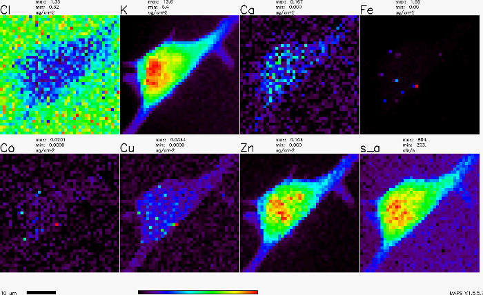
付着性のセルの x 線蛍光マップを図 1に示します。各パネルは、セルの上の特定の要素 (例えば、銅、鉄、亜鉛など) の分布を示します。パネルは、's_a' ショーの x 線の吸収をラベル付けします。
申請書と概要
タグ
スキップ先...
このコレクションのビデオ:

Now Playing
蛍光 x 線 (XRF)
Analytical Chemistry
25.4K 閲覧数

試料分析の準備のため
Analytical Chemistry
84.8K 閲覧数

社内基準
Analytical Chemistry
204.9K 閲覧数

標準添加法
Analytical Chemistry
320.3K 閲覧数

検量線
Analytical Chemistry
797.3K 閲覧数

(紫外-可視) 紫外可視分光法
Analytical Chemistry
624.0K 閲覧数

ラマン分光を用いた化学分析
Analytical Chemistry
51.2K 閲覧数

炎イオン化検出ガスクロマトグラフィー (GC)
Analytical Chemistry
282.2K 閲覧数

高速液体クロマトグラフィー (HPLC)
Analytical Chemistry
384.9K 閲覧数

イオン交換クロマトグラフィー
Analytical Chemistry
264.7K 閲覧数

キャピラリー電気泳動 (CE)
Analytical Chemistry
94.0K 閲覧数

質量分析への紹介
Analytical Chemistry
112.3K 閲覧数

走査型電子顕微鏡 (SEM)
Analytical Chemistry
87.3K 閲覧数

ポテンショスタット/Galvanostat を使用して担持触媒の電気化学測定
Analytical Chemistry
51.4K 閲覧数

サイクリックボルタンメトリー (CV)
Analytical Chemistry
125.4K 閲覧数
Copyright © 2023 MyJoVE Corporation. All rights reserved
