
Method Article
에 대한 기능성 연구를위한 3D 인간의 폐 조직 모델
요약
Human tuberculosis infection is a complex process, which is difficult to model in vitro. Here we describe a novel 3D human lung tissue model that recapitulates the dynamics that occur during infection, including the migration of immune cells and early granuloma formation in a physiological environment.
초록
결핵 (TB)는 여전히 전세계 사람들의 건강에 큰 위협을 보유하고, 우리는 새로운 치료 방법의 발견을 질병 메커니즘을 이해 미리 돕는 비용 효율적이지만 신뢰성을 위해 필요하다. 단층의 시험관 내 세포 배양을 또는 공동 배양은 3 차원 환경과 조직 반응이 부족하다. 여기서, 우리는 결핵균 (M. 결핵) 감염시 발생하는 복잡한 이벤트를 연구하는데 효과적인 도구로 유망 인간 폐 조직의 시험 관내 모델에 혁신을 설명한다. 3D 조직 모델은 다공성 막 위에 콜라겐 매트릭스에서 배양 조직 특이 상피 세포와 섬유 아세포로 구성된다. 공기에 노출시, 상피 세포 및 계층화 근단 측에서 점액 분비. 일차 인간 대 식세포를 도입하여 M. 감염된 티슈 모드 결핵난, 우리는 면역 세포가 감염된-조직으로 마이그레이션 및 결핵 육아종의 초기 단계를 형성하는 것으로 나타났습니다. 이러한 구조는 근본적으로 다른 또는 일반적으로 널리 사용되는 실험 동물 모델에서 관찰되지 인간 결핵, 육아종의 고유 한 기능을 요점을 되풀이. 이것의 Organotypic 배양법은 숙주 세포 병원체 상호 작용의 시공간적 특성에 중요한 정보를 제공하는 3 차원 시각화 및 강력한 정량 분석을 가능하게한다. 함께 촬영, 폐 조직 모델은 결핵에 대한 연구에 대한 생리 학적으로 관련 조직 미세 환경을 제공합니다. 따라서, 폐 조직 모델은 기본 기전 연구 및 응용에 대한 가능성을 모두 의미를 갖는다. 중요한 것은, 모형하여 폐에 영향을주는 감염성 질환의 다양한 모델링의 사용을 넓혀 개별 세포 유형의 추가 또는 조정할 수있다.
서문
인간에서, 감염, 염증 조직, 세포 모집, 조직 리모델링 및 조직 항상성의 조절에 대한 응답은 다른 유형의 세포를 포함하는 복합 이벤트이다. 따라서, 이러한 프로세스는 최상의 로컬 조직 환경에서 연구한다. 이전에는, 이것은 주로 실험 동물 모델을 이용 가능했다. 그들은 종종 인간보다 다른 방식으로 병원체에 응답하고 또한 병 (1)의 서로 다른 코스를 표시하지만, 광범위하게 사용되는 실험 동물은 많은 제한을 잡아. 체외 폐 조직 모델에서 인간은 인간의 폐의 특이 적 면역 반응을 연구하기 위해 가능성을 보유하고있다.
인간의 결핵 (TB)은. 결핵균 (M. 결핵), TB의 원인 물질은 세균 폐 dendri 의해 잠겼되는 폐포 공간으로 이송되는 에어로졸 방울 통해 폐에 도달 주로 폐에 영향을 미치는 질환TIC 세포를 감염 2,3에 대한 선천성 면역 반응의 일환으로 폐포 대 식세포. 병원체의 식균 작용은 phagosome 내 버그의 구획화에 이르게 이상적 식세포에 의해 중화 및 병원균의 사멸을 초래한다. M.에 노출 된 개인의 50 % 이하 결핵은 선천성 면역 반응 (4)를 통해 감염을 취소 할 수있을 것으로 생각된다. 감염의 다른 결과는 나중에, 잠재 감염이나 최악의 경우 만성 활동성 질환 5의 적응 면역 시스템에 의해 통관 있습니다.
이전에 인간의 결핵의 연구에 더 체외 조직 모델이 없었다. 인간 대 식세포 또는 다른 말초 혈액 세포의 단일 세포 배양 물은 종종 6,7 사용되고있다. 이 방법의 단점은 M. 노출 폐 조직에서 함께 작동하는 다른 세포 유형의 역학을 반영 할 수 없다는 것이다 결핵 . 따라서, TB에 기능적 역학적 연구를 수행 할 수 있도록하기위한 시험 관내 모델이 필요하다. 셀 기반 여기에 설명 된 시험 관내 인간의 폐 조직 모델에서 원래 수지상 세포의 기능 (8)에 대한 연구에 대한 우리의 그룹에 의해 설립되었습니다. 우리는 결핵의 연구에이 방법을 적용하고있다.
여기에 제시된 인간의 폐 조직 모델은 조직 특이 상피 세포와 섬유 아세포 (8)로 구성되어 있습니다. 이러한 세포는 트랜스 웰 인서트 및 정상적인 인간 폐 조직 (도 1)과 유사한 형태의 구조로 다공성 막 위에 콜라겐 매트릭스에서 배양된다. 공기에 노출 될 때 세포는 정점면 (8)에서 점액을 분비하기 시작. 인간의 차 대식 세포를 주입하여 M. 감염 면역 세포가 조직에 마이그레이션 및 결핵 육아종 (9)의 초기 단계를 형성하는 방법 모델에 결핵은, 우리가 관찰하고있다. 이것은 제 인체 조직 모델이다 대하여 descr결핵에 대한 ibed과 결핵 및 폐의 다른 질환에 대한 선천성 면역 반응을 연구하기위한 유망한 도구를 포즈. 지금까지, 우리 모델의 면역 세포로서 만 단핵구 및 대 식세포를 사용하고 있지만, 복잡도의 레벨은 추가적인 중요한 세포 유형의 포함에 의해 증가 될 수있다.

폐 조직 모델의 도식 개요도. (A) 모델이 M., 인간 폐 관련 상피 세포로 구성되어있다 결핵은 차 대식 세포를 -infected과 붉은 염료 표시 단핵구는 트랜스 웰 필터를 준비 콜라겐 포함 된 섬유 아 세포에 접종. 공기 조직 모델의 노출은 상피에 의해 세포 외 기질 단백질의 생산, 점액 분비와 계층화를 시작합니다. 이렇게 개발 된 3 차원 조직 모델은 엠을 연구하는 유용한 도구입니다 CLO 환경에서 결핵 감염sely 인간 폐. (B) 조직 모델의 제조에서 상이한 단계의 대표 현미경 사진. (C) 폐 모델 조직 절편의 완전한 구조를 닮았다. 스케일 -. 100 μm의 이 그림의 더 큰 버전을 보려면 여기를 클릭하십시오.
프로토콜
참고 : 린 셰핑 대학 병원의 혈액 은행에서 구입 건강한 익명의 혈액 기증자로부터 인간의 말초 혈액이, 스웨덴이 연구를위한 면역 세포의 소스로 사용되었다. 이 프로토콜은 24mm 6 웰 플레이트 삽입을 위해 설계되었습니다. 다른 잘 형식에 직접 적응은 개발 기간 동안 모두 수직 및 수평 조직 모델 계약을하기 때문에 사용하지 않는 것이 좋습니다.
박테리아 / 세포주의 재료, 미디어 및 문화 1. 준비
- 박테리아의 문화 :
- 마이코 박테리아 균주 M. 성장 0.05 %를 함유하는 미들 7H9 배지의 구성 적 녹색 형광 단백질 (GFP)을 발현하는 pFPV2 플라스미드를 운반 결핵균 H37Rv, 트윈 80, 0.5 % 글리세롤, 가나 마이신 (20 μg의 / ㎖) (미들 알부민, 덱 스트로스 및 카탈라제 농축 보충 미들 ADC 농축) 7-10 일 5 % CO 2와 37 ° C에서.
참고 : 모든 실험 단계를라이브 악성 M.를 포함하는의 결핵 균주 BSL -3- 설비에서 수행되어야한다.
- 마이코 박테리아 균주 M. 성장 0.05 %를 함유하는 미들 7H9 배지의 구성 적 녹색 형광 단백질 (GFP)을 발현하는 pFPV2 플라스미드를 운반 결핵균 H37Rv, 트윈 80, 0.5 % 글리세롤, 가나 마이신 (20 μg의 / ㎖) (미들 알부민, 덱 스트로스 및 카탈라제 농축 보충 미들 ADC 농축) 7-10 일 5 % CO 2와 37 ° C에서.
- 1X 둘 베코 변형 이글 배지 (DMEM) 완전 배지를 제조 (1 mM 피루브산 나트륨, 2mM L- 글루타민, 100 U가 / ㎖ 페니실린, 100 μg의 / ㎖ 스트렙토 마이신, 10 mM의 HEPES, 0.1 mM의 비 필수 아미노산 및 10로 보충 %의 소 태아 혈청 (FBS))를 열 불 활성화. 또한 항생제없는 DMEM 완전 배지를 준비합니다.
- 1X 최소 필수 매체 (MEM) 완전 배지를 제조 (1 mM 피루브산 나트륨, 2mM L- 글루타민, 100 U / ㎖ 페니실린, 100 μg의 / ㎖ 스트렙토 마이신, 10 mM의 HEPES, 0.1 mM 비 필수 아미노산, 10 % 열 - 불 활성화 된 소 태아 혈청 (FBS)).
- 피브로넥틴 / 콜라겐 코팅 된 플라스크 (총 10 ㎖)의 제조 :
- 깨끗한 튜브에 피펫 8.8 ml의 멸균 1X 인산 완충 식염수 (PBS). 1 ml의 소 혈청 알부민 (1 ㎎ / ㎖) 100 ㎕를 타입 I 콜라겐 (3 ㎎ / ㎖) 100 ㎕를 소 재조합 주석을 추가남자 피브로넥틴 (/ ㎖ 1 mg)을 얻었다.
- 거꾸로 5 번 튜브를 돌려 솔루션을 섞는다. 플라스크 (T-25, T-75 플라스크에 2 ml의 1 ml)에 피브로넥틴 / 콜라겐 용액으로 코팅된다. 37 ° C에서 / O N을 맡겨. 배양 후, 용액을 제거하고 실온에서 코팅 된 플라스크를 저장한다.
주 : 수거 용액을 4 ℃에서 저장하고 3 회 재사용 될 수있다. 2 주 이상 보관 액은 폐기해야에 따라 갈색 (결정의 형성)을 설정하는 원인이 될 수 있습니다.
- 섬유 아세포의 문화 :
- 37 ℃에서 5 % CO 2에서 완전 DMEM에서 성장 및 MRC-5 (14 주령 남성 태아의 정상적인 폐 조직 유래의 인간 폐 섬유 아세포 세포주)를 유지한다. 구절 24-26에서 섬유 아세포를 사용 70~80% 합류 할 때까지 성장한다.
참고 : 경로> (30)에서 MRC-5 세포 라인 형태를 잃게하는 경향이 조직 모델에서 사용하지 않는 것이 좋습니다.
- 37 ℃에서 5 % CO 2에서 완전 DMEM에서 성장 및 MRC-5 (14 주령 남성 태아의 정상적인 폐 조직 유래의 인간 폐 섬유 아세포 세포주)를 유지한다. 구절 24-26에서 섬유 아세포를 사용 70~80% 합류 할 때까지 성장한다.
- 상피 세포의 문화 :
- 16HBE14o- (16HBE), 정상적인 인간의기도 상피 세포의 분화 형태와 기능을 유지 불후의 인간 기관지 상피 세포 라인을, (이 박사 디터 Gruenert, 시온 산 암 센터, 캘리포니아 대학, 샌 프란 시스 코에서 선물했다 확보 미국 10.). 피브로넥틴 / 콜라겐 코팅 된 플라스크에서 배양 16HBE 세포는 37 ℃에서 5 % CO 2에서 완전한 MEM에서 세포를 유지한다.
- 5 배 DMEM의 준비
- DMEM 분말 13.4 g 및 멸균 증류수 150 mL의 탄산 수소 나트륨 3.7 g을 용해시켜 5 배 DMEM을 준비한다. , 7.3 매체의 pH를 조정 200ml의에 볼륨을 구성하고 0.22 μm의 멤브레인 필터를 사용하여 필터링 할 수 있습니다. 멸균 용기에 여과 매체를 수집하고 실온에서 사용할 때까지 저장됩니다.
콜라겐이 포함 된 섬유 아세포 2. 준비
- 37 ° C의 물을 욕조에 FBS와 L- 글루타민의 해동 냉동 분취.해동 후 얼음에 샘플을 유지. 4 ℃, 중탄산 나트륨 (71.2 ㎎ / ㎖) 및 겐타 마이신 (50 ㎎ / ㎖)을 배치했다. 50 ㎖ 원심 분리기 튜브, 4 ° C에서 10 ml의 멸균 피펫을 사전 멋진.
참고 : (5 배 DMEM 제외)에 사용되는 모든 재료는 사용 전에 얼음에 차게하고 모든 단계가 얼음에서 수행됩니다. 이 콜라겐의 응고를 방지 나는 소 콜라겐 (1.1 ㎎ / ㎖)을, 차가운 유지해야 입력합니다. - 섬유 모세포를 제조 :
- 따뜻한 37 ° C의 물을 욕조에 트립신, 37 ° C에서 5 % CO 2에서 10 분 동안 폐 섬유 아 세포 (MRC-5) 세포에 충분한 양을 품어. 1X DMEM이 완료 추가하여 트립신을 중화. 5 분 동안 300 XG에 세포 현탁액을 원심 분리 대기음. 상등액을 흡인하고 완전 DMEM에 2.3 × 10⁵ 세포 / ml에서 세포를 재현 탁. 사용할 준비가 될 때까지 얼음 세포를 놓습니다.
- 사전 믹스를 준비합니다
- "미리 혼합"이라는 튜브에 다음을 추가; (395)5 배 DMEM의 μL, 40 μL L 글루타민, 120 μL을 NaHCO3 (71.2 ㎎ / ㎖), 440 μL의 FBS, 5 μL 젠타 마이신 (50 ㎎ / ㎖), 전체 볼륨 1,000 μL, 다음 잘 미리 섞어 넣어 소용돌이 얼음.
참고 : 주어진 볼륨은 하나의 24mm 6 웰 배양 삽입에 대한 것입니다. 인서트의 총 수에 필요한 특정 량을 계산하지만, 반드시 충분한 예비 - 혼합물이 준비 될 하나 더 추가한다.
- "미리 혼합"이라는 튜브에 다음을 추가; (395)5 배 DMEM의 μL, 40 μL L 글루타민, 120 μL을 NaHCO3 (71.2 ㎎ / ㎖), 440 μL의 FBS, 5 μL 젠타 마이신 (50 ㎎ / ㎖), 전체 볼륨 1,000 μL, 다음 잘 미리 섞어 넣어 소용돌이 얼음.
- 무 세포 콜라겐 혼합물을 제조 :
- 각각의 문화에 무 세포 콜라겐 액 1 ㎖를 추가합니다. 주어진 순서에 얼음에 50 ML 원뿔 튜브에 다음을 추가; 1.1 ㎎ / ㎖의 콜라겐 686 μL, 250 μL 예비 - 혼합물과 1000 μL의 총 부피가 64 μL의 1X 완전 DMEM. 잘 기포를 확보하지 액을 혼합한다. 빠른 작동 및 공기 방울을 방지하기 위해 상기 튜브의 벽에 콜라겐을 추가한다.
- 6 잘 접시에 배치 삽입에 무 세포 층 혼합물의 1 ML을 추가합니다. 삽입 외부에 잘 어떤 매체를 추가하지 마십시오. 에37 ° C 배양기에서 30 분 동안 cubate. 무 세포 혼합물이 공기 방울없이 전체 삽입을 포함해야합니다.
- 휴대 콜라겐 혼합물을 제조 :
- 다음 순서로 얼음에 보관 50 ㎖ 원뿔형 튜브에 세포 층의 성분을 혼합; 2 ml의 콜라겐, 615 μL 사전 믹스, 1X 완전한 DMEM 58 μL와 327 μL 세포 현탁액 폐 섬유 아 세포 (MRC-5) 3000 μL에 총 볼륨을 확인합니다. 각각의 문화는 세포의 콜라겐 혼합물의 3 ㎖를 필요로한다.
중요한 단계 : 조심스럽게 세포 현탁액을 첨가하기 전에 콜라겐과 프리믹스를 혼합해야합니다. 이 섬유 아세포에 독성 효과를 피하기 위해 콜라겐의 pH를 중화한다. - 무 세포 콜라겐 층 위에 셀룰러 층 (3 ㎖)를 첨가하고, 37 ℃ 배양기에서 2 시간 동안 배양한다. 빠른 작동 및 공기 방울을 방지하기 위해 상기 튜브의 벽에 콜라겐을 추가한다.
- 중합 후, t 완전한 DMEM 2 ㎖를 추가그는 (삽입 아래) 6 웰 플레이트의 바닥과 24 시간 동안 배양한다.
참고 : 중합 반응은 발생하지 않은 경우, 섬유 아세포의 콜라겐 매트릭스를 포함하는 플레이트 삽입을 무시하고 시작 다시. 원인은 추가 또는 위 시약 중 하나의 잘못된 볼륨에서 오류가 발생합니다.
- 다음 순서로 얼음에 보관 50 ㎖ 원뿔형 튜브에 세포 층의 성분을 혼합; 2 ml의 콜라겐, 615 μL 사전 믹스, 1X 완전한 DMEM 58 μL와 327 μL 세포 현탁액 폐 섬유 아 세포 (MRC-5) 3000 μL에 총 볼륨을 확인합니다. 각각의 문화는 세포의 콜라겐 혼합물의 3 ㎖를 필요로한다.
섬유 아세포의 콜라겐 매트릭스의 3 연속 문화
- 조심스럽게 깨끗한 집게를 사용하여 삽입을 들어 올려 우물의 바닥에서 문화 매체를 대기음. 삽입 내에서 2 ml의 완전한 DMEM에 의해 잘 다음의 바닥에 2 ml의 완전한 DMEM을 추가합니다. 이는 외부 및 내부 챔버 사이의 영양소의 확산을 방지하는 바와 같이, 삽입 하에서 기포를 도입하는 피. 마이크로 피펫 팁과 공기 방울을 제거합니다.
- 약 5~7일위한 배양 배지 (내부 및 삽입 아래)마다 둘째 날과 문화를 변경합니다. 삽입에서 용지를 제거에주의하십시오. 접점을 방지하려면섬유 아세포의 콜라겐 매트릭스와 CT는 약간 깨끗한 집게를 사용하여 삽입을 기울 삽입 벽에서 미디어를 대기음.
중요한 단계 : 콜라겐 매트릭스의 섬유 아세포가 연장 된 표현형을 얻고, 콜라겐, 다음 계약을 개조해야한다. 5-7 일 후, 매트릭스는 인서트의 중심에서 플랫폼 (직경 10-14 mm)를 형성하도록 수축했다. 수축 행렬은 다음 단계에 사용하기위한 준비가되어있다. 섬유 아세포 전에 (단계 2.6.1) 시드에 콜라겐과 잘 혼합된다는 사실을 중요 행렬의 균일 한 수축을 구하십시오.
면역 세포 (감염 / 감염되지 않은 단핵구 - 대식 세포 혼합물) 4. 시드
참고 : 다음 실험 단계는 악성 마이코 박테리아를 포함하기 때문에 BSL-3 시설에서 수행해야합니다.
- 차 단핵 세포와 대 식세포의 준비 :
- 의 establi를 사용하여 기증자의 혈액에서 말초 혈액 단핵 세포를 분리프로토콜을 흘렸다. 조직 모델과 문화를 설정하는 것과 같은 날에 단핵 세포를 분리 및 M. 감염이 약 7 일간 대 식세포로 분화 결핵.
참고 :이 보장합니다 모두 식세포와 계약 된 섬유 아세포의 콜라겐 매트릭스 7 일 후 사용할 수 있습니다. 또한 감염된 대 식세포와 함께 추가됩니다 신선한 단핵 세포를 분리.
- 의 establi를 사용하여 기증자의 혈액에서 말초 혈액 단핵 세포를 분리프로토콜을 흘렸다. 조직 모델과 문화를 설정하는 것과 같은 날에 단핵 세포를 분리 및 M. 감염이 약 7 일간 대 식세포로 분화 결핵.
- 엠의 준비 결핵 -infected 대 식세포
- , 1X PBS는 항생제가없는 완전한 DMEM에서 0.05 % 트윈 80, 재현 탁를 포함하는 세척, 배양 된 박테리아를 수확 세균 덩어리를 분산 광학 밀도를 측정하는 절단 멸균 27 G 바늘을 통과.
참고 : M. 사전 결정 실험실에서 광학 밀도의 단위 등가물을 형성 결핵 식민지. 이는 박테리아의 추정 감염에 사용될 줄 것이다. - 4 시간 동안 식세포를 품어 M. 감염의 다양성 (MOI) 10)에서 결핵. 감염 후 세포 외 박테리아를 제거하는 1X PBS로 3 배 씻는다. M. 같은 방법으로하지만하지 않고 배양 감염되지 않은 대 식세포를 사용하여 결핵, 컨트롤 등.
- 37 ° C에서 10 분 동안 2 mM의 EDTA로 처리하여 배양 플레이트에서 대 식세포를 분리하고 항생제가없는 완전 DMEM에 세포를 재현 탁.
- , 1X PBS는 항생제가없는 완전한 DMEM에서 0.05 % 트윈 80, 재현 탁를 포함하는 세척, 배양 된 박테리아를 수확 세균 덩어리를 분산 광학 밀도를 측정하는 절단 멸균 27 G 바늘을 통과.
- 단핵 세포의 라벨링
주 : 단핵 세포의 분리 및 라벨링 BSL -2- 벤치에서 수행 한 후, 추가 처리를 위해 BSL -3- 설비로 수행 될 수있다.- 제조업체의 지시에 따라, 5 분간 2 μM의 PKH26 적색 염료의 최종 농도 갓 준비한 단핵구 (2 × 10⁷ 세포)를 염색. 워시 3X는 1 × 10⁷ 세포 / ml의 밀도로 항생제없이 DMEM으로 전체 세포를 재현 탁하고.
- 면역 세포의 첨가는 섬유 아세포 콜라겐 매트릭스
- 따고는 섬유 아세포의 콜라겐 매트릭스의 배양 5-7 일 외측 및 내측 챔버로부터 배지를 흡인하고 외부 챔버 완전한 신선한 무 항생제 DMEM 1.5 ㎖에 추가한다.
- 미주리의 비율로 표시 단핵구 - 대식 세포 (감염되지 않은 / 감염) 혼합물을 준비 : MQ (5 : 1) 50 μL의 DMEM에 완료. 50,000 대 식세포를 들어, 25 만 표시된 단핵구을.
- 섬유 아세포의 콜라겐 매트릭스에 MQ 혼합물을 37 ℃에서 5 % CO 2에서 1 시간 동안 품어 : 50 μL의 MO를 추가합니다. 배양 후, 부드럽게 삽입에 배지 2 ㎖를 추가하고 37 ° C에서 5 % CO 2에서 추가로 24 시간 동안 배양한다.
주의 : 첨가 세포를 느슨하게 부착 된 바와 같이, 매체의 첨가가 부드럽게 삽입 체의 벽에 첨가함으로써 저하 될 것이다.
폐 상피 세포의 5. 시드 (16HBE)
주 : 다음 단계는 BSL-3 시설에서 수행해야합니다.
- 시드 폐 EMQ-섬유 아세포의 콜라겐 층 : MO의 상단에 pithelial 세포 (16HBE). 이를 수행하기 위해, 제 (. 2.2.1 에서처럼) 트립신 처리하여 플라스크로부터 세포를 해리 16HBE 항생제없이 DMEM에서 4 × 10⁶ 세포 / ml로 재현 탁.
- 내부와 외부의 삽입 배양 배지를 흡인. 다음 삽입 외부 우물의 바닥에서 완전한 1.5 ml의 항생제없는 DMEM를 추가합니다.
- 면역 세포 섬유 아세포의 콜라겐 매트릭스의 상단에 16HBE의 50 μl를 추가합니다. 후드에서 2 분 동안 그대로두고 5 % CO 2와 37 ° C 배양기에서 1 시간 동안 배양한다. 부화 후 3 일 동안 37 ° C에서 삽입과 문화 내에서 전체 항생제없는 DMEM의 부드럽게 2ml를 추가합니다. 배양 공정은 조직 모델에서 상피 세포의 증식을 촉진한다.
주 : 추가 세포가 느슨하게 부착으로, 미디어의 추가는 삽입물의 벽을 밀어 천천히 부드럽게해야한다.
3D 폐 6. 공기 노출모델
주 : 감염된 대 식세포 5 일 후 첨가 한 후, 조직 모델은 공기에 노출되고 다음 단계 BSL -3- 설비에서 수행되어야한다.
- 내부와 외부의 삽입 배양 배지를 흡인.
참고 :이 단계에서는 상층 액 분비 인자의 검출을 위해 수집 할 수 있습니다. -70 ° C에서 원심 분리기, 살균 필터 및 저장 상층 액. - 외부 챔버에 1.8 ml의 항생제없는 DMEM 완성하고 2 일 동안 37 ° C 배양기에서 5 % CO 2에서 배양한다. 삽입 내에서 문화 매체를 추가하지 마십시오.
참고 : 조직 모델의 에어 리프팅 인간의 폐 조직에 대한 조직 및 생리 학적 유사성에 강도를 제공 계층화 상피 세포와 점액 분비의 형성을 용이하게한다.
7. 수확 및 3D 폐 조직 모델을 장착
주 : 다음 단계는 BSL-3 시설에서 수행해야합니다.
- 감염된 대 식세포의 7 일 후 이식에서 조직 모델은 수확을위한 준비가되어 있습니다. 조직 모델에서 완전히 배양 배지를 제거합니다. RT에서 어둠 속에서 30 분 동안 4 % 파라 포름 알데하이드 조직 모델을 수정. 이 단계는 박테리아를 죽이고, 추가 처리를위한 조직 / 세포 형태를 고정.
- 메스를 사용하여, 잘 인서트로부터 막 분리. 잘 함유 1X PBS로 조직을 포함하는 멤브레인을 전송.
- 잘라 깨끗한 메스를 사용하여 조직 모델의 측면을 제거합니다. 그리고 4와 거의 동일한 사각형 조각으로 조직 모델을 조각. superfrost 유리 슬라이드에 조직의 조각을 전송합니다. 4 ° C에서 1X PBS에서 조직 조각을 저장합니다.
- 5 분의 조직을 건조 및 DAPI와 coverslip에와 연장 골드 antifade를 사용하여 탑재합니다. 건조 될 때까지 실온에서 어둠 속에서 방해하지 않고 슬라이드를 둡니다.
참고 : 조직의 두께는 약간의 tilti의 원인, 중심과 주변 사이에 다를 수 있습니다커버 슬립의 NG. 이를 피하기 위해, (예를 파라 필름의 경우) 스페이서는 커버 슬립의 모퉁이에 배치 될 수있다. - 커버 슬립의 가장자리에 매니큐어를 적용하고 건조 할 수 있습니다. BSL-3 시설에서 가지고 그들을 안전하게 70 % 에탄올에 슬라이드를 담가.
8. 시각화, 수집 및 정량 3D 분석
- 레이저는 각각 GFP (녹색 채널) PKH26로 표지 단핵구 대 DAPI (블루)에 대한 420 nm 내지 555 nm의 (적색)의 자극에 대한 488 nm에서의 방출에 공 초점 현미경 시스템을 이용하여 조직 슬라이드를 시각화.
- 1.5 μm의 스택 사이의 분리 - Z - 스택은 20 μm의 두께의 최소 피복 1을 갖는 512 × 512 해상도에서 3 차원 이미지를 획득. 조직의 전체 조각을 덮고 5 ~ 10 다른 분야를 획득.
참고 : 광학 해상도 (파장, 레이저 파워 / 노출, 픽셀 크기 및 줌)의 최적의 설정을 같은 Nyqvist으로 구성을 사용합니다. 유엔을 피데르 나 픽셀의 채도. - 3D 이미지 처리 소프트웨어와 공 촛점 이미지를 분석한다. 세포 클러스터의 3D 정량화를 들어, 다음 단계는 최적의 분석을 위해 권장됩니다.
- 3D 이미지 처리 소프트웨어를 열고 이미지를로드합니다. 예를 들어, 이미지 분석 될 물체의 크기를 측정, 핵, 개별 단핵구 및 단일 박테리아의 크기. 이러한 관찰은 객체를 정의하거나 필터링하는 데 유용합니다.
- 디스플레이 조정 공구를 사용하여, 화상 콘트라스트, 밝기 및 불투명도를 블렌드 각 채널을 조정하여 볼륨 렌더링을 최적화한다. 이 단계는 볼륨 렌더링 노이즈의 간섭을 최소화하는 것이다.
참고 : 감마 보정은 피해야한다 따라서 이미지의 조작으로 이어질 수 있습니다. - 임계 값 (자동 또는 수동 선택) 빨강 채널 (단핵구)을 선택하여 표면을 생성하고 설정합니다. 필요한 경우, 적색 단핵구의 선택을 제한하는 필터를 사용하거나, (B)를 제외 할ackground. 마찬가지로, 녹색 (결핵균) 전술 한 바와 같이 블루 (핵) 채널의 표면을 만들 수 있습니다.
- MS-Excel 파일에 데이터를 보냅니다. 그것은 특정 매개 변수 또는 모든 데이터를 내보낼 수 있습니다. 셀 클러스터링의 분석을위한 관련이 매개 변수는 볼륨, 강도, 개체 수, 복셀과 구형의 수입니다.
- 저장하고 적절한 그림 형식 바람직 TIFF에서 이미지를 내보낼 수 있습니다.
주 : 애니메이션 또한 애니메이션 메뉴를 사용하여 제조 및 미디어 파일로 저장 될 수있다. - 추가 매개 변수 옵션을 사용하여 각 채널의 분석 설정을 저장하고이 기능을 다시 사용하여 나중에 검색 할 수 있습니다. 동시에 배치 프로세싱 도구를 사용하여 더 많은 파일을 분석한다.
참고 : 취득 처리와 동일한 방식으로 분석해야 비교 될 모든 이미지. 예를 들어 8 비트 이미지 (256 정수)는 12 비트 이미지 (4096 정수)과 비교해서는 안됩니다.
결과
인간의 TB에 대한 3D 폐 조직 모델에서 효과적으로 M. 호스트 병원체 상호 작용을 연구하는데 사용될 수있다 결핵 감염. 이 방법의 기본 단계는 다른 단계와 조직 절편의 전체적인 미세 구조를 나타내는 현미경 사진은도 1에 요약되어있다. 모델은 폐 섬유 아세포, 기관지 상피 세포와 주요 단핵 세포 / 대식 세포를 포함하여 인간 폐 조직의 여러 구성 요소를 갖는다 3D 조직 환경에 매립. 인간의 폐 조직의 구성 요소를 통합하는 외에,이 모델은 생리적 조건을 상피 세포와 점액 분비, 즉 계층화와 비슷합니다.
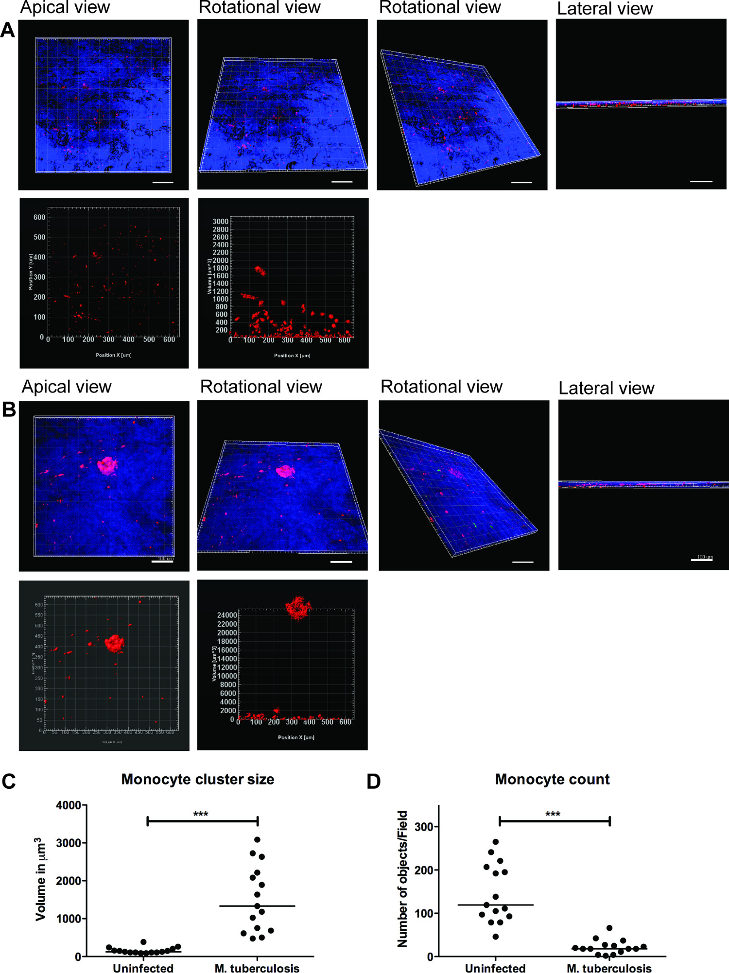
TB 감염을 모니터링하는 폐 조직 모델의 사용에 대한 예는도 2에 제시되어있다. M.를 시각화 결핵 -immune 세포의 이동과 상호 작용, 우리는 M.에 감염된 대 식세포를 도입 GFP를 표현 결핵(녹색) 함께 조직 모델에 갓 격리 PKH26 표지 단핵 세포 (빨간색)와 (파란색, DAPI는 핵에 대한 스테인드). 엠의 7 일 후 추가에 조직 모델에 결핵 -infected 세포, 공 초점 현미경은 인간의 결핵 9의 특징 병변을 모방 감염 (녹색)의 위치 (그림 2), 적색 단핵구의 클러스터링을 알 수있다.
M.의 3 차원 시각화를위한 대표 이미지 일련 결핵은 조직 모델과 세포 클러스터의 정량화가 그림 3에 표시됩니다. 3D 시각화, 상호 작용을 검토하고 3D 이미지에 여러 가지 기능을 정량화 할 수있는 사용자의 유연성을 제공 -infected. M.의 사이트에있는 단핵의 클러스터링을 보여준다도 3b에 도시 된 바와 같이, 녹색 및 적색 박테리아 단핵구 클러스터의 공간적 배치는, 정점 회전 및 측면도에서 알 수있는 결핵. 클러스터 OB되지 않았습니다감염되지 않은 조직 (그림 3A)에 제공합니다. 우리는 단핵구 세포 클러스터의 크기와 수를 정량 개별 단핵구의 수 M.에서 (p <0.01) 감소하면서 셀 클러스터의 크기 (부피), (p <0.001) 향상되는 것을 발견 감염되지 않은 조직의 모델 (도 3C 및 3D)에 비하여 결핵 감염 조직. 이 데이터는 M. 초기 육아종 형성의 이전 연구 결과의 유효성을 검사 결핵 감염 차원 조직 섹션 (9)에서 분석 폐 조직 모델에서 관찰했다.
우리의 데이타는 조직 모델이 복잡한 호스트 M.를 조사하기 위해 셀 - 3D 자연 서식처를 제공하는 것을 제안 결핵 통신망. 우리는 또한 3D 시각화 및 정량 분석은 조직 모델 (그림 3) 기능을 연구하기위한 더 나은 도구가 있음을 발견했다. 셀 클러스터의 정량 (예를 들어 육아종) 자주 스트레치 여러 세포층에 HES와 완전히 3D 정량 분석에 의해 포착 될 수있다. 또한, 개별 셀 또는 모델의 박테리아의 정확한 공간과 시간 기능의 시각화 지정된 실험실에서 라이브 영상, 마이그레이션 및 추적 연구를 할 수 있습니다.

병원성 M. 주위 조직 모델 클러스터도 2 단핵구 결핵. 감염되지 않은 및 엠의 대표 공 촛점 이미지 결핵에 감염된 조직 모델이 제시된다. 감염되지 않은 조직에 비해 패널 녹색에서 (M. tuberculosis- GFP), 빨간색 (PKH26 표지 단구), 블루 (DAPI 염색 핵)과 합병 채널은 감염된 조직에서 단핵 세포의 채용을 보여줍니다. 스케일 - 100 ㎛.large.jpg "스타일 ="글꼴 크기 : 14px; 라인 - 높이 : 28px; "대상 ="_ 빈 ">이 그림의 더 큰 버전을 보려면 여기를 클릭하십시오.

도 3 차원 시각화 및 조직 모델의 정량 분석은 유용한 정보를 제공한다. M. 감염된 조직 전체 모델 (A) 감염되지 않은 조직, (B)의 3 차원 시각화의 대표적인 이미지를 결핵, Imaris 화상 처리 소프트웨어 (버전 7.6.8)에 의해 혈구 LSM700 공 촛점 현미경을 사용하여 정량 분석을 통해 광학 단면. 이러한 이미지는 20X 배율로 취득하고, 정점, 회전, 수평, 회전의 수직 및 측면도 (A와 B)에서 시각화를 허용 1.5 ㎛의 간격으로 19.5 ㎛의 조직 두께를 덮고 14 Z-스택. (C) 쿠아M. 후 초기 육아종 클러스터의 단핵구 세포 클러스터 ntitative 분석 강화 모서리 받침 (p <0.0001) 크기 감염된 조직에 더 많은 클러스터를 되풀이, 감염되지 않은 조직에 비해 감염의 유무에 비해 결핵 감염. (D) 단핵 세포의 수의 정량은 감염된 조직의 감소 (P <0.01)를 보였다. 녹색 - M. 결핵 -GFP, 레드 - PKH26 표지 단핵구, 블루 - 세포 핵, 스케일 -. 100 μm의 이 그림의 더 큰 버전을 보려면 여기를 클릭하십시오.
토론
The ability to recruit and form organized cell clusters at the site of infection is the hallmark of human TB 11. These dynamic structures known as tubercle granulomas primarily consist of immune cells (macrophages, monocytes, T-cells and B-cells) and multi-nucleated giant cells surrounding M. tuberculosis. The role of the granuloma has long been considered to wall off the infection, preventing local spread of bacteria. However, more recent studies show that granuloma formation is critical for early bacterial survival, growth and dissemination 12. A strategy of new studies is to identify molecules or pathways that could efficiently be targeted to inhibit the cellular migration in granuloma formation and/or TB dissemination.
A caveat for novel studies on TB is the lack of models that recapitulate human TB. The most widely used experimental animals do not form true granuloma upon M. tuberculosis infection, and are therefore not appropriate choices for studies of TB 13-16. Non-human primates have the closest resemblance to human TB 17, but are not the preferred choice owing to high operational costs and ethical issues. Human TB is a complex immunological process and is difficult to model in vitro. Cell cultures of monolayers or co-cultures lack the 3D environment and tissue responses. Therefore, we have developed an innovative lung tissue model based on human primary immune cells and human lung-specific cell lines 8,9. The model displays characteristic features of human lung tissue, including epithelia with evenly integrated macrophages, formation of extracellular matrix, stratified epithelia and mucus secretion 9.
The 3D human lung tissue model has several benefits over the in vitro single or co-cultures seeded on tissue culture plates or transwell inserts. First, the human lung-specific cells (fibroblasts and epithelial cells) are not commonly included in the in vitro single or co-cultures. Second, the immune cells and lung-specific cells are embedded in a 3D physiological context (collagen rich extra-cellular matrix products). The response of cells to a stimulus/infection and the migratory behaviour of cells, for instance formation of a granuloma, differ significantly between a 2D and 3D environment. Furthermore, the described method enables the 3D visualization and robust 3D quantitative analysis that provides pivotal information on spatial distribution and intricate cellular interactions.
Experimental infection in the model tissue with M. tuberculosis resulted in clustering of macrophages at the site of infection, reminiscent of early TB granuloma (Figure 2 and 3). We have recently demonstrated that mutant strains defective in the ability to secrete the virulence factor ESAT-6 or Mycobacterium bovis BCG that lacks ESAT-6 did not induce the clustering of monocytes (no early granuloma), in contrast to the virulent M. tuberculosis 9. These data are consistent with the observations made from Mycobacterium marinum-infected zebrafish embryos, whose transparency allows for elegant live imaging of granuloma formation 12. As there is no gold-standard model for TB, we took advantage of the surgically resected tissue biopsies from TB patients for validation of the method 9. Our in vitro tissue model shares several characteristics with the lung and lymph node biopsies from TB patients, including the aggregation of macrophages in granuloma, the presence of both intra- and extracellular bacteria 18 and induction of necrosis 11.
Although the described model has physiological relevance to human TB and has several advantages over other in vitro models, it has some limitations. For instance, out of more than 20 collagen proteins identified in humans, only type I is included to the model to mimic the extra-cellular matrix. However, type I collagen is a complex mixture of extra-cellular matrix products and is the most abundant collagen in the human body. Further, we have demonstrated the presence of collagen IV and several extra-cellular matrix proteins such as tropoelastin, vimentin and laminin, which are produced by the epithelial cells and fibroblasts in the tissue model, indicating the synthesis of new collagen 8. Presently, the lung tissue model only has monocytes and macrophages, besides lung-specific cells. It lacks neutrophils and lymphocytes that are also known to be present in the granuloma. Remarkably the model is not limited to the introduction of additional immune cells and is of interest to explore how they contribute to the complex cellular interactions in human TB. Implantation of primary alveolar macrophages, skin-specific cells and lung carcinoma cells has already been tested in the model. Since our objective was to use a model that closely resembles human TB, introduction of mouse cells have not been attempted.
In summary, the lung tissue model has implications for both basic mechanistic and applied studies. Potential applications of the lung model include the study of innate immunity, investigating mechanistic aspects of host defences such as phagosomal maturation, autophagy, production of cytokines, chemokines and anti-microbial peptides, and functional characterization of individual cell types. Strikingly, the in vitro tissue model allows manipulation of one or more cells types and provides a relevant tissue micro-environment, not only for studies on TB, but for a variety of infectious and non-infectious diseases that affect the lungs.
공개
The authors declare no competing financial interests.
감사의 말
The authors acknowledge the Microscopy core facility at the Faculty of Health Sciences, Linköping University for providing access to advanced imaging systems; Karl-Eric Magnusson (Emeritus Scientist) at the Dept. of Clinical and Experimental Medicine, Linköping University for providing access to Imaris 3D/4D image processing software (Bitplane, Switzerland); and S. Braian for his help with the lung model cartoon. This work was supported by funds from the Swedish Research Council (Alternatives to animal research, 2012-1951) and Swedish Research Council (2012-3349) to M.L. and Swedish Foundation for Strategic Research to S.B. S.B. receive grants from the Karolinska Institutet, Swedish Research Council, the Swedish International Development Cooperation Agency (Sida) and the Swedish Civil Contingencies Agency (MSB), and the Swedish Heart and Lung Foundation (HLF). M.S. received grants from the Karolinska Institutet and Stockholm County Council.
자료
| Name | Company | Catalog Number | Comments |
| Cell culture inserts | BD Falcon | 353092 | |
| 6-well culture plates | BD Falcon | 353046 | |
| MRC-5 cells, lung fibroblasts | ATCC#CCL-171 | ||
| 16HBE cells, lung epithelial cells | Gift from Dr. Dieter Gruenert, Mt. Zion Cancer Center, University of California, San Fransisco, USA | ||
| 5 x Dulbecco’s modified Eagle’s medium (5 x DMEM) | Gibco | 12800-082 | Made from powder but add 5 times less water. Adjust pH to 7.3 and filter it using a 0.2 µm filter. |
| Dulbecco’s modified Eagle’s medium with glucose (DMEM) 1x | Gibco | 41965-039 | |
| Minimum Essential Medium (MEM) 1x with Earle’s salts | Sigma | M4655 | |
| Non-Essential Amino Acids Solution, 100x | Life Technologies | 11140-035 | |
| L-glutamine 200 mM (100x) | Gibco | 25030-024 | |
| Sodium Pyruvate | Life Technologies | 11360-039 | |
| NaHCO3 (71.2 mg/ml) | Prepared in house | ||
| Heat inactivated Fetal Bovine Serum (FBS) | Gibco | 10270-106 | Heat inactivated for 30 min, 56 °C |
| Gentamicin (50 mg/ml) | Gibco | 15750-060 | |
| Hepes buffer solution 1M | Gibco | 15630-056 | |
| Penicillin Streptomycin (Pen Strep) | Gibco | 15140-122 | |
| Lymphoprep | Axis-Shield | 7801 | |
| Ultrapure 0.5 M EDTA | Gibco | 15575 | |
| Bovine Collagen PA treated (500 ml) | Organogenesis | 200-055 | |
| Pure col purified Bovine Collagen solution (100 ml) | Advanced biomatrix | 5005-B | |
| Extracellular matrix protein, Fibronectin (1 mg) | BD | 354008 | |
| Primary human monocytes/macrophages | Isolated from human whole blood or buffy coats. | ||
| PKH26 Red fluorescent cell linker | Sigma | MINI26 | |
| Mycobacterium tuberculosis H37Rv expressing green fluorescent protein | M. tuberculosis H37Rv wild type was transformed with the pFPV2 plasmid constitutively expressing GFP. | ||
| Middlebrook 7H9 medium | Difco | 271310 | |
| BBL Middlebrook ADC Enrichment | BBL | 211887 | |
| Tween-80 | |||
| Glycerol | |||
| Kanamycin B sulfate (20 µg/ml) | Sigma | B5264 | |
| Prolong Gold anti=-fade reagent with DAPI | Invitrogen | P36935 | |
| Trypsin -EDTA | |||
| Bovine serum albumin | |||
| Paraformaldehyde | |||
| DAPI | |||
| LSM700 Confocal microscope | Zeiss | ||
| iMaris Scientific 3D/4D image processing software, version 7.6.8 | Bitplane AG |
참고문헌
- Sakamoto, K. The pathology of Mycobacterium tuberculosis infection. Veterinary pathology. 49, 423-439 (2012).
- Saunders, B. M., Britton, W. J. Life and death in the granuloma: immunopathology of tuberculosis. Immunol Cell Biol. 85, 103-111 (2007).
- Kaufmann, S. H. New issues in tuberculosis. Ann Rheum Dis. Ann Rheum Dis. 63, ii50-ii56 (2004).
- Morrison, J., Pai, M., Hopewell, P. C. Tuberculosis and latent tuberculosis infection in close contacts of people with pulmonary tuberculosis in low-income and middle-income countries: a systematic review and meta-analysis. Lancet Infect Dis. 8, 359-368 (2008).
- Barry 3rd, C. E., et al. The spectrum of latent tuberculosis: rethinking the biology and intervention strategies. Nat Rev Microbiol. 7, 845-855 (2009).
- Puissegur, M. P., et al. An in vitro dual model of mycobacterial granulomas to investigate the molecular interactions between mycobacteria and human host cells. Cell Microbiol. 6, 423-433 (2004).
- Kapoor, N., et al. Human Granuloma In Vitro Model, for TB Dormancy and Resuscitation. PLoS One. 8, e53657 (2013).
- Nguyen Hoang, ., T, A., et al. Dendritic cell functional properties in a three-dimensional tissue model of human lung mucosa. Am J Physiol Lung Cell Mol Physiol. 302, L226-L237 (2012).
- Parasa, V. R., et al. Modeling Mycobacterium tuberculosis early granuloma formation in experimental human lung tissue. Dis Model Mech. 7, 281-288 (2014).
- Cozens, A. L., et al. CFTR expression and chloride secretion in polarized immortal human bronchial epithelial cells. Am J Respir Cell Mol Biol. 10, 38-47 (1994).
- Brighenti, S., Andersson, J. Local immune responses in human tuberculosis: learning from the site of infection. J Infect Dis. 205, S316-S324 (2012).
- Davis, J. M., Ramakrishnan, L. The role of the granuloma in expansion and dissemination of early tuberculous infection. Cell. 136, 37-49 (2009).
- Gupta, U. D., Katoch, V. M. Animal models of tuberculosis. Tuberculosis (Edinb). 85, 277-293 (2005).
- Kashino, S. S., Napolitano, D. R., Skobe, Z., Campos-Neto, A. Guinea pig model of Mycobacterium tuberculosis latent/dormant infection. Microbes Infect. 10, 1469-1476 (2008).
- Singhal, A., et al. Experimental tuberculosis in the Wistar rat: a model for protective immunity and control of infection. PLoS One. 6, e18632 (2011).
- Subbian, S., et al. Phosphodiesterase-4 inhibition alters gene expression and improves isoniazid-mediated clearance of Mycobacterium tuberculosis in rabbit lungs. PLoS Pathog. 7, e1002262 (2011).
- Lin, P. L., et al. Tumor necrosis factor neutralization results in disseminated disease in acute and latent Mycobacterium tuberculosis infection with normal granuloma structure in a cynomolgus macaque model. Arthritis Rheum. 62, 340-350 (2010).
- Rahman, S., et al. Compartmentalization of immune responses in human tuberculosis: few CD8+ effector T cells but elevated levels of FoxP3+ regulatory t cells in the granulomatous lesions. Am J Pathol. 174, 2211-2224 (2009).
재인쇄 및 허가
JoVE'article의 텍스트 или 그림을 다시 사용하시려면 허가 살펴보기
허가 살펴보기This article has been published
Video Coming Soon
Copyright © 2025 MyJoVE Corporation. 판권 소유