Method Article
Multimodal Cross-Device 및 Marker-Free Co-registration of Preclinical Imaging Modalities (전임상 이미징 양식의 Multimodal Cross-Device 및 Marker-Free Co-Registration)
요약
병태생리학에 대한 포괄적인 이해를 얻기 위해 여러 이미징 양식의 조합이 필요한 경우가 많습니다. 이 접근 방식은 팬텀을 사용하여 두 양식의 좌표계 간에 차등 변환을 생성한 다음 공동 정합에 적용됩니다. 이 방법을 사용하면 생산 스캔에서 기준점이 필요하지 않습니다.
초록
양전자 방출 단층 촬영(PET)과 결합된 X선 컴퓨터 단층 촬영(CT) 또는 PET와 결합된 자기 공명 영상(MRI)과 같은 통합 전임상 복합 이미징 시스템은 널리 사용 가능하며 일반적으로 강력한 공동 등록 볼륨을 제공합니다. 그러나 독립형 MRI를 기존 PET-CT와 결합하거나 광학 단층 촬영 또는 고해상도 X선 마이크로단층 촬영의 추가 데이터를 통합하기 위해 별도의 장치가 필요한 경우가 많습니다. 이를 위해서는 이미지 공동 정합이 필요하며, 여기에는 다중 모드 마우스 베드 설계, 기준 마커 포함, 이미지 재구성 및 소프트웨어 기반 이미지 융합과 같은 복잡한 측면이 포함됩니다. 기준 마커는 동적 범위 문제, 이미징 시야의 제한, 마커 배치의 어려움 또는 시간 경과에 따른 마커 신호 손실(예: 건조 또는 붕괴로 인한)로 인해 생체 내 데이터에 문제를 제기하는 경우가 많습니다. 이러한 과제는 이미지 공동 등록이 필요한 각 연구 그룹에서 이해하고 해결해야 하며, 관련 세부 사항은 기존 출판물에서 거의 설명되지 않기 때문에 반복적인 노력이 필요합니다.
이 프로토콜은 이러한 문제를 극복하는 일반적인 워크플로를 간략하게 설명합니다. 차등 변환은 처음에 기준 마커 또는 시각적 구조를 사용하여 생성되지만 이러한 마커는 생산 스캔에 필요하지 않습니다. 재구성 소프트웨어에서 생성된 볼륨 데이터 및 메타데이터에 대한 요구 사항이 자세히 설명되어 있습니다. 이 논의에서는 각 양식에 대해 개별적으로 요구 사항을 달성하고 확인하는 방법을 다룹니다. 팬텀 기반 접근 방식은 두 이미징 양식의 좌표계 간에 차등 변환을 생성하는 데 설명되어 있습니다. 이 방법은 기준 마커 없이 생산 스캔을 공동 등록하는 방법을 보여줍니다. 각 단계는 사용 가능한 소프트웨어를 사용하여 설명되며 상업적으로 사용 가능한 팬텀에 대한 권장 사항이 있습니다. 다양한 현장에 설치된 다양한 이미징 기법의 조합에 대한 이 접근 방식의 실현 가능성이 입증되었습니다.
서문
다양한 전임상 이미징 방식에는 뚜렷한 장점과 단점이 있습니다. 예를 들어, X선 컴퓨터 단층 촬영(CT)은 뼈와 폐와 같은 다양한 전파 밀도를 가진 해부학적 구조를 검사하는 데 적합합니다. 빠른 획득 속도, 높은 3차원 해상도, 이미지 평가의 상대적 용이성, 조영제 1,2,3의 유무에 관계없이 다양한 용도로 인해 널리 사용됩니다. 자기공명영상(MRI)은 전리 방사선 없이 가장 다재다능한 연조직 조영제를 제공합니다4. 반면, 양전자 방출 단층 촬영(PET), 단일 광자 방출 컴퓨터 단층 촬영(SPECT), 형광 매개 단층 촬영(FMT) 및 자기 입자 이미징(MPI)과 같은 추적자 기반 양식은 분자 과정, 대사 및 고감도로 방사성 표지 진단 또는 치료 화합물의 생체 분포를 정량적으로 평가하기 위한 확립된 도구입니다. 그러나 해상도와 해부학적 정보가 부족합니다 5,6. 따라서, 보다 해부학 지향적인 양식은 일반적으로 추적자 검출에 강점을 가진 매우 민감한 양식과 쌍을 이룬다(7). 이러한 조합은 특정 관심 영역 내에서 추적자 농도의 정량화를 가능하게 합니다 8,9. 결합된 이미징 장치의 경우, modality co-registration은 일반적으로 기본 제공 기능입니다. 그러나 다른 장치에서 스캔을 공동 등록하는 것도 유용합니다(예: 장치를 별도로 구입했거나 하이브리드 장치를 사용할 수 없는 경우).
이 기사에서는 기초 연구 및 약물 개발에 필수적인 소동물 이미징의 교차 양식 융합에 중점을 둡니다. 선행 연구(10 )에서는 이것이 특징 인식(feature recognition), 윤곽 매핑(contour mapping) 또는 기준점 마커(fiducials)를 사용하여 달성될 수 있다고 지적한다. 기준점은 다양한 이미징 양식의 이미지를 정확하게 정렬하고 상관시키기 위한 기준점입니다. 특별한 경우에, 기준점은 누드 마우스의 피부에 중국 잉크의 점일 수도 있다11; 그러나 종종 기준 마커가 내장된 이미징 카트리지가 사용됩니다. 이것은 강력하고 잘 개발된 방법(10)이지만, 모든 스캔에 이를 사용하는 것은 실제적인 문제를 야기한다. MRI로 감지할 수 있는 기준점은 액체 기반인 경우가 많으며 보관 중에 건조되는 경향이 있습니다. PET에는 방사성 마커가 필요하며, 방사성 마커의 신호는 방출체의 반감기에 따라 붕괴되며, 이는 일반적으로 생물 의학 응용 분야에서는 짧기 때문에 스캔 직전에 준비가 필요합니다. 기준 마커와 검사된 물체의 신호의 동적 범위의 불일치와 같은 다른 문제는 in vivo imaging에 큰 영향을 미칩니다. 광범위한 동적 대비를 위해서는 마커 신호 강도를 검사 대상에 맞게 자주 조정해야 합니다. 결과적으로, 약한 마커 신호는 분석에서 감지되지 않을 수 있지만 강한 마커 신호는 이미지 품질을 손상시키는 아티팩트를 생성할 수 있습니다. 또한 마커를 일관되게 포함하려면 많은 응용 분야에서 시야가 불필요하게 커야 하며, 이로 인해 방사선 노출이 높아지고, 데이터 볼륨이 커지고, 스캔 시간이 길어지고, 경우에 따라 해상도가 낮아질 수 있습니다. 이는 실험실 동물의 건강과 생성된 데이터의 품질에 영향을 미칠 수 있습니다.
변환 및 미분 변환
이미지 데이터 세트는 복셀 데이터와 메타데이터로 구성됩니다. 각 복셀은 강도 값과 연결되어 있습니다(그림 1A). 메타데이터에는 이미징 장치의 좌표계에서 데이터셋 배치(그림 1B)와 좌표계의 배율을 조정하는 데 사용되는 복셀 크기를 지정하는 변환이 포함되어 있습니다. 장치 유형 또는 스캔 날짜와 같은 추가 정보는 메타데이터에 선택적으로 저장할 수 있습니다. 언급된 변환은 수학적으로 강체 변환이라고 합니다. 강체 변형은 이미지 또는 기하학적 공간에서 오브젝트의 방향이나 위치를 변경하는 동시에 각 점 쌍 사이의 거리를 유지하는 데 사용되며, 이는 변환된 오브젝트가 공간에서 회전 및 평행 이동하는 동안 크기와 모양을 유지한다는 것을 의미합니다. 이러한 일련의 변환은 회전과 변환으로 구성된 단일 변환으로 설명할 수 있습니다. 데이터 좌표에서 메트릭 목표 좌표로 이동하기 위해 소프트웨어에서 사용하는 공식은 그림 1C에 나와 있으며, 여기서 R은 정규 직교 회전 행렬, d 및 v는 복셀 인덱스 및 크기, t는 3 x 1 변환 벡터12입니다. 회전은 그림 1D에 자세히 설명되어 있습니다.
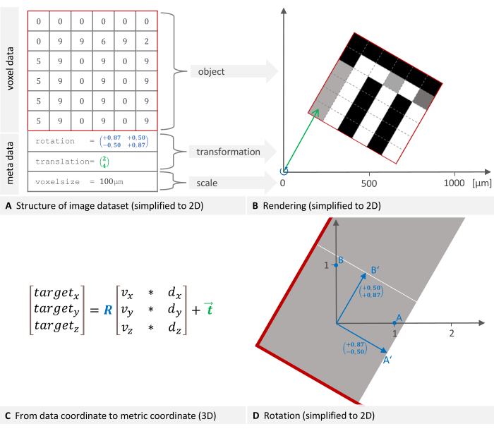
그림 1: 이미지 데이터셋 구조의 2D 표현 및 글로벌 좌표계에서의 배치. (A) 이미지 데이터셋은 복셀 데이터와 메타데이터로 구성됩니다. 배치 및 복셀 크기를 지정하는 변환은 필수 메타데이터 구성 요소입니다. (B) 이미지가 장치의 좌표계로 렌더링됩니다. 오브젝트를 배치하는 데 필요한 변환은 회전(파란색)과 평행 이동(녹색)으로 구성됩니다. (C) 데이터 좌표에서 목표 좌표로 이동하기 위해 소프트웨어는 이 공식을 사용합니다. 여기서 R은 정규 직교 회전 행렬, d, v는 복셀 인덱스 및 크기, t는 3 x 1 평행 이동 벡터입니다. (D) 회전 행렬(평면 A의 파란색)은 회전 점의 선형 변환을 나타냅니다. 점의 좌표에 이 행렬을 곱하면 새로운 회전 좌표가 생성됩니다. 이 그림의 더 큰 버전을 보려면 여기를 클릭하십시오.
차등 변환은 한 좌표계에서 다른 좌표계로(예: PET에서 X선 마이크로단층촬영(μCT)으로) 좌표를 변환하는 강체 변환이며, 기준 마커를 사용하여 계산할 수 있습니다. 적어도 세 개의 공통점(기준점)이 두 좌표계에서 모두 선택됩니다. 좌표에서 좌표를 변환하는 수학적 변환을 파생할 수 있습니다. 이 소프트웨어는 최소 제곱법을 사용하며, 이 방법은 측정된 데이터에 오류 또는 노이즈가 있는 연립방정식에 가장 적합한 솔루션을 제공합니다. 이를 프로크루스테스 문제13 이라고 하며 특이값 분해를 사용하여 해결됩니다. 이 방법은 독특하고 잘 정의된 솔루션으로 이어지기 때문에 신뢰할 수 있고 견고합니다(적어도 3개의 비동일 선상에 있는 마커가 제공되는 경우). 6개의 자유 매개변수가 계산되며, 3개는 변환용이고 3개는 회전용입니다. 다음에서는 기술적으로 회전 행렬과 평행 이동 벡터로 구성되지만 변환 행렬이라는 용어를 사용합니다.
각 이미징 장치에는 고유한 좌표계가 있으며, 소프트웨어는 좌표계를 정렬하기 위해 차등 변환을 계산합니다. 그림 2A,B는 미분 변환이 결정되는 방법을 설명하고, 그림 2C,D는 미분 변환이 적용되는 방법을 설명합니다. 그림 2E의 CT와 PET가 융합된 예시 이미지에서 볼 수 있듯이 두 양식의 이미지는 서로 다른 치수를 가질 수 있으며 공정 중에 유지될 수 있습니다.
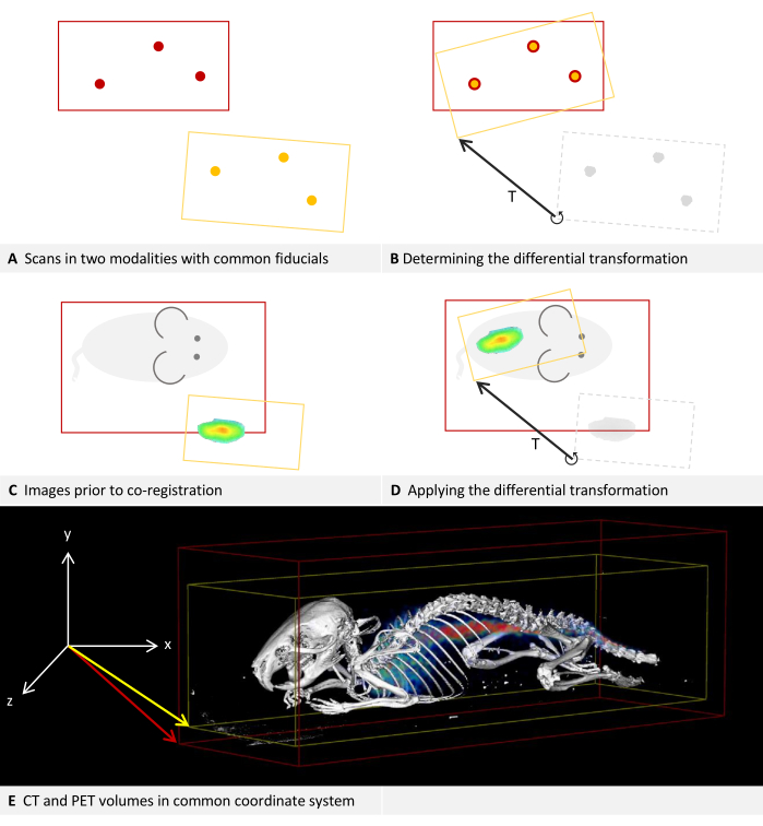
그림 2: 미분 변환. (A-D) 2D로 단순화. 다른 모달리티에도 적용 가능하지만, 이 예에서는 모달리티가 CT 및 PET라고 가정합니다. (답,씨) 빨간색 경계 상자가 있는 CT 이미지는 좌표계에 배치됩니다. 동일한 좌표계에 적용하면 노란색 경계 상자가 있는 PET 이미지가 편차로 배치됩니다. (B) CT와 PET 모두에 위치할 수 있는 기준 마커를 사용하여 미분 변환 T를 결정할 수 있습니다. 이것은 화살표로 상징됩니다. 미분 변환 행렬이 저장됩니다. (D) 그런 다음 이전에 저장된 미분 변환 매트릭스 T를 각 PET 이미지에 적용할 수 있습니다. 이렇게 하면 메타데이터의 원래 변환을 대체하는 새 변환이 생성됩니다. (E) PET 영상과 융합된 CT 영상. 두 이미지의 메타데이터에 있는 변환은 동일한 좌표계를 참조합니다. 이 그림의 더 큰 버전을 보려면 여기를 클릭하십시오.
방법 및 요구 사항
제시된 방법의 경우, 두 모달리티에서 모두 볼 수 있는 마커를 포함하는 팬텀이 두 장치에서 모두 스캔됩니다. 그런 다음 제안된 소프트웨어에서 이러한 기준점을 표시하여 두 양식 간의 미분 변환을 계산하는 것으로 충분합니다. 차등 변환은 각 장치 쌍에 대해 개별적으로 만들어야 합니다. 저장하고 나중에 새 이미지에 적용할 수 있으므로 후속 스캔에서 기준 마커가 필요하지 않습니다. 다른 장치의 좌표계에서 이미지의 최종 배치는 다시 변환으로 설명될 수 있으며 이미지의 메타데이터에 저장되어 원래 변환을 대체할 수 있습니다.
이 방법에 대한 네 가지 요구 사항을 공식화할 수 있습니다: (1) 멀티모달 팬텀: 두 모달리티에서 모두 볼 수 있는 마커를 포함하는 팬텀을 사용할 수 있어야 합니다. 다양한 팬텀이 상업적으로 구할 수 있으며, 팬텀 제작을 위한 3D 프린팅의 사용은 널리 설명되어 왔으며14 심지어 방사성 이형성(radioisitopes)15의 통합도 포함된다. 다음 예에서 사용된 팬텀은 재료 표에 나열되어 있습니다. 적어도 3개의 동일 선상에 있지 않은 점이 필요합니다16. 마커는 적절한 트레이서로 채울 수 있는 캐비티, 각 모달리티에서 쉽게 감지할 수 있는 재료로 만들어진 작은 물체 또는 두 모달리티에서 식별할 수 있는 한 팬텀 자체의 구멍, 절단 또는 가장자리일 수 있습니다. (2) 멀티모달 캐리어: 마우스 베드와 같은 캐리어가 필요하며, 이 캐리어는 두 장치 모두에서 재현 가능한 위치에 고정할 수 있어야 합니다. 이상적으로는 오류를 피하기 위해 역방향으로 사용할 수 없어야 합니다. 캐리어는 진정된 동물을 위치를 변경하지 않고 한 이미징 장치에서 다른 이미징 장치로 운반해야 하기 때문에 생체 내 이미징에 특히 중요합니다. 우리의 경험에 비추어 볼 때, 진정제를 투여한 쥐는 오목한 모양의 쥐 침대에 비해 평평한 쥐 침대에서 위치를 변경할 가능성이 더 높습니다. 또한, 움직임을 최소화하기 위해 마우스의 경골을 고정하기 위한 맞춤형 3D 프린팅 지그가 이전에 제안되었습니다17. (3) 자기 일관성(Self-consistency): 각 이미징 장치는 재현 가능하고 일관성 있는 방식으로 참조 프레임에서 복원된 볼륨의 회전 및 이동을 제공해야 합니다. 이는 또한 작은 영역만 스캔할 때 전체 장치의 좌표계가 유지됨을 의미합니다. 이는 이미징 장치의 자체 일관성을 테스트하는 프로토콜의 일부입니다. (4) 소프트웨어 지원: 제안된 소프트웨어는 장치에서 제공하는 재구성된 볼륨에 저장된 메타데이터(복셀 크기, 변환, 방향)를 해석할 수 있어야 합니다. 볼륨은 DICOM, NIfTI, Analyze 또는 GFF 파일 형식일 수 있습니다. 다양한 파일 형식에 대한 개요는 Yamoah et al.12를 참조하십시오.
두 양식의 공동 등록이 설명되는 동안, 절차는 예를 들어, 두 개의 양식을 하나의 참조 양식에 공동 등록함으로써, 세 개 이상의 양식에도 적용 가능하다.
프로토콜
프로토콜의 소프트웨어 단계는 "분석 소프트웨어"라고 하는 Imalytics 전임상에서 수행되어야 합니다( 재료 표 참조). 볼륨을 "underlay"와 "overlay"18라고 하는 두 개의 다른 레이어로 로드할 수 있습니다. 언더레이 렌더링은 일반적으로 세분화의 기반이 될 수 있는 해부학적으로 상세한 데이터 세트를 검사하는 데 사용됩니다. 투명하게 렌더링할 수 있는 오버레이는 이미지 내의 추가 정보를 시각화하는 데 사용할 수 있습니다. 일반적으로 트레이서 기반 모달리티의 신호 분포가 오버레이에 표시됩니다. 이 프로토콜을 사용하려면 선택한 계층을 여러 번 전환해야 합니다. 이 레이어는 편집 작업의 영향을 받습니다. 현재 선택한 레이어는 마우스 아이콘과 창 아이콘 사이의 상단 도구 모음에 있는 드롭다운 목록에 표시됩니다. Tab 키를 눌러 언더레이와 오버레이 사이를 전환하거나 드롭다운 목록에서 원하는 레이어를 직접 선택할 수 있습니다. 이 프로토콜은 자체 일관성을 테스트하고 차등 변환을 결정하는 데 사용되는 스캔(또는 이미지)을 "보정 스캔"이라고 하며, 이는 이후에 콘텐츠 생성 이미징에 사용되는 "생산 스캔"과 대조됩니다. 프로토콜에 사용되는 양식은 CT와 PET입니다. 그러나 앞서 설명한 바와 같이 이 방법은 부피 데이터를 획득할 수 있는 모든 전임상 이미징 양식에 적용됩니다.
1. 캐리어와 팬텀 조립
알림: 팬텀을 고정할 수 있는 적절한 다중 모드 캐리어(예: 마우스 베드)를 사용할 수 있어야 합니다. 이 어셈블리와 관련된 제안 사항, 자주 발생하는 문제 및 문제 해결에 대한 설명을 참조하십시오.
- 팬텀에서 기준 마커를 준비합니다.
참고: 필요한 특정 준비는 사용된 양식과 트레이서에 따라 다릅니다. 예를 들어, 많은 MRI 팬텀에는 물로 채워야 하는 구멍이 있는 반면, PET에는 또 다른 예로 방사성 추적자가 필요합니다. - 팬텀을 캐리어에 넣고 이미지 품질을 손상시키지 않는 테이프와 같은 재료로 고정합니다.
참고: 팬텀에 대한 요구 사항은 소개 섹션에 자세히 설명되어 있습니다.
2. 캘리브레이션 스캔 수행 및 자체 일관성 확인
참고: 각 이미징 장치에 대해 이 단계를 반복해야 합니다.
- 서로 다른 시야를 가진 두 개의 스캔을 획득합니다.
- 이미징 장치에 캐리어를 놓습니다. 신뢰할 수 있고 재현 가능한 방식으로 배치되었는지 확인하십시오.
- 장치 제조업체의 지침에 따라 전체 팬텀을 덮는 넓은 시야를 사용하여 스캔합니다. 이 이미지는 다음 단계에서 "이미지 A"라고 합니다.
참고: 이 스캔은 미분 변환 매트릭스를 계산하는 데에도 사용되므로 모든 기준점을 포함하는 것이 중요합니다. - 이미징 장치에서 캐리어를 제거하고 교체합니다.
알림: 이 단계는 장치에 대한 이동통신사의 배치가 신뢰할 수 있는지 확인합니다. - 이미징 장치가 제한된 시야를 지원하지 않는 경우, 즉 항상 전체 시야를 스캔하는 경우, 자기 일관성을 합리적으로 가정할 수 있습니다. 3단계로 바로 진행합니다.
- 장치 제조업체의 지침에 따라 두 번째 스캔을 수행하되 이번에는 훨씬 더 작은 시야를 사용합니다. 이 이미지는 다음 단계에서 "이미지 B"라고 합니다.
알림: 서로 다른 시야로 두 번 스캔하는 것이 중요합니다. 시야의 정확한 위치는 팬텀 구조 또는 가능한 한 많은 기준점과 같은 일부 가시 정보가 포함되는 한 이미지 B에 중요하지 않습니다.
- 언더레이를 로드합니다.
- 해석 소프트웨어를 엽니다.
- 이미지 A를 언더레이로 로드: 메뉴 파일 > 언더레이 > 언더레이 로드. 다음 대화 상자에서 이미지 파일을 선택하고 열기를 클릭합니다.
- 3D 뷰가 없는 경우 [Alt + 3] 을 눌러 활성화합니다.
- 윈도잉 조정: [Ctrl + W] 를 누르고 다음 대화 상자에서 왼쪽 및 오른쪽 세로 막대를 조정하여 팬텀 또는 양식에 따라 트레이서를 명확하게 구별할 수 있도록 합니다. 확인을 클릭하여 대화 상자를 닫습니다.
- 오버레이를 로드합니다.
- 이미지 B를 오버레이로 로드: 오버레이 로드 > 메뉴 파일 > 오버레이. 다음 대화 상자에서 이미지 파일을 선택하고 열기를 클릭합니다.
- 렌더링 방법 변경: 메뉴 3D-Rendering > 오버레이 모드 > ISO 렌더링을 확인합니다.
참고: PET 또는 SPECT와 같은 추적자 기반 양식은 일반적으로 볼륨 렌더링으로 표시되지만 이 경우 ISO 렌더링을 사용하면 위치를 더 쉽게 비교할 수 있습니다. 언더레이는 기본적으로 ISO 렌더링에서 열렸습니다. - 바운딩 박스 보기 활성화: 메뉴 보기 > 심볼 표시 > 바운딩 박스 표시 > 언더레이 바운딩 박스 표시; 메뉴 보기 > 심볼 표시 > 경계 상자 표시 > 오버레이 경계 상자 표시.
- 이미지 정렬을 확인합니다.
- 3D 뷰에 마우스 포인터를 놓고 [Ctrl + 마우스 휠] 을 사용하여 뷰를 확대/축소하여 두 바운딩 박스가 모두 보이도록 합니다. [Alt + 마우스 왼쪽 버튼] 을 누른 상태에서 마우스 포인터를 움직여 뷰를 회전합니다.
- 선택한 레이어를 오버레이로 전환합니다.
- 창 및 색상표 조정: [Ctrl + w]를 누릅니다. 다음 대화 상자의 왼쪽에 있는 드롭다운 목록에서 노란색을 선택합니다. 다음 대화상자의 범위를 언더레이에 대해 선택한 유사한 범위로 조정한 다음, 노란색 렌더링이 흰색 렌더링 내에서만 보일 때까지 작은 단계로 설정을 변경합니다. 확인을 클릭하여 대화 상자를 닫습니다.
참고: 이미지 A(언더레이)의 렌더링은 이제 흰색으로 표시되며 빨간색 경계 상자로 둘러싸여 있습니다. 이미지 B(오버레이)의 렌더링은 노란색으로 표시되며 노란색 경계 상자로 둘러싸여 있습니다. - 이미징 장치와 팬텀을 배치하는 방법이 필요에 따라 자체 일관성이 있는지 육안으로 확인하십시오. 팬텀(또는 양식에 따라 트레이서)은 언더레이와 오버레이에서 완전히 정렬되어야 합니다. 노란색 렌더링은 흰색 렌더링의 하위 집합이어야 합니다.
참고: 노란색 바운딩 박스는 더 작아야 하며 빨간색 테두리 박스 안에 있어야 합니다. 시각적 예는 Representative Results 섹션을 참조하십시오. 정렬이 일치하지 않으면 일반적인 배치 문제 및 문제 해결에 대한 설명을 참조하십시오.
3. 미분 변환의 계산
- 두 양식의 이미지를 불러옵니다.
- 해석 소프트웨어를 엽니다.
- CT 영상 A를 언더레이로 로드: 메뉴 파일 > 언더레이 > 언더레이 로드. 다음 대화 상자에서 이미지 파일을 선택하고 열기를 누릅니다.
- PET 이미지 A를 오버레이로 불러오기: 오버레이 불러오기> 메뉴 파일 > 오버레이를 선택합니다. 다음 대화 상자에서 이미지 파일을 선택하고 열기를 누릅니다.
- 여러 슬라이스 뷰 표시: [Alt + A], [Alt + S], [Alt + C] 를 눌러 축, 궁상, 코로나 슬라이스 뷰를 표시합니다.
알림: 기술적으로는 하나의 평면으로 기준점을 찾기에 충분하지만 모든 평면을 동시에 볼 수 있으므로 더 나은 방향과 더 빠른 탐색이 가능합니다.
- 마커 기반 융합을 수행합니다.
참고: 3.2단계 및 3.3단계는 언더레이와 오버레이를 정렬하는 다른 방법입니다. 3.2단계를 먼저 시도하면 재현하기 쉽고 잠재적으로 더 정확할 수 있습니다. 3.3단계는 명확하게 식별할 수 있는 마커가 충분하지 않은 경우 대체됩니다.- 언더레이만 표시하도록 뷰 전환: 메뉴 보기 > 레이어 설정 > 레이어 가시성 > 오버레이 체크를 해제하십시오. 메뉴 View > Layer Settings > Layer Visibility > 언더레이를 확인합니다.
- 선택한 도면층을 언더레이로 전환합니다.
- 필요한 경우 창 조정: [Ctrl + W] 를 누르고 다음 대화 상자에서 왼쪽 및 오른쪽 세로 막대를 조정하여 기준점을 더 잘 볼 수 있습니다. 확인을 클릭하여 대화 상자를 닫습니다.
- 마우스 동작 모드 "create marker"를 활성화하려면 왼쪽의 수직 도구 모음에서 마커 기호를 클릭합니다. 마우스 포인터에 마커 기호가 표시됩니다.
- 팬텀의 각 기준점에 대해 수행: 기준으로 이동합니다. 이를 위해 평면 뷰 위에 마우스 포인터를 놓고 [Alt + 마우스 휠] 을 사용하여 평면을 절단합니다. 마우스 포인터를 기준점의 중앙에 놓고 마우스 왼쪽 버튼을 클릭합니다.
- 그러면 소프트웨어가 연속된 숫자가 있는 이름을 제안하는 대화 상자가 열립니다. 제안된 이름(예: "Marker001")을 유지하고 확인을 클릭하여 마커를 저장합니다.
참고: 오버레이에 대해 동일한 마커 이름을 다시 사용하는 경우 다른 이름을 사용할 수 있습니다.
- 그러면 소프트웨어가 연속된 숫자가 있는 이름을 제안하는 대화 상자가 열립니다. 제안된 이름(예: "Marker001")을 유지하고 확인을 클릭하여 마커를 저장합니다.
- 오버레이를 표시하도록 보기 설정 조정: 메뉴 보기 > 레이어 설정 > 레이어 가시성 > 오버레이를 확인합니다.
알림: 언더레이의 시야를 활성화된 상태로 유지하는 것이 좋습니다., 방향을 유지하고 두 양식 모두에서 올바른 마커를 식별하는 데 도움이 됩니다. 두 양식이 동기화되지 않았거나 오버레이가 혼동되는 경우 비활성화하십시오: 메뉴 뷰 > 레이어 설정 > 레이어 가시성 > 언더레이 체크를 해제합니다. - 선택한 레이어를 오버레이로 전환합니다.
- 윈도잉 조정: 기준 마커가 명확하게 보이지 않는 경우 [Ctrl + W] 를 누르고 다음 대화 상자에서 왼쪽 및 오른쪽 세로 막대를 조정하여 기준점이 가능한 한 잘 배치될 수 있도록 합니다. 확인을 클릭하여 대화 상자를 닫습니다.
- 팬텀의 각 기준점에 대해 수행: 기준으로 이동합니다. 이를 위해 평면 뷰 위에 마우스 포인터를 놓고 [Alt + 마우스 휠] 을 사용하여 평면을 절단합니다. 마우스 포인터를 기준점의 중앙에 놓고 마우스 왼쪽 버튼을 클릭합니다.
- 그러면 소프트웨어가 연속된 숫자가 있는 이름을 제안하는 대화 상자가 열립니다. 제안된 이름을 유지하고 확인을 클릭하여 마커를 저장합니다.
참고: 언더레이와 오버레이에서 일치하는 소프트웨어 마커에 대해 동일한 이름을 갖는 것이 중요합니다. 제안된 이름을 유지하고 동일한 순서를 사용하여 두 모달리티에서 마커를 만드는 경우 이 작업이 보장됩니다. 이름을 변경하는 경우 이름이 일치하는지 확인합니다.
- 그러면 소프트웨어가 연속된 숫자가 있는 이름을 제안하는 대화 상자가 열립니다. 제안된 이름을 유지하고 확인을 클릭하여 마커를 저장합니다.
- 두 레이어의 뷰를 활성화합니다: 메뉴 보기 > 레이어 설정 > 레이어 가시성 > 언더레이를 확인합니다. 메뉴 보기 > 레이어 설정 > 레이어 가시성 > 오버레이를 확인합니다.
- 언더레이와 오버레이의 마커 정렬: Menu Fusion > 오버레이를 언더레이에 등록> 회전 및 평행 이동(마커)을 계산합니다. 다음 대화 상자는 융합의 잔차를 보여줍니다. 이 측정을 기록하고 확인을 클릭하십시오.
- 정렬 결과 확인: 언더레이와 오버레이의 표식기가 시각적으로 일치해야 합니다. 토론 섹션에서 문제 해결 및 융합 잔류에 대한 정확도에 대한 참고 사항을 확인하십시오.
참고: 오버레이의 변형이 변경되었습니다. 새 오버레이 변환의 세부 정보를 표시하려면 [Ctrl + I]를 누릅니다.
- 마커 기반 융합이 불가능한 경우 대화형 융합을 수행합니다. 3.2단계가 완료되면 3.4단계로 바로 진행합니다.
- 두 레이어의 뷰를 활성화합니다: 메뉴 보기 > 레이어 설정 > 레이어 가시성 > 언더레이를 확인합니다. 메뉴 보기 > 레이어 설정 > 레이어 가시성 > 오버레이를 확인합니다.
- 마우스 모드 "interactive image fusion"을 활성화하려면 왼쪽의 수직 도구 모음에 있는 기호를 클릭합니다. 기호는 공통 중심에 점이 있는 세 개의 오프셋 타원으로 구성됩니다. 이제 마우스 포인터에 이 기호가 표시됩니다.
- 마우스 모드에 대한 설정 도구 모음이 영구 도구 모음 아래의 위쪽 영역에 나타나는지 확인합니다. 언더레이(underlay), 오버레이(overlay) 및 세그먼트화(segmentation)에 대한 세 가지 확인란이 있습니다. 오버레이를 확인합니다. 언더레이 및 세그먼트화를 선택 취소합니다.
- 중첩을 언더레이에 대화식으로 정렬: 언더레이와 오버레이가 가능한 한 잘 정렬될 때까지 서로 다른 뷰에서 회전 및 변환을 수행합니다.
- 회전: 마우스 포인터를 뷰의 가장자리 근처(축, 코로나 또는 시수)에 놓습니다. 이제 마우스 포인터 기호가 화살표로 둘러싸여 있습니다. 마우스 왼쪽 버튼을 누른 상태에서 마우스를 움직여 오버레이를 회전합니다.
- 변환 : 마우스 포인터를보기의 중심 근처에 놓습니다. 마우스 포인터가 동그라미 모양으로 둘러싸여 있지 않습니다. 마우스 왼쪽 버튼을 누른 상태에서 마우스를 움직여 오버레이를 이동합니다.
- 차등 변환 생성 및 저장: Menu Fusion > 오버레이 변환 > 차등 변환 생성 및 저장. 다음 대화 상자에서 원본 오버레이 파일을 선택하고 열기를 클릭합니다. 두 번째 대화 상자에서 차등 변환의 파일 이름을 입력하고 저장을 누릅니다.
참고: 소프트웨어는 원래 변환을 읽은 다음 미분 변환을 계산하기 위해 원본 오버레이 파일이 필요합니다. 사용되는 이미징 장치를 지정하는 파일 이름으로 differential transformation matrix를 저장하는 것이 좋습니다.
4. 생산 이미징
- 두 이미징 장치에서 모두 스캔합니다.
- 샘플(예: 진정제를 투여한 실험실 동물)을 캐리어에 고정합니다.
알림: 캐리어 내 샘플의 위치가 두 스캔 간에 변경되지 않도록 하는 것이 중요합니다. - 캐리어를 CT 장치에 놓습니다. 보정 스캔 중에 수행한 것과 동일한 방식으로 캐리어를 배치해야 합니다.
- 장치 제조업체의 지침에 따라 스캔하십시오.
- 캐리어를 PET 장치에 넣습니다. 보정 스캔 중에 수행한 것과 동일한 방식으로 캐리어를 배치해야 합니다.
- 장치 제조업체의 지침에 따라 스캔하십시오.
- 샘플(예: 진정제를 투여한 실험실 동물)을 캐리어에 고정합니다.
- 미분 변환의 적용을 수행합니다.
- 해석 소프트웨어를 엽니다.
- CT 파일을 언더레이로 로드: 메뉴 파일 > 언더레이 > 언더레이 로드. 다음 대화 상자에서 CT 이미지 파일을 선택하고 확인을 누릅니다.
- PET 파일을 오버레이로 로드: 오버레이 > 로드 오버레이 > 메뉴 파일. 다음 대화 상자에서 PET 이미지 파일을 선택하고 확인을 누릅니다.
- 두 레이어의 뷰를 활성화하십시오 : 메뉴 뷰 > 레이어 설정 > 레이어 가시성 > 언더레이를 확인하십시오. 메뉴 보기 > 레이어 설정 > 레이어 가시성 > 오버레이를 확인합니다.
- 이전에 저장한 차등 변환 매트릭스 로드 및 적용: Fusion > 오버레이 변환 > 변환 로드 및 적용> 메뉴를 선택합니다. 캘리브레이션 과정에서 저장한 differential transformation matrix가 포함된 파일을 선택하고 open을 누릅니다.
참고: 이 단계에서는 오버레이의 메타데이터를 변경합니다. - 변경된 오버레이 저장: 메뉴 > 파일 > 오버레이 > 오버레이 저장. 다음 대화 상자에서 이름을 입력하고 저장을 클릭합니다.
참고: 변경되지 않은 원본 데이터를 유지하는 것이 좋으므로 오버레이를 새 이름으로 저장하는 것이 좋습니다.
결과
그림 3과 그림 4 는 CT에서 볼 수 있는 팬텀의 예를 제공하며 이 경우 SPECT용 트레이서로 채워진 관형 공동이 포함되어 있습니다. 사용된 팬텀과 트레이서는 재료 표에 나열되어 있습니다.
프로토콜의 2단계에서는 보정, 스캔 및 각 이미징 장치의 자체 일관성을 간략하게 설명합니다. 시야각이 다른 두 스캔의 렌더링은 각 장치에 대해 일치해야 합니다. 따라서 노란색으로 표시된 이미지 B는 흰색으로 표시된 이미지 A의 하위 집합이어야 합니다. CT를 사용한 예가 그림 3A에 나와 있습니다. PET 또는 SPECT와 같은 트레이서 기반 모달리티는 일반적으로 볼륨 렌더링으로 시각화됩니다(그림 3B). 그러나 ISO 렌더링을 사용하면 위치를 쉽게 비교할 수 있습니다. 따라서 프로토콜은 사용된 양식에 관계없이 언더레이와 오버레이를 ISO 렌더링으로 전환하도록 사용자에게 지시합니다. 따라서 SPECT 예제에서 노란색 렌더링은 흰색 렌더링의 하위 집합이기도 해야 합니다(그림 3C). 모든 경우에 노란색 바운딩 박스는 더 작아야 하며 빨간색 바운딩 박스 내에 위치해야 합니다. 정렬이 일치하지 않는 경우 토론은 일반적인 배치 문제를 강조하고 문제 해결 제안을 제공합니다.
프로토콜의 3단계에서는 기준 마커를 사용하여 두 양식 간의 차등 변환을 결정하는 방법을 설명합니다. 트레이서 기반 양식의 트레이서는 볼륨으로 존재하기 때문에 사용자는 (점 모양) 기준 마커로 사용할 적절한 포인트를 결정해야 합니다. 그림 4에서 팬텀의 CT 이미지는 언더레이로 로드되고 SPECT 이미지는 언더레이로 로드됩니다. 팬텀 내부의 튜브 곡선 중심은 그림 4A-C와 같이 축방향, 관상식 및 시상 보기에서 볼 수 있듯이 CT 언더레이의 기준 마커로 선택됩니다. 해당 지점은 오버레이에 표시해야 하며, 이는 그림 4D-F와 같이 축방향, 코로나 및 시상 보기에서 표시됩니다. 이제 소프트웨어가 차등 변환을 계산하여 오버레이에 적용할 수 있습니다. 이렇게 하면 그림 4G,H와 같이 두 양식의 마커가 정렬됩니다.
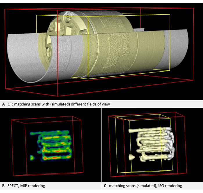
그림 3: 자기 일관성을 보여주는 이미지. (A) CT 볼륨. 프로토콜의 2.4단계에서는 이미지 정렬을 확인해야 합니다. 프로토콜의 단계에 따라 언더레이는 흰색으로 렌더링되고 오버레이의 오버레이와 경계 상자는 노란색으로 렌더링됩니다. 두 레이어가 모두 정렬되어 있습니다(여기서 두 번째 스캔은 첫 번째 스캔의 잘린 복사본에 의해 시뮬레이션됨). (B) 트레이서로 채워진 튜브를 사용한 팬텀의 SPECT 이미징. NIH 색상표를 사용한 볼륨 렌더링. (C) ISO 렌더링의 SPECT 이미지. 언더레이는 흰색으로 렌더링되고 오버레이와 오버레이의 경계 상자는 노란색으로 렌더링됩니다. 두 레이어가 모두 정렬되어 있습니다(여기서 두 번째 스캔은 첫 번째 스캔의 잘린 복사본에 의해 시뮬레이션됨). 이 그림의 더 큰 버전을 보려면 여기를 클릭하십시오.

그림 4: CT 및 SPECT 이미지의 마커 배치. 팬텀의 CT 이미지가 언더레이로 로드됩니다. SPECT 이미지는 오버레이로 로드되고 NIH 색상표를 사용하여 렌더링됩니다. (A-C) 프로토콜의 3.2단계에서는 언더레이에 마커를 배치해야 합니다. 팬텀 내부의 튜브 곡선 중심이 기준점으로 선택되고 Marker001이 여기에 배치되며, 이는 축방향, 관상식, 시상도에서 빨간색 점으로 표시됩니다. (D-F) 일치하는 마커가 오버레이에 배치됩니다. (G) 변환 후의 축 방향 보기. (H) 융합 양식의 3D 보기. 최대 인텐션 프로젝션 렌더링은 팬텀 내에서 SPECT 트레이서를 볼 수 있도록 하는 데 사용됩니다. 이 그림의 더 큰 버전을 보려면 여기를 클릭하십시오.
토론
생산 스캔을 위한 기준 마커가 필요하지 않은 다중 모드 이미지 공동 정합 방법을 제시합니다. 팬텀 기반 접근 방식은 두 이미징 양식의 좌표계 간에 차등 변환을 생성합니다.
융합의 잔차와 미분 변환의 검증
미분 변환을 계산할 때, 소프트웨어는 융합의 잔차를 밀리미터 단위로 표시하며, 이는 변환의 평균 제곱근 오차19 를 나타냅니다. 이 잔차가 복셀 크기의 크기 순서를 초과하는 경우 데이터 세트에서 일반적인 문제를 검사하는 것이 좋습니다. 그러나 모든 이미지에는 약간의 왜곡이 있으므로 잔차가 임의로 작아질 수 없습니다. 사용된 마커의 맞춤만 반영합니다. 예를 들어, 3개의 마커가 있는 공동 정합은 4개의 잘 분산된 마커가 있는 변환보다 동일한 데이터 세트에 대한 잔차가 더 작을 수 있습니다. 이는 더 적은 수의 기준점이 사용될 때 마커 자체가 과도하게 장착될 수 있기 때문에 발생합니다. 마커 수가 많을수록 전체 데이터 세트의 정확도가 향상됩니다.
이 방법의 정량적 정확도는 사용되는 특정 장치 쌍에 따라 다릅니다. 두 장치의 좌표계 간에 계산된 차동 변환은 다음 단계에 따라 검증할 수 있습니다. 프로토콜의 4단계를 준수하지만 기준 마커가 있는 팬텀을 다시 "샘플"로 사용합니다. 팬텀을 어느 위치에나 배치하여 미분 변환을 추정하는 데 사용되는 것과 다른지 확인합니다. 사용 가능한 경우 해당 양식에 적합한 다른 팬텀을 사용할 수도 있습니다. 다음으로, 앞에서 결정한 미분 변환(4.2.5단계)을 적용하여 두 모달리티를 정렬합니다. 그런 다음 프로토콜의 3.2단계에 따라 두 양식의 이미지에 마커를 배치합니다. 이러한 마커의 융합 잔차를 계산하려면 Fusion > Register Overlay to Underlay > Showing Residual Score를 클릭합니다.
잔차 오차는 신호의 평균 오배치를 설명하며 복셀 크기 순서여야 합니다. 콘크리트 수용 임계값은 응용 분야에 따라 다르며 이미징 시스템의 강성 및 정확도와 같은 여러 요인에 따라 달라질 수 있지만 이미지 재구성 아티팩트의 영향을 받을 수도 있습니다.
자체 일관성 문제 해결
종종 자기 일관성의 어려움은 신뢰할 수 없는 배치로 인해 발생합니다. 일반적인 오류는 캐리어를 측면으로 반전된 위치에 배치하는 것입니다. 이상적으로는 이미징 장치에 한 방향으로만 기계적으로 삽입해야 합니다. 이것이 가능하지 않은 경우 사용자를 위해 이해할 수 있는 표시를 추가해야 합니다. 또 다른 빈번한 문제는 세로 축에서 움직일 가능성이 있어 축 방향 위치 지정을 신뢰할 수 없다는 것입니다. 마우스 베드를 제자리에 고정하기 위해 한쪽 끝에 부착할 수 있는 스페이서를 사용하는 것이 좋습니다. 예를 들어, 맞춤형 스페이서는 3D 프린팅으로 빠르고 쉽게 만들 수 있습니다. 그러나 일부 장치는 다양한 시야에서 자체 일관성을 제공할 수 없습니다. 이러한 경우 공급업체에 문의하는 것이 좋으며, 공급업체는 비호환성을 확인하고 향후 업데이트에서 잠재적으로 해결해야 합니다. 그렇지 않으면, 보정 및 생산 이미징을 포함한 모든 스캔에 대해 동일한 시야가 유지되는 경우 이 방법은 신뢰할 수 있습니다.
배치가 벗어난 일부 생산 스캔의 경우, 충분한 캐리어 구조를 식별할 수 있는 경우 보정된 위치로 변환할 수 있습니다. in vivo imaging 의 경우, 진정된 동물은 하나의 캐리어에 있어야 하며, 두 장치에 모두 안전하게 맞는 단일 캐리어를 구성하는 것이 항상 가능한 것은 아닙니다. 종종 추적자 기반 모달리티를 위한 마우스 베드가 사용되며, 그런 다음 CT 장치에 즉석으로 배치됩니다. 예를 들어, 그림 5A에서 MPI 마우스 베드는 기계적 제약으로 인해 CT 마우스 베드 위에 배치되었습니다. 축 방향 여유 공간과 롤링 가능성으로 인해 이 포지셔닝은 신뢰할 수 없습니다. 이러한 경우 하단 마우스 베드를 대체하고 연동이 가능한 어댑터를 설계하는 것이 좋습니다. 예를 들어, 하부에 부착된 트러니언과 상부 마우스 베드의 바닥에 있는 추가 구멍을 사용할 수 있습니다.
그러나 CT 영상에서 마우스 베드를 감지할 수 있으므로 기존 이미지에 대한 후향적 보정이 가능합니다. 이 프로토콜은 캘리브레이션 스캔을 필요로 하며, 그 후에 언더레이에 대한 오버레이의 차등 변환을 계산해야 합니다. 절차는 유사하지만 마우스 베드 구조를 기준점으로 사용하여 각 개별 생산 CT 스캔을 보정 스캔에 매핑해야 합니다.
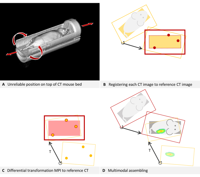
그림 5: 배치 문제 해결. (A) MPI 마우스 베드가 CT 마우스 베드 위에 배치됩니다. 따라서 CT의 위치를 안정적으로 재현할 수 없습니다. 자기 일관성은 각 CT 이미지를 미분 변환을 추정하는 데 사용되는 참조 CT 이미지에 융합하여 달성할 수 있습니다. (비-디) 2D로 단순화. (B) 각 프로덕션 CT 이미지는 오버레이로 로드되고 CT에서 볼 수 있는 마우스 베드의 구조를 사용하여 참조 CT 이미지(언더레이)에 등록됩니다. 보정된 생산 CT 영상은 이제 참조 CT와 일치하며 미분 변환 T와 함께 사용할 수 있습니다. (C) MPI 오버레이는 팬텀의 기준 마커를 사용하여 참조 CT 영상에 등록됩니다. (D) 멀티모달 이미지가 조립됩니다. 이를 위해 각 CT 이미지는 개별 차동 변환을 통해 기준 위치에 매핑됩니다. 그 후, MPI 오버레이는 장치의 모든 이미지에 유효한 차동 변환을 사용하여 참조 위치에 등록됩니다. 이 그림의 더 큰 버전을 보려면 여기를 클릭하십시오.
생산 CT 스캔을 보정 스캔에 매핑하려면 프로토콜의 섹션 3을 참조하여 다음 수정 사항을 통합하십시오. 명확성을 위해 설명은 CT 언더레이 및 MPI 오버레이의 예를 계속 사용합니다. 3.1단계에서 CT 보정 스캔(이미지 A)을 언더레이로 로드하고 보정할 CT 스캔을 오버레이로 로드합니다. MPI 마우스 베드의 구조를 3.2단계의 마커 또는 3.3단계의 시각적 참조로 활용합니다. 3.4단계를 건너뛰고 오버레이를 저장하면 수정된 CT 볼륨이 표시됩니다(메뉴 파일 > 오버레이 > 다른 이름으로 오버레이 저장). 다음 대화 상자에서 새 이름을 입력하고 저장을 클릭합니다. Menu File > Overlay > Closing overlay로 이동하여 오버레이를 닫습니다. 수정이 필요한 다음 CT 스캔을 오버레이로 로드하고 프로토콜의 3.2단계부터 절차를 재개합니다. 이 단계의 기본 개념은 그림 5B에 나와 있습니다.
이제 마우스 베드는 최근에 저장된 모든 CT 볼륨의 보정 스캔과 거의 동일하게 정렬됩니다. 표준 절차의 일부로, 캘리브레이션 스캔은 차동 변환 T를 사용하여 MPI 이미지에 등록됩니다(그림 5C). 이후에 CT 이미지를 MPI와 병합하려면 항상 보정된 CT 볼륨을 사용하십시오(그림 5D).
대칭 이동된 이미지 및 크기 조정 문제 해결
여기에 소개된 등록 방법은 상당히 정확한 이미지 품질을 가정하고 회전과 평행 이동만 조정합니다. 뒤집힌 이미지나 잘못된 배율은 수정되지 않습니다. 그러나 이 두 가지 문제는 차등 변환을 계산하기 전에 수동으로 해결할 수 있습니다.
서로 다른 제조업체의 데이터 형식 간의 불일치로 인해 일부 데이터 세트, 특히 DICOM 형식의 데이터 세트가 소프트웨어에서 미러 반전으로 표시될 수 있습니다. 팬텀 침대와 마우스 침대는 종종 대칭이기 때문에 이 문제는 즉시 드러나지 않을 수 있습니다. 뒤집힌 이미지를 감지하는 것은 그림 3H의 팬텀에서 볼 수 있는 올바른 방향의 돌출된 글자와 같이 각 양식에 인식 가능한 글자가 포함되어 있을 때 더 쉽습니다. 그림 6의 예에서 CT 데이터는 언더레이로 로드되고 MPI 데이터는 오버레이로 로드됩니다. 이것은 기준 마커가 부착된 MPI 마우스 베드에 배치된 마우스의 생체 내 스캔입니다. MPI 마우스 베드는 μCT 마우스 베드 위에 위치합니다(그림 6A). 프로토콜을 준수하고 언더레이와 오버레이 모두에서 일관된 회전 방향으로 기준점을 표시하면 눈에 띄게 일치하지 않는 결과가 생성됩니다(그림 6B). 그러나 자세히 살펴보면 문제를 식별할 수 있습니다. 기준점은 비대칭 삼각형을 형성합니다. 축 방향 보기(그림 6C, D)에서 삼각형의 변을 가장 짧은 쪽에서 중간 쪽에서 가장 긴 쪽으로 관찰하면 CT 데이터에서는 시계 방향 회전이 분명하고 MPI 데이터에서는 시계 반대 방향 회전이 분명합니다. 이것은 이미지 중 하나가 측면으로 반전되어 있음을 보여줍니다. 이 경우 CT 데이터가 정확하다고 가정합니다. MPI 오버레이를 수정하려면 이미지가 뒤집힙니다: 그렇게 하려면 선택한 레이어를 오버레이로 전환하고 메뉴 편집 > Flip > Flip X를 클릭합니다. 소프트웨어에 의해 계산된 미분 변환은 필요한 모든 회전을 포함하므로 이미지가 다른 방향으로 뒤집힌 것처럼 보이더라도 "Flip X"로 충분합니다.
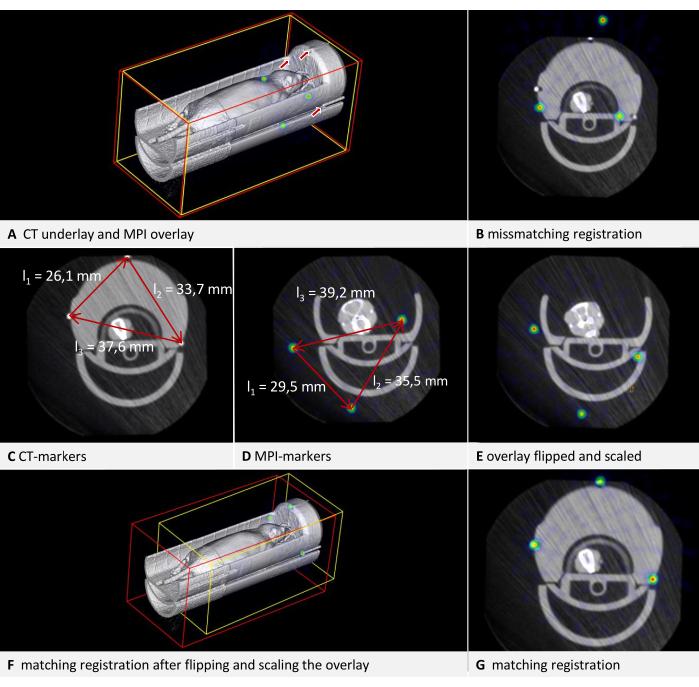
그림 6: 변환 문제 해결. CT 데이터는 복셀 크기가 0.240mm인 언더레이로 로드되고 MPI 데이터는 복셀 크기가 0.249mm인 오버레이로 로드됩니다. 마우스 베드에는 기준 마커가 포함되어 있습니다. (A) 수정되지 않은 오버레이 이미지의 3D 보기. CT 언더레이의 기준점은 화살표로 표시됩니다. MPI 오버레이의 기준점은 NIH 색상표에서 구로 표시됩니다. (B) 적절한 수정 없이 수행된 변환의 일치하지 않는 결과. 융합 잔류 = 6.94mm. (C) CT에서 기준점 사이의 거리 측정. 가장 짧은 거리에서 가장 긴 거리까지 시계 방향 회전. (D) MPI에서 기준점 사이의 거리 측정. 가장 짧은 거리에서 가장 먼 거리까지 시계 반대 방향으로 회전합니다. CT 측정과 비교하면 0.928774의 스케일링 계수가 나옵니다. (E) 대칭 이동 및 크기 조정 후 오버레이가 수정되었습니다. (F) 3D 뷰에서 일치하는 결과를 사용한 변환. (G) 축 보기에서 일치하는 결과를 가진 변환. 융합 잔류 = 0.528mm. 이 그림의 더 큰 버전을 보려면 여기를 클릭하십시오.
잘못된 복셀 크기가 있는 데이터 세트도 수동으로 수정할 수 있습니다. 팬텀의 치수를 알아야 하기 때문에 이미지에서 확인할 수 있습니다. 가장 간단한 방법은 알려진 길이의 모서리를 사용하는 것입니다. 가장자리의 한쪽 끝에서 [Ctrl + 마우스 오른쪽 버튼]을 누르고 버튼을 누른 상태에서 마우스 포인터를 가장자리의 다른 쪽 끝으로 이동한 후 버튼을 놓습니다. 후속 대화 상자에서, 소프트웨어는 이미지에서 측정된 거리의 길이를 표시합니다. 그림 6에 표시된 예에서 두 양식에서 기준점 사이의 거리를 비교할 때 크기가 일치하지 않는다는 것이 분명합니다(그림 6C,D). 다시 말하지만, CT 데이터는 정확하다고 가정합니다. 배율을 수정하기 위해 배율 인수(SF)가 계산됩니다. 길이의 비율(CT/MPI)이 삼각형의 각 변에 대해 정확히 동일하지 않기 때문에 평균 몫이 계산됩니다: SF = ((l1CT/l1MPI) + (l2CT/l2MPI) + (l2CT/l2MPI)) / 3.
그런 다음 각 차원에 SF를 곱하여 오버레이의 복셀 크기를 조정합니다. 이렇게 하려면 선택한 레이어를 오버레이로 전환하고 메뉴 편집 > 복셀 크기 변경을 엽니다. 각 차원을 계산하고 값을 입력한 다음 확인을 클릭합니다. 두 보정의 결과는 그림 6E에 나와 있습니다. 그런 다음 오버레이는 프로토콜에 따라 언더레이에 등록됩니다. 결과 정렬은 그림 6F,G에 표시되어 있습니다. 이는 기존 스캔을 수정하기 위한 빠른 솔루션을 제공하지만 프로덕션 사용을 위해 이미징 장치를 보정하는 것이 좋습니다.
제한
이 방법은 큐브 모양의 복셀로 구성된 기존 체적 데이터의 공간 공동 정합으로 제한됩니다. 이미징 장치에 의해 생성된 원시 데이터(예: CT의 투영)에서 부피를 계산하는 재구성 프로세스는 포함되지 않습니다. 이 단계에는 반복 방법(20,21) 및 인공 지능(21)의 응용과 같은 다양한 이미지 향상 기술이 관련되어 있다. 설명된 방법은, 원칙적으로, 정육면체 형상의 복셀로 3D 이미지를 생성하는 모든 양식에 적용 가능하지만, 2D 적외선 서모그래피(22) 또는 형광 이미징과 결합된 MRI 볼륨과 같이 3D 데이터를 2D 데이터와 융합하는 데 사용할 수 없으며, 이는 이미지 유도 수술 응용에 관련될 수 있다. 3D 데이터의 정합은 코일 가장자리의 MRI 이미지에서 발생하는 것과 같은 왜곡을 수정하지 않습니다. 필수는 아니지만 재구성 과정에서 왜곡을 수정할 때 최적의 결과를 얻을 수 있습니다. 또한 자동화된 변환은 대칭 이동된 이미지나 잘못된 크기 조정을 해결하지 않습니다. 그러나 이러한 두 가지 문제는 문제 해결 섹션에 설명된 대로 수동으로 해결할 수 있습니다.
이 방법의 의의
제안된 방법은 생산 스캔에서 기준 마커의 필요성을 제거하여 몇 가지 이점을 제공합니다. 마커 유지 보수 또는 빈번한 교체가 필요한 양식에 이점이 있습니다. 예를 들어, 대부분의 MRI 마커는 수분을 기반으로 하지만 시간이 지남에 따라 건조되는 경향이 있으며 방사성 PET 마커는 붕괴합니다. 생산 스캔에서 기준점의 필요성을 제거함으로써 시야를 줄여 획득 시간을 단축할 수 있습니다. 이는 고처리량 설정에서 비용을 절감하고 CT 스캔에서 X선 선량을 최소화하는 데 도움이 됩니다. 방사선은 종단 영상 연구에서 시험 동물의 생물학적 경로에 영향을 미칠 수 있기 때문에 선량을 줄이는 것이 바람직하다23.
또한, 이 방법은 특정 양식에 국한되지 않습니다. 이러한 다양성의 단점은 더 적은 단계가 자동화된다는 것입니다. μCT 및 FMT 데이터를 융합하기 위해 이전에 발표된 방법은 모든 스캔에 대해 마우스 베드에 내장된 마커를 사용하며 재구성 중에 자동화된 마커 감지 및 왜곡 보정을 수행할 수 있습니다24. 다른 방법은 이미지 유사성을 활용하여 마커의 필요성을 제거합니다. 이 접근법은 좋은 결과를 낳고 왜곡을 교정할 수도 있지만(25), 이는 두 모달리티가 충분히 유사한 이미지를 제공하는 경우에만 적용할 수 있다. 이것은 일반적으로 해부학적으로 상세한 양식과 추적자 기반 양식의 조합에서는 그렇지 않습니다. 그러나, 이러한 조합은 항암 나노요법(27,28)과 같은 분야에서 응용되는 표적 제제(26)의 약동학을 평가하기 위해 필요하다.
품질 관리는 임상 응용 분야에 비해 전임상에서 덜 엄격하기 때문에 결합 이미징 장치의 정렬 불량은 인식된 문제입니다29. 이러한 정렬 불량의 영향을 받는 데이터는 팬텀을 스캔하고 차등 변환을 결정하여 소급적으로 개선할 수 있으며, 이를 통해 잠재적으로 비용을 절감하고 동물에게 미치는 피해를 최소화할 수 있습니다. 기준 마커를 사용하여 미분 변환을 계산한 다음 생산 스캔에 적용하는 시연된 방법 외에도 이미지 융합의 추가 가능성이 설명되고 사용됩니다. 사용 가능한 다양한 소프트웨어에 대한 참조를 포함하는 개요는 Birkfellner et al.30에서 찾을 수 있습니다.
결론적으로, 제시된 방법은 다중 모드 이미지 공동 정합을 위한 효과적인 솔루션을 제공합니다. 이 프로토콜은 다양한 이미징 양식에 쉽게 적용할 수 있으며, 제공된 문제 해결 기술은 일반적인 문제에 대한 분석법의 견고성을 향상시킵니다.
공개
FG는 생체 의학 이미지 분석용 소프트웨어를 상용화하는 RWTH Aachen University의 분사체인 Gremse-IT GmbH의 소유주입니다. J. J는 분자 이미징용 팬텀을 상용화하는 Phantech LLC의 공동 소유자입니다. 나머지 저자들은 연구가 잠재적인 이해 상충으로 해석될 수 있는 상업적 또는 재정적 관계가 없는 상태에서 수행되었다고 선언합니다. M. T가 원래 원고를 썼다. J. J는 기사에 표시된 바와 같이 모범적인 CT/SPECT 스캔을 수행했습니다. B. S와 Y. Z는 이 기사에서 예시적인 CT/MPI 스캔을 수행했습니다. F. G는 연구를 감독하고 논문을 수정했다. 모든 저자가 논문에 기여하고 제출된 버전을 승인했습니다.
감사의 말
저자들은 노르트라인베스트팔렌 연방정부, 유럽연합(EFRE), 독일연구재단(프로젝트 ID 403224013 - SFB 1382, 프로젝트 Q1 CRC1382)에 자금을 지원해 준 것에 대해 감사의 뜻을 전한다.
자료
| Name | Company | Catalog Number | Comments |
| 177Lu | radiotracer | ||
| Custom-build MPI mousebed | |||
| Hot Rod Derenzo | Phantech LLC. Madison, WI, USA | D271626 | linearly-filled channel derenzo phantom |
| Imalytics Preclinical 3.0 | Gremse-IT GmbH, Aachen, Germany | Analysis software | |
| Magnetic Insight | Magnetic Insight Inc., Alameda, CA, USA | MPI Imaging device | |
| Quantum GX microCT | PerkinElmer | µCT Imaging device | |
| U-SPECT/CT-UHR | MILabs B.V., CD Houten, The Netherlands | CT/SPECT Imaging device | |
| VivoTrax (5.5 Fe mg/mL) | Magnetic Insight Inc., Alameda, CA, USA | MIVT01-LOT00004 | MPI Markers |
참고문헌
- Hage, C., et al. Characterizing responsive and refractory orthotopic mouse models of hepatocellular carcinoma in cancer immunotherapy. PLOS ONE. 14 (7), (2019).
- Mannheim, J. G., et al. Comparison of small animal CT contrast agents. Contrast Media & Molecular Imaging. 11 (4), 272-284 (2016).
- Kampschulte, M., et al. Nano-computed tomography: technique and applications. RöFo - Fortschritte auf dem Gebiet der Röntgenstrahlen und der bildgebenden Verfahren. 188 (2), 146-154 (2016).
- Wang, X., Jacobs, M., Fayad, L. Therapeutic response in musculoskeletal soft tissue sarcomas: evaluation by magnetic resonance imaging. NMR in Biomedicine. 24 (6), 750-763 (2011).
- Hage, C., et al. Comparison of the accuracy of FMT/CT and PET/MRI for the assessment of Antibody biodistribution in squamous cell carcinoma xenografts. Journal of Nuclear Medicine: Official Publication, Society of Nuclear Medicine. 59 (1), 44-50 (2018).
- Borgert, J., et al. Fundamentals and applications of magnetic particle imaging. Journal of Cardiovascular Computed Tomography. 6 (3), 149-153 (2012).
- Vermeulen, I., Isin, E. M., Barton, P., Cillero-Pastor, B., Heeren, R. M. A. Multimodal molecular imaging in drug discovery and development. Drug Discovery Today. 27 (8), 2086-2099 (2022).
- Liu, Y. -. H., et al. Accuracy and reproducibility of absolute quantification of myocardial focal tracer uptake from molecularly targeted SPECT/CT: A canine validation. Journal of Nuclear Medicine Official Publication, Society of Nuclear Medicine. 52 (3), 453-460 (2011).
- Zhang, Y. -. D., et al. Advances in multimodal data fusion in neuroimaging: Overview, challenges, and novel orientation. An International Journal on Information Fusion. 64, 149-187 (2020).
- Nahrendorf, M., et al. Hybrid PET-optical imaging using targeted probes. Proceedings of the National Academy of Sciences. 107 (17), 7910-7915 (2010).
- Zhang, S., et al. In vivo co-registered hybrid-contrast imaging by successive photoacoustic tomography and magnetic resonance imaging. Photoacoustics. 31, 100506 (2023).
- Yamoah, G. G., et al. Data curation for preclinical and clinical multimodal imaging studies. Molecular Imaging and Biology. 21 (6), 1034-1043 (2019).
- Schönemann, P. H. A generalized solution of the orthogonal procrustes problem. Psychometrika. 31 (1), 1-10 (1966).
- Filippou, V., Tsoumpas, C. Recent advances on the development of phantoms using 3D printing for imaging with CT, MRI, PET, SPECT, and ultrasound. Medical Physics. 45 (9), e740-e760 (2018).
- Gear, J. I., et al. Radioactive 3D printing for the production of molecular imaging phantoms. Physics in Medicine and Biology. 65 (17), 175019 (2020).
- Sra, J. Cardiac image integration implications for atrial fibrillation ablation. Journal of Interventional Cardiac Electrophysiology: An International Journal of Arrhythmias and Pacing. 22 (2), 145-154 (2008).
- Zhao, H., et al. Reproducibility and radiation effect of high-resolution in vivo micro computed tomography imaging of the mouse lumbar vertebra and long bone. Annals of Biomedical Engineering. 48 (1), 157-168 (2020).
- Gremse, F., et al. Imalytics preclinical: interactive analysis of biomedical volume data. Theranostics. 6 (3), 328-341 (2016).
- Willmott, C. J., Matsuura, K. On the use of dimensioned measures of error to evaluate the performance of spatial interpolators. International Journal of Geographical Information Science. 20 (1), 89-102 (2006).
- Thamm, M., et al. Intrinsic respiratory gating for simultaneous multi-mouse µCT imaging to assess liver tumors. Frontiers in Medicine. 9, 878966 (2022).
- La Riviere, P. J., Crawford, C. R. From EMI to AI: a brief history of commercial CT reconstruction algorithms. Journal of Medical Imaging. 8 (5), 052111 (2021).
- Hoffmann, N., et al. Framework for 2D-3D image fusion of infrared thermography with preoperative MRI. Biomedical Engineering / Biomedizinische Technik. 62 (6), 599-607 (2017).
- Boone, J. M., Velazquez, O., Cherry, S. R. Small-animal X-ray dose from micro-CT. Molecular Imaging. 3 (3), 149-158 (2004).
- Gremse, F., et al. Hybrid µCt-Fmt imaging and image analysis. Journal of Visualized Experiments. 100, e52770 (2015).
- Bhushan, C., et al. Co-registration and distortion correction of diffusion and anatomical images based on inverse contrast normalization. NeuroImage. 115, 269-280 (2015).
- Lee, S. Y., Jeon, S. I., Jung, S., Chung, I. J., Ahn, C. -. H. Targeted multimodal imaging modalities. Advanced Drug Delivery Reviews. 76, 60-78 (2014).
- Dasgupta, A., Biancacci, I., Kiessling, F., Lammers, T. Imaging-assisted anticancer nanotherapy. Theranostics. 10 (3), 956-967 (2020).
- Zhu, X., Li, J., Peng, P., Hosseini Nassab, N., Smith, B. R. Quantitative drug release monitoring in tumors of living subjects by magnetic particle imaging nanocomposite. Nano Letters. 19 (10), 6725-6733 (2019).
- McDougald, W. A., Mannheim, J. G. Understanding the importance of quality control and quality assurance in preclinical PET/CT imaging. EJNMMI Physics. 9 (1), 77 (2022).
- Birkfellner, W., et al. Multi-modality imaging: a software fusion and image-guided therapy perspective. Frontiers in Physics. 6, 00066 (2018).
재인쇄 및 허가
JoVE'article의 텍스트 или 그림을 다시 사용하시려면 허가 살펴보기
허가 살펴보기This article has been published
Video Coming Soon
Copyright © 2025 MyJoVE Corporation. 판권 소유