
Motor Nerve Transection and Time-lapse Imaging of Glial Cell Behaviors in Live Zebrafish
June 20th, 2013
•Although the peripheral nervous system (PNS) is capable of significant repair after injury, little is known about the cellular and molecular mechanisms that govern this phenomenon. Using live, transgenic zebrafish and a reproducible nerve transection assay, we can study dynamic glial cell behaviors during nerve degeneration and regeneration.
Related Videos

Methods for Experimental Manipulations after Optic Nerve Transection in the Mammalian CNS

Time-lapse Live Imaging of Clonally Related Neural Progenitor Cells in the Developing Zebrafish Forebrain

Time-lapse Imaging of Neuroblast Migration in Acute Slices of the Adult Mouse Forebrain

Spinal Cord Transection in the Larval Zebrafish

Long-term Time Lapse Imaging of Mouse Cochlear Explants

Ex Vivo Oculomotor Slice Culture from Embryonic GFP-Expressing Mice for Time-Lapse Imaging of Oculomotor Nerve Outgrowth
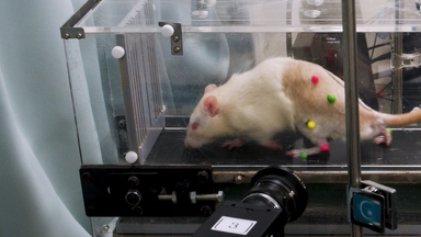
3D Kinematic Analysis for the Functional Evaluation in the Rat Model of Sciatic Nerve Crush Injury

Time-lapse Live Imaging and Quantification of Fast Dendritic Branch Dynamics in Developing Drosophila Neurons
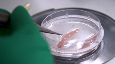
Purification of Fibroblasts and Schwann Cells from Sensory and Motor Nerves in Vitro

Three-Dimensional Motor Nerve Organoid Generation
Copyright © 2024 MyJoVE Corporation. 판권 소유