
Setting Up a Simple Light Sheet Microscope for In Toto Imaging of C. elegans Development
May 5th, 2014
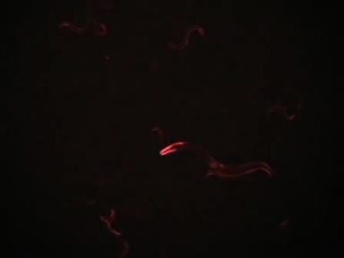
•This protocol describes the setup of a light sheet microscope and its implementation for in vivo imaging of C. elegans embryos.
Related Videos
Generation of Stable Transgenic C. elegans Using Microinjection

Major Components of the Light Microscope

Generation of Transgenic C. elegans by Biolistic Transformation

Imaging C. elegans Embryos using an Epifluorescent Microscope and Open Source Software
RNAi Screening to Identify Postembryonic Phenotypes in C. elegans
A Method for Culturing Embryonic C. elegans Cells

Measurements of Physiological Stress Responses in C. Elegans
Measuring RAN Peptide Toxicity in C. elegans

Simultaneous Live Imaging of Multiple Insect Embryos in Sample Chamber-Based Light Sheet Fluorescence Microscopes

Light Sheet Microscopy of Fast Cardiac Dynamics in Zebrafish Embryos
Copyright © 2024 MyJoVE Corporation. 판권 소유