
Induction of Cellular Differentiation and Single Cell Imaging of Vibrio parahaemolyticus Swimmer and Swarmer Cells
May 15th, 2017
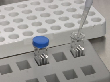
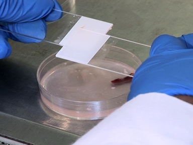
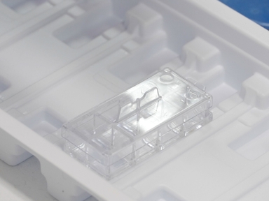
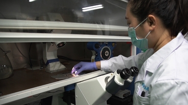
•This protocol enables single cell microscopy of the differentially distinct Vibrio parahaemolyticus swimmer and swarmer cells. The method produces a population of swarmer cells easily available for single cell analysis and covers preparation of cell cultures, induction of swarmer differentiation, sample preparation, and image analysis.
Related Videos

Colonization of Euprymna scolopes Squid by Vibrio fischeri

Quantitative High-throughput Single-cell Cytotoxicity Assay For T Cells
Clinical Application of Sleeping Beauty and Artificial Antigen Presenting Cells to Genetically Modify T Cells from Peripheral and Umbilical Cord Blood
Isolation and Th17 Differentiation of Naïve CD4 T Lymphocytes

Cortical Actin Flow in T Cells Quantified by Spatio-temporal Image Correlation Spectroscopy of Structured Illumination Microscopy Data

Development of a More Sensitive and Specific Chromogenic Agar Medium for the Detection of Vibrio parahaemolyticus and Other Vibrio Species

Induction of an Inflammatory Response in Primary Hepatocyte Cultures from Mice
Highly Multiplexed, Super-resolution Imaging of T Cells Using madSTORM

Assessing Cellular Stress and Inflammation in Discrete Oxytocin-secreting Brain Nuclei in the Neonatal Rat Before and After First Colostrum Feeding
Functionalization of Atomic Force Microscope Cantilevers with Single-T Cells or Single-Particle for Immunological Single-Cell Force Spectroscopy
Copyright © 2024 MyJoVE Corporation. 판권 소유