Aby wyświetlić tę treść, wymagana jest subskrypcja JoVE. Zaloguj się lub rozpocznij bezpłatny okres próbny.
Method Article
Surgical Tips and Tricks for Performing Porcine Pancreas Transplantation
W tym Artykule
Podsumowanie
The video article summarizes the technique of pancreatectomy and pancreas allotransplantation in a porcine 3-day survival model with a step-by-step description of the method and emphasis on the surgical tips and tricks to deal with the precarious and delicate porcine visceral anatomy.
Streszczenie
Despite the promising results of pancreas transplantation in type 1 diabetes mellitus and metabolic syndrome, the biggest concern around this state-of-the-art technique remains the paucity of organs deemed fit for transplantation. High intravascular resistance, delicate intraparenchymal capillary framework, and complex lobular anatomy around the mesenteric vasculature are what make this organ more susceptible to injury and less tolerant to trivial trauma compared to organs such as the liver and kidney. Meticulous surgical dissection and judicious tissue handling form the cornerstone of the entire exercise of pancreas transplantation. Owing to morphological similarity between the anatomy of the porcine pancreas to the surrounding mesenteric vessels and the organs when compared to the human anatomy, demonstration of the technique in the porcine model could help to most accurately extrapolate this to a human setting. The present article aims to outline the essential surgical tips and tricks that need to be followed, in order to ensure a higher success rate of transplantation of this highly susceptible organ in a porcine 3-day survival model.
Wprowadzenie
Over the past several decades, there has been significant progress in perioperative management strategies and surgical techniques leading to the evolution of pancreas transplantation into one of the most promising strategies for the treatment of diabetes mellitus with end-stage renal disease (usually in conjunction with kidney transplantation)1. However, complications such as graft pancreatitis, ischemia-reperfusion injury, and vascular thrombosis remain the biggest challenges to overcome to ensure successful outcomes, more so in the more damaged extended criteria grafts2. In addition, pancreas grafts are the most commonly discarded grafts from procurement and have the lowest utilization rates (9%) for any organ3. Therefore, machine perfusion aims to provide an optimum homeostatic milieu to the pancreas graft with the goal of increasing graft utilization rate, similar to what has been achieved in liver, kidney, and lung transplantation4. The porcine pancreatic anatomy is complex in terms of its lobular architecture (comprising three lobes), its extensions all around the mesenterico-portal axis, its mesenteric vascular variations (in 40%-50%), and its delicate vascular channels along the C loop of the duodenum5. These anatomical attributes contribute to a challenging dissection in both retrieval of the pancreato-duodenal graft and recipient pancreatectomy to induce an iatrogenic apancreatic diabetes state, i.e., surgically induced state of diabetes mellitus with a fasting glucose level above 8 mmol/L. Based on these features, porcine pancreatectomy with transplantation provides the closest possible replication of the technique that could be performed in humans as a definitive treatment against end-stage diabetes mellitus. The present article aims at covering the following aspects: (i) outline of the peri-operative porcine care during recipient pancreatectomy and pancreas graft implantation; (ii) technical step by step details of the recipient pancreatectomy and implantation of the pancreato-duodenal graft and (iii) tips and tricks of donor and recipient pancreatic operation in porcine models to minimize graft and recipient injury.
Protokół
The protocol received ethical approval from the Animal Care Committee, Toronto general research institute. Animals received humane care in compliance with the National Society of Medical Research and Guide for the care of laboratory animals, National Institute of Health (NIH), Ontario, Canada. For this study, 15-week-old unrelated Yorkshire male swine, weighing 40-50 kg were used.
NOTE: The entire protocol of the study is divided into the following major steps: (i) Organ retrieval and back-table preparation; (ii) Recipient total pancreatectomy and (iii) Graft implantation. The entire surgery of donor and recipient is done in 1 day.
1. Donor organ retrieval and back-table preparation
NOTE: The method for organ retrieval has been described in a separate protocol6. However, the protocol, in brief, is described here along with some additional take-home points specific to the surgical technique (surgical tips relevant to donor operation).
- In brief, anesthetize the donor pig, and secure the airway and central venous catheter (described below in the recipient pancreatectomy section). Incise and expose the viscera. Perform aortocaval dissection and mobilization till the bifurcation into iliac vessels.
- Bowel handling: Porcine small bowel is long and tortuous, and the large bowel is mostly distended. Make sure to reposition the bowel loops in their anatomical position after every step of donor operation to ensure adequate perfusion and avoid the possibility of mesenteric torsion.
- Hilar dissection: Ligate and divide the arterial branches and bile duct to expose the portal vein. Dissect the hilum (hepato-duodenal ligament) as high/distal as possible to avoid the risk of injuring the small vascular channels that perfuse the superior aspect of the pancreato-duodenal region. Start skeletonizing the vessels in a right to left sequence, till the anterior surface of the portal vein lies bare.
- Aorto-caval dissection: Expose the supra-hepatic infra-diaphragmatic part of the aorta by dividing the diaphragmatic crura. Avoid dissection of the inferior vena cava around the pancreato-duodenal grove. Use the renal arteries on both sides as the marked upper limit of dissection.
- Expose the pancreas by opening the lesser sac. Ligate and divide the renal arteries. Cannulate the infrarenal aorta and connect with the University of Wisconsin (UW) solution for flushing.
- Ligate the supra-hepatic aorta and clamp. Flush with 1.5 L-2 L of UW solution to ensure adequate flushing of the bowel and pancreas (evident by the visual appearance of pallor). During in situ flushing of the organ, reposition the small bowel and large bowel in sequence to the midline ensuring no mesenteric twist. This step ensures uniform flushing of the bowel and pancreas with the UW (University of Wisconsin) solution and is important to remember.
- Incise the portal vein and infrahepatic vena cava simultaneously after starting the flush to let out the venous effluent. Cover the abdomen with ice to ensure cold perfusion. Dissect the organ (pancreatoduodenal graft with parts of retroperitoneum, psoas, spleen, and adrenal) en masse.
- Cold dissection: Start mobilization of the graft from lateral to medial direction, from the tail of the pancreas to the junction between the head and the corpus, by sharp dissection of the fascia between the pancreas and large bowel. While performing this step, hold the pancreas by its retroperitoneal covering with forceps and ask the assistant to give counter traction on the large bowel toward the foot.
- Mesenteric clamping: After dividing the thin tissue around the corpus of the pancreas, duodenojejunal loop, and large bowel, ask the assistant to loop around the mesenteric pedicle and pull the large bowel loop caudally. This will ensure delineation of the mesentery and safe placement of the clamp to divide the mesenteric major vessels in sequence without injuring the pancreatic parenchyma.
- Complete extra-peritoneal dissection of the graft: After dividing the mesenteric vessels, start retrieving the graft by dissection from medial to lateral in a counterclockwise direction, dividing the extrapancreatic tissues, including the pararenal fascia, adrenal gland, cuff of IVC, psoas muscle, and diaphragmatic crus. Ensure an adequate plane away from the aorta to avoid injuring the superior mesenteric and coeliac arteries.
- Perform back table preparation on ice. Store the organ in an ice box (cold ischemia time of 5 h).
2. Recipient pancreatectomy
- Pre-operative preparation
- Fast the animal for at least 6 h before the stipulated induction time. Administer an injection containing midazolam (0.15 mg/kg), atropine (0.04 mg/kg), and ketamine (25 mg/kg) subcutaneously (SC), 15 min before transporting the animal to the operating room (OR). Administer buprenorphine 0.3 mg sustained-release injection SC 15 min prior to transport to the OR.
- Positioning: Place the animal supine on the operating table and harness the forelimbs to ensure a stable position during the surgery.
- Airway and induction: Ventilate the animal using a bag and mask with 3%-5% isoflurane and 2-3 L oxygen per min, while connecting the pulse oximeter probe and monitoring the heart rate and O2 saturation.
- After adequate relaxation, confirmed by relaxation of the jaws and stable oxygen saturation and heart rate, ask the assistant to hold open the mouth with adequate traction on the relaxed upper and lower jaws of the animal, and visualize the vocal cords using the laryngoscope. Spray 2% lidocaine to relax the vocal cords to prevent spasms induced by intubation (the porcine vocal cords are highly vascular and fragile!).
- Intubate using a 7 mm endotracheal tube and inflate the cuff using 3-5 mL of air. Ensure the position of the tube using the end-tidal CO2 (ETCO2) probe and connect the tube to the ventilator, with 14-16 breaths per min to achieve a tidal volume of 10-15 mL/kg. Lower the isoflurane to 2.5% as a maintenance dose of inhalational anesthesia.
- Intravenous line: Aseptically prepare the surgical site with betadine and place a sterile drape. Then, identify the landmark to place the central venous catheter, which is the centroid of the triangle formed between the mastoid process, acromion process, and head of the clavicle. Use a 16 G needle to puncture the vein; after ensuring free flow of venous blood, apparent by the color and lack of pulsatility, introduce a guidewire using the Seldinger technique.
- Dilate the tract using the dilator provided. Replace the guidewire and dilator with an 8.5 x 10 Fr catheter and secure in place by suturing with a 0-0 silk suture cutting needle. Connect the IV line to the intravenous antibiotics containing cefazolin (1 gm) and metronidazole (500 mg) followed by intravenous anesthesia done by propofol infusion at the rate of 10 mL/h.
- Invasive blood pressure monitoring: After aseptically preparing the surgical site with betadine, place a sterile drape and make an incision parallel to the trachea 2 cm from the midline. Dissect the fibers of sternomastoid muscle and paratracheal fascia by blunt dissection using Lahey's right-angled forceps and hemostatic forceps.
- Identify the jugular vein lateral to the fibers of the strap muscles and dissect along the muscle compartmentalizing it from the carotid artery medially using blunt dissection as described above. Identify the artery by its pulsation, texture (cord-like), and perivascular plexus. Mobilize the vessel and sling it using two silk 2-0 ties.
- Puncture the vessel with a 16 G angio-catheter with a beveled tip pointing upward and withdraw the inner stylet once inside the vessel. Slide the outer catheter sheath using the Seldinger technique and connect the outlet to the arterial blood pressure (BP) measuring system. Prior to connecting the invasive BP monitor, calibrate the reading to zero to ensure accurate recording and secure the catheter in place using the silk ties across.
- Temperature monitoring and warming: Place the temperature recording probe orally. Cover the animal with the Bair-hugger heating duvet and ensure a temperature of 37-38 °C. Lubricate the eyes with a neutral eye lubricant gel. Paint and drape the surgical field
- Surgical procedure
NOTE: The skin is aseptically prepared for all surgical procedures using betadine scrubs followed by the placement of sterile drapes.- Incision and exposure: Make a midline incision from the xiphisternum to the symphysis pubis using the pure cutting mode of Bovie's electrocautery. Ensure to keep lateral to the urethra in the lower part of the incision beyond the phallus to avoid injury. Place the self-retaining abdominal wall retractor taking care not to injure the spleen and liver, to optimize exposure of the surgical field.
- Mobilization of the head of the pancreas: With the assistant providing counter-traction to the pancreato-duodenal grove, start by dissecting along the avascular fascial plane between the pancreatic parenchyma (duodenal lobe) and the infra-hepatic IVC.
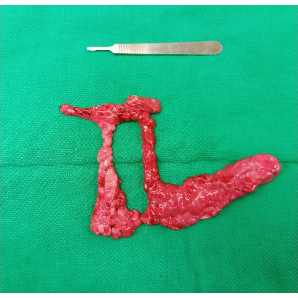
- Pancreatic ring mobilization: The porcine pancreas forms a ring around the mesenterico-portal axis (connecting lobe). Dissect along the fascia on either aspect of the portal vein (PV) to ensure complete mobilization of the connecting lobe from the overlying PV. The safest way is to start the dissection along either side of the portal vein and separate the tissues circumferentially in a clockwise or anticlockwise direction.
- Pancreatic tail mobilization: Next, identify the junction between the pancreatic and pararenal tissues (seen as a white line) and start dissecting the tail off the underlying splenic vein staying close to the parenchyma. Ligate and divide the small venous tributaries to the parenchyma, taking care not to injure the splenic vein using silk 3/0 ties.
- Pancreatic corpus mobilization: Dissect along the thin layer of tissue separating the parenchyma from the large bowel and the stomach, taking care to preserve the thin vascular arcade along the infra-pyloric region.
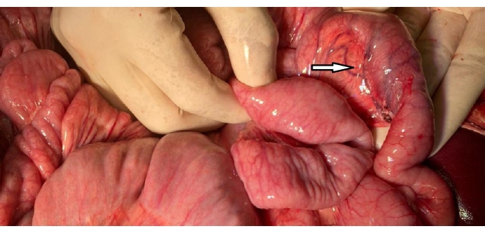
- Pancreato-duodenal mobilization: Next, separate the parenchyma from the C loop of the duodenum by sharp dissection in the pancreato-duodenal grove by traction-counter traction, taking care to preserve the thin vascular arcade along the C loop of the duodenum.
- Division of the pancreatic duct: Identify the pancreatic duct, ligate with silk 2-0 ties and divide keeping around 3-5 mm stump of the duct on the duodenal C loop, staying away from the duodenal vascular arcade (in close relation to the duct).
- Removal of the specimen: Complete the last part of the mobilization by dissecting the parenchyma from the junction between the connecting lobe, duodenum, and colon on either side of the portal vein. Extract the specimen en-masse (Figure 1) by dividing the connecting lobe on the anterior aspect of the PV. Division of the parenchyma is required to extract the specimen out because of its circumferential location around the PV.
- Ensure hemostasis along the areas of dissection and inspect the duodenum for any injury and vascular congestion (Figure 2).
- Implantation of the pancreato-duodenal graft
- Preparation of the graft: Prepare the graft PV and proximal aortic end by trimming the edges for anastomosis. Place the prepared graft in the organ bag with UW solution on an ice bath. Take core needle biopsies from the pancreatic tail -store in formalin, snap freeze, and extract RNA at a later stage. Suture the biopsy site using Prolene 6-0 in a figure of 8 fashion.
- Aorto-caval dissection: Begin by identifying the right ureter and dissect along the fascia separating the ureter from the retroperitoneal covering over the inferior vena cava. Dissect along the lateral border of the aorta to expose it from the underlying psoas muscle. Next, dissect along the interaortocaval grove to separate the aorta from the IVC.
- Clamp test: Ensure adequate mobilization of the aorta and IVC by a trial of Satinsky's vascular clamp placement. Switch position with the first assistant and prepare the surgical field by retracting the bowel loops on the left side to expose the major vessels for anastomosis. Place the trial clamps on IVC and aorta to re-assess an adequate mobilization of both the vessels for anastomosis.
- Venous anastomosis: After placing the vascular side-biting clamp in the IVC, make a small opening in the anterior wall of the vessel and extend it to an adequate size (matched with the graft vessel) cranially and caudally using Pott's scissors. Flush the lumen with heparinized saline using the flexible sheath of an IV cannula.
- Using Prolene 6-0 double-needle, make corner sutures on the IVC (inside-out) and graft portal vein (inside-out). Secure the caudal corner suture and run one of the needles along the posterior wall of the graft and recipient vein in a continuous fashion. Secure the anterior wall in a similar (outside-in continuous) fashion and secure the knot in the center of the anterior wall after flushing the lumen of the anastomosis with heparinized saline.
- De-clamping the vein: Place a Bulldog's vascular clamp on the graft portal vein, away from the anastomosis, and slowly release the side-biting clamp from the recipient IVC. Check for venous refilling and any major bleed from the anastomosis.
- Arterial anastomosis: Place the side-biting clamp on the aorta taking care not to injure the lumbar branches along the posteromedial wall of the vessel. Instruct the anesthetist to inject heparin (100 U/kg BW) and methylprednisolone (500 mg) through the central venous catheter.
- Make an opening on the anterior wall of the vessel and extend it cranially and caudally taking care not to dissect along the vessel wall. Flush the lumen with heparinized saline and using Prolene 6-0 double-needle, suture the proximal end of the graft aorta to the recipient aortic opening in a continuous fashion by the Parachute technique. Flush the lumen with heparinized saline before tying the final knot on the anterior wall of the vessel.
- Reperfusion: Take care of the following two aspects while performing this step. First, the hemodynamic alterations where a drastic fall in the mean BP occurs. Monitor the invasive BP minute to minute and perform a dose titration of the vasopressor (norepinephrine infusion), and fluid preload to keep the target mean BP between 45-50 mmHg.
- Second, maintain the hemostasis. After removal of the bulldog clamp from the vein, assess for any major bleed from the parenchyma, para-aortic region, and peri-portal area; then, release the aortic clamp and check for bleeding from the arterial anastomotic site. Secure the bleeding points with ligatures and hemostatic suturing. Ask the anesthetist to inject a vial of tranexamic acid through the central venous catheter.
NOTE: An important rule is to minimize aggressive handling of the pancreas during this phase of reperfusion (to avoid graft oedema and hematoma). - Bowel anastomosis: After ensuring no mesenteric torsion, isolate a loop of jejunum, 40-50 cm away from the duodenojeunal junction, and bring it close to the graft. Anastomose the graft duodenum to the recipient jejunum in a side-to-side continuous fashion using Polydioxanone 4-0 suture, taking care to ensure an adequate luminal diameter of 1.5-2 cm.
- Hemostasis and post-reperfusion biopsy: Ensure hemostasis around the graft and at the sites of anastomosis. Take three core needle biopsies from the pancreatic tail1 h after reperfusion-store in formalin, snap freeze, and extract RNA at a later stage. Suture the biopsy site with Prolene 6-0 in a figure of 8 fashion.
- Abdominal wall closure: After assessing for retained mops and securing hemostasis, suture the rectus abdominis in a continuous fashion using Polydioxanone 0 needle taking care in the lower midline to keep away from the urethra. Close the skin using silk 0 suture in a continuous fashion.
NOTE: Ideally, monofilament sutures (Nylon) are recommended; however, since this is a 3-day survival model, silk is acceptable in these experimental models. - Strapping the central venous catheter: Create a subcutaneous tunnel on the neck and secure the central venous catheter by burying it under the tunnel and suturing the overlying skin with a silk 0-0 cutting needle.
- Removal of the arterial catheter: After ensuring a stable mean BP (40-50 mmHg) and an improving trend of pH and lactate levels on the blood gas analysis, remove the arterial catheter and ligate the vessel to secure hemostasis in the neck. Close the overlying skin with silk 0 suture in a continuous fashion.
- Position the animal and extubation: Under all necessary precautions, turn the animal sternal on a transporting cart. Observe for the O2 saturation and reversal of anesthesia (movement of limbs, spontaneous breathing efforts, SpO2 >95% off O2, and ventilator support) and extubate the animal. Transport to the pen and position the animal sternal and monitor the hemodynamic status till stable.

Figure 1: Pancreatectomy specimen resected en masse. Note the ring of pancreatic tissue surrounding the portal vein in vivo. Please click here to view a larger version of this figure.

Figure 2: Image of the duodenum. The C loop of the duodenum with a preserved vascular arcade (arrowhead) compared with the surrounding jejunal loop and assessed for congestion. Please click here to view a larger version of this figure.
Wyniki
The peri-operative and post-operative, up to 3-day, biochemical parameters from the five pancreas transplant survival models are summarized below (numbered as PTX 1 to 5 in chronology). Of the five total pancreas transplants, all fared well during the 3-day survival period, as evident by their general well-being and the pancreatic injury and endocrine function tests. The results depicted below are representative of the experience of a 3-day survival model of recipient pancreatectomy followed by pancreas allotransplantati...
Dyskusje
The current protocol has been performed to demonstrate the technique and the feasibility of pancreatectomy and pancreas allotransplantation in porcine models. The animals were observed for a period of 3 days after transplantation to demonstrate the reliability of the technique of pancreatectomy and the allotransplantation. All animals were monitored and nursed for 3 days after the surgery using a standardized animal care protocol of antibiotics, fluids, analgesics, supplemental nutrition, and immunosuppressants (i.e., cy...
Ujawnienia
The authors have no conflicts of interest to disclose.
Podziękowania
None.
Materiały
| Name | Company | Catalog Number | Comments |
| Belzer UW Cold storage solution | Bridge to life Ltd (Columbia, SC, USA) | 4055 | |
| Calcium gluconate (10%) | Fresenius Kabi Canada Ltd (Toronto, ON) | C360019 | |
| Composelect (blood collection bags) | Fresenius Kabi Canada Ltd (Toronto, ON) | PQ31555 | |
| Heparin (10000 IU/10 ml) | Fresenius Kabi Canada Ltd (Toronto, ON) | C504710 | |
| Lactated Ringer's | Baxter (Mississauga, ON, Canada) | JB2324 | |
| Percutaneous Sheath Introducer Set with Integral Hemostasis Valve/side Port for use with 7-7.5 Fr Catheters | Arrow International LLC | SI-09880 | |
| Sodium bicarbonate (8.4%) | Fresenius Kabi Canada Ltd (Toronto, ON) | C908950 | |
| Solu-Medrol | Pfizer Canada Inc. | 52246-14-2 | |
| Surgical retreival and transplant instrument set |
Odniesienia
- Gruessner, R. W., Gruessner, A. C. The current state of pancreas transplantation. Nature Reviews. Endocrinology. 9 (9), 555-562 (2013).
- Redfield, R. R., Rickels, M. R., Naji, A., Odorico, J. S. Pancreas transplantation in the modern era. Gastroenterology Clinics of North America. 45 (1), 145-166 (2016).
- Canadian Institute for Health Information. Annual Statistics on Organ Replacement in Canada: Dialysis, Tranplantation and Donation, 2009 to 2018. Canadian Institute for Health Information. , (2019).
- Prudhomme, T., et al. Ex-situ perfusion of pancreas for whole-organ transplantation: Is it safe and feasible?A systematic review. Journal of Diabetes Science and Technology. 14 (1), 120-134 (2020).
- Ferrer, J., et al. Pig pancreas anatomy: Implications for pancreas procurement, preservation, and islet isolation. Transplantation. 86 (11), 1503-1510 (2008).
- Parmentier, C., et al. Normothermic ex vivo pancreas perfusion for the preservation of pancreas allografts before transplantation. Journal of Visualized Experiments. , (2022).
- Chaib, E., et al. Total pancreatectomy: Porcine model for inducing diabetes - Anatomical assessment and surgical aspects. European Surgical Research. 46 (1), 52-55 (2011).
- Prudhomme, T., et al. Total pancreatectomy and pancreatic allotransplant in a porcine experimental model. Experimental and Clinical Transplantation. 18 (3), 353-358 (2022).
- Grussner, R., et al. Streptozotocin-induced diabetes mellitus in pigs. Hormone and Metabolic Research. 25 (4), 199-203 (1993).
- Mazilescu, L. I., et al. Normothermic ex situ pancreas perfusion for the preservation of porcine pancreas grafts. American Journal of Transplantation. 22 (5), 1339-1349 (2022).
- Kumar, R., et al. Ex vivo normothermic porcine pancreas: A physiological model for preservation and transplant study. International Journal of Surgery. 54, 206-215 (2018).
- Kaths, J. M., et al. Normothermic ex vivo kidney perfusion for the preservation of kidney grafts prior to transplantation. Journal of Visualized Experiments. (101), e52909 (2015).
Przedruki i uprawnienia
Zapytaj o uprawnienia na użycie tekstu lub obrazów z tego artykułu JoVE
Zapytaj o uprawnieniaPrzeglądaj więcej artyków
This article has been published
Video Coming Soon
Copyright © 2025 MyJoVE Corporation. Wszelkie prawa zastrzeżone