
High-Accuracy Correction of 3D Chromatic Shifts in the Age of Super-Resolution Biological Imaging Using Chromagnon
June 16th, 2020
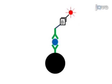
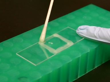
•Correction of chromatic shifts in three-dimensional (3D) multicolor fluorescence microscopy images is crucial for quantitative data analyses. This protocol is developed to measure and correct chromatic shifts in biological samples through acquisition of suitable reference images and processing with the open-source software, Chromagnon.
Tags
Vídeos Relacionados

A Novel RFP Reporter to Aid in the Visualization of the Eye Imaginal Disc in Drosophila
Rapid Homogeneous Detection of Biological Assays Using Magnetic Modulation Biosensing System

Studying Age-dependent Genomic Instability using the S. cerevisiae Chronological Lifespan Model

Super-resolution Imaging of the Bacterial Division Machinery

Test Samples for Optimizing STORM Super-Resolution Microscopy

Sample Preparation for Single Virion Atomic Force Microscopy and Super-resolution Fluorescence Imaging

Super-resolution Imaging of the Cytokinetic Z Ring in Live Bacteria Using Fast 3D-Structured Illumination Microscopy (f3D-SIM)
High-resolution Time-lapse Imaging and Automated Analysis of Microtubule Dynamics in Living Human Umbilical Vein Endothelial Cells

Open-source Single-particle Analysis for Super-resolution Microscopy with VirusMapper

Ground State Depletion Super-resolution Imaging in Mammalian Cells
SOBRE A JoVE
Copyright © 2024 MyJoVE Corporation. Todos os direitos reservados