A subscription to JoVE is required to view this content. Sign in or start your free trial.
Method Article
Dissection and Culture of Mouse Dopaminergic and Striatal Explants in Three-Dimensional Collagen Matrix Assays
In This Article
Summary
Explants from the midbrain dopamine system and striatum are used in a collagen matrix assay for the in vitro analysis of mesostriatal and striatonigral pathway development. In this assay axonal outgrowth and guidance can be manipulated and quantified. It can also be modified for assessing other regions or molecular cues.
Abstract
Midbrain dopamine (mdDA) neurons project via the medial forebrain bundle towards several areas in the telencephalon, including the striatum1. Reciprocally, medium spiny neurons in the striatum that give rise to the striatonigral (direct) pathway innervate the substantia nigra2. The development of these axon tracts is dependent upon the combinatorial actions of a plethora of axon growth and guidance cues including molecules that are released by neurites or by (intermediate) target regions3,4. These soluble factors can be studied in vitro by culturing mdDA and/or striatal explants in a collagen matrix which provides a three-dimensional substrate for the axons mimicking the extracellular environment. In addition, the collagen matrix allows for the formation of relatively stable gradients of proteins released by other explants or cells placed in the vicinity (e.g. see references 5 and 6). Here we describe methods for the purification of rat tail collagen, microdissection of dopaminergic and striatal explants, their culture in collagen gels and subsequent immunohistochemical and quantitative analysis. First, the brains of E14.5 mouse embryos are isolated and dopaminergic and striatal explants are microdissected. These explants are then (co)cultured in collagen gels on coverslips for 48 to 72 hours in vitro. Subsequently, axonal projections are visualized using neuronal markers (e.g. tyrosine hydroxylase, DARPP32, or βIII tubulin) and axon growth and attractive or repulsive axon responses are quantified. This neuronal preparation is a useful tool for in vitro studies of the cellular and molecular mechanisms of mesostriatal and striatonigral axon growth and guidance during development. Using this assay, it is also possible to assess other (intermediate) targets for dopaminergic and striatal axons or to test specific molecular cues.
Protocol
1. Preparation of Rat Tail Collagen
- Collect 6-10 adult rat tails (it is possible to store the tails at -20 °C until use).
- Soak the tails in 95% ethanol overnight at room temperature (RT).
Dissection of rat tails (in tissue culture hood):
(keep tools in 70% ethanol when not using them and make sure that all solutions, tools and glassware used throughout this procedure are sterile)
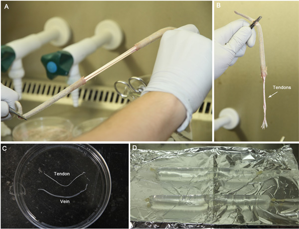
- To collect tendons from the tails, cut off the tip of the tail. Hold large end of the tail with a pair of forceps. Use another pair of forceps to hold, bend and break the tail close to the other forceps. Pull apart and the tendons will be left behind (Figure 1A,B).
- Collect the tendons in sterile H2O and after collecting all tendons move them to a new petridish with sterile H2O.
- Take 2-3 tendons and shred them using 2 pairs of forceps in a new petridish with sterile H2O. Remove non-tendon tissue such as veins (tendon tissue is shiny and reflective; Figure 1C). After removal of all non-tendon tissue, each tail yields approximately 100-150 mg of tendons.
- Transfer the tendons to 300 ml of sterile 3% acetic acid and stir slowly overnight at 4 °C.
- Add an additional 200 ml of sterile 3% acetic acid and stir slowly overnight at 4 °C.
- Centrifuge the acetic acid solution containing the tendons at ~2700 xg for 120 min at 4 °C to pellet non-dissolved tendons and non-tendon tissue.
- During centrifugation prepare dialysis tubing:
- cut pieces of 40 cm
- boil in 500 μM EDTA for 5 min
- cool in sterile H2O
- Tie a knot at one end of the dialysis tubing and fill the tubing with the supernatant of the centrifuged tendon solution on ice in the tissue culture hood using an automatic pipette and a 25 ml pipette. Avoid producing bubbles. Knot the other end of the tubing and store tubing on sterile aluminum foil on ice until all pieces of tubing are filled (Figure 1D).
- Prepare 10 l of sterile 0.1X MEM, pH 4.0, in the tissue culture hood. Add floater to pieces of tubing and add these to the pre-cooled 0.1X MEM solution in a 10 l beaker. Dialyze overnight at 4 °C while slowly stirring. This dialysis removes excess acid while keeping the pH low.
- Replace with new pre-cooled 10 l 0.1X MEM and stir overnight at 4 °C.
- Repeat the previous step by replacing with new pre-cooled 10 l 0.1X MEM and stir overnight at 4 °C.
- Aliquot collagen solution into sterile 50 ml tubes. Store aliquots at 4 °C. Keep 6-12 months, only handle the collagen stock in the tissue culture hood.
- It is important to test whether the purified collagen needs to be diluted (in 0.1x MEM) to obtain optimal results in the collagen matrix assay. Therefore, generate a collagen dilution series (undiluted collagen, 1:1, 1:2 and 1:5 diluted) and perform collagen matrix assays as described below to determine the optimal collagen dilution.
2. Dissection of the Dopaminergic Midbrain7,8
- Dissect E14.5 mouse embryos from the uterus of the mother and keep in L15 medium on ice until needed.
All subsequent dissection steps are performed in L15 medium on ice.
- Dissect out the brain.
- Remove the telencephalon by cutting along the medial part of each telencephalic vesicle (use the telencephalic vesicles for striatal explants).
- Remove the meningeal sheath.
- Make a dorsoventral cut just rostral of the mesencephalic flexure.
- Make another dorsoventral cut just caudal of the mesencephalic flexure.
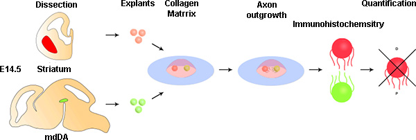
- Make a rostrocaudal cut along the dorsal midline using a microdissection knife to expose the underlying ventral midbrain tissue. Be careful not to hit the ventral midbrain tissue as it contains the dopaminergic neurons (Figure 2).
- Make two rostrocaudal cuts lateral and parallel to the ventral midline to remove dorsal midbrain tissue.
- Divide the remaining ventral midbrain tissue into explants using a microdissection knife.
- Store the explants in L15 medium containing 5% FBS on ice until use.
3. Dissection of the Striatum
All dissection steps are performed in L15 medium on ice.
- Use telencephalic vesicles dissected during midbrain dissection.
- Remove the thalamus by cutting in between the thalamus and the striatum using forceps.
- Make a mediolateral cut rostral to the striatum to remove rostral structures such as the olfactory bulb. Repeat this step for tissue located caudally to the striatum.
- Position the remaining slice to obtain a coronal view of the striatum (Figure 2).
- The striatum can be recognized as a slightly less dense (more transparent) part of tissue. Dissect out the striatum and cut into explants using a microdissection knife. Avoid the slightly darker tissue close to the midline, which contains migrating neurons of the lateral and medial ganglionic eminence.
- Store the explants in L15 medium containing 5% FBS on ice until use.
4. Assembly Collagen Matrix Assays
Preparation of the collagen (all steps on ice, use cooled pipette tips)
- Mix 860 μl of diluted collagen with 100 μl of 10x MEM and 40 μl of 1M sodium bicarbonate (NaHCO3) and keep on ice. If at this point the collagen warms up it will solidify.
- Add a drop of 20 μl (approximately 5 mm in diameter) of prepared collagen to a coverslip in a well of a 4-well Nunc dish and let it stand in a CO2 incubator at 37 °C and 5%CO2 for 30 min (ambient O2 concentration is sufficient). During this incubation the collagen will gelatinize.
Setup co-culture in collagen gel
- After the collagen has gelatinized, use a pipette with a 200 μl tip to transfer a dopaminergic or striatal explant to the collagen.
- Move the explants in close proximity to each other using a needle. Keep them apart at a distance of approximately the diameter of one explant (~200-300 μm) (Figure 2).
- Remove excess medium and add 20 μl of prepared collagen on top of the explants. This will often cause the explants to move around. Reposition the explants using a needle as described in 4.4.
- Let the collagen solidify for 15 min at RT followed by 30 min at 37 °C and 5% CO2.
- After the collagen has set, add 400 μl of explant medium and grow for 2-3 days in a CO2 incubator at 37 °C and 5% CO2.
5. Analysis by Immunohistochemistry and Quantification
Immunohistochemistry
- To fix explants, gently add 400 μl of 8% paraformaldehyde (PFA) in PBS to 400 μl of medium (thereby diluting the PFA to 4%) and let stand for 1 hour at RT.
- Wash 3x 15 min in PBS at RT.
- Incubate in blocking buffer (BB; PBS + 1% FBS + 0.1% Triton X-100) for 2 hours.
- Incubate overnight with primary antibody in BB at 4 °C. For dopaminergic axons use anti-tyrosine hydroxylase antibodies (1:1000)7, for striatal axons anti-DARPP32 antibodies (1:500)9 and for visualizing all axons anti-βIII tubulin (1:3000)7.
- Wash 5x 1 hour in PBS at RT.
- Incubate with secondary antibody conjugated to the appropriate fluorophore (1:500) in BB overnight at 4 °C. From this step on minimize exposure of the explants to light (e.g. cover them with aluminum foil).
- Wash overnight in PBS at 4 °C followed by several washes during the next day.
- Mount explants on microscope slides using Prolong Antifade reagent mounting medium. Add a drop of ~10 μl on a glass microscope slide. Take the coverslip and gently place it on the drop of mounting medium with the explant side facing down. Avoid trapping any air under de coverslip by very gently lowering it on the drop of mounting medium.
Quantification by calculating P/D ratio
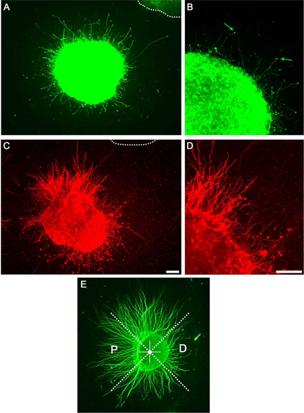
- Acquire digital images of the explants using an epifluorescence microscope (Figure 3).
- Using these images, divide each explant into quadrants to generate a proximal quadrant (i.e. part of the explant closest to the adjacent explant) and a distal quadrant (part of the explant farthest away from the adjacent explant) (Figure 2, 4).
- Measure the length of 20 longest neurites emerging from the explant both in the proximal and distal quadrants.
- Use the average length of the 20 longest neurites to calculate the P/D ratio for each individual explant. A P/D ratio > 1 indicates axon attraction, while a ratio < 1 indicates axon repulsion.
- In some cases, the dense growth of neurites prevents the assessment of individual neurite length. In these situations, quantification can be performed by measuring the distance between the edge of the explant and the leading front of axon growth in the proximal and distal quadrants.
6. Representative Images
After successful culture of dopaminergic and striatal explants a large number of axons are visible using bright field microscopy or as visualized by anti-βIII tubulin immunocytochemistry (not shown). A subset of these axons will be dopaminergic or striatal axons as visualized using immunocytochemistry (Figure 3). By dividing the explants into quadrants as described and determining P/D ratios, a putative axon attractive or repulsive effect can be quantified (Figure 3E).

Figure 1. Photos illustrating the preparation of collagen from rat tail tendons. A) Pulling apart the rat tail exposes the tendons. B) Tendons are visible as rope-like bundles. C) The tendons are easy to distinguish from veins by their shiny white appearance. D) After dissolving the tendons in acetic acid, the solution is transferred to dialysis tubing. To keep the dissolved collagen cold, tubes are placed on sterile aluminum foil on ice.

Figure 2. Schematic indicating the different steps of the procedure. Appropriate brain regions are dissected and cut to generate explants. A single midbrain and striatal explant are positioned in close proximity in a collagen matrix and left to grow for 48-72 hours at 37 °C. Axons are visualized by fluorescence immunohistochemistry and a P/D ratio is calculated to quantify chemotropic responses.

Figure 3. Representative results showing axon growth in a collagen matrix culture. A) Midbrain explant stained with anti-tyrosine hydroxylase antibody revealing axon outgrowth. Dotted line indicates adjacent explant. B) Magnification of A showing individual neurites and growth cones (arrows). C) Striatal explant stained with anti-DARPP32. D) Magnification of C showing individual neurites. E) Example of P/D ratio quantification. Explants are divided into equal quadrants. The proximal quadrant is facing the adjacent explant while the distal quadrant is facing away from it. Scale bars indicate 100 μm.
Discussion
The collagen matrix assay described here has been used and improved by many different labs in the past decades to investigate a variety of axon guidance molecules and neuronal systems (e.g. see references 5-8). These studies have shown that this assay is a powerful tool for studying the effects and regulation of axon guidance molecules secreted by different (intermediate) target tissues.
However, it should be noted that the collagen matrix is a substitute for the extracellular environment (EC...
Disclosures
We have nothing to disclose.
Acknowledgements
The collagen matrix assay has been developed and improved by the work of many different research groups during the past two to three decades. The approaches described here for dopaminergic and striatal explants greatly benefit from these studies. In addition, the authors would like to thank Asheeta Prasad for her help in setting up striatal explant cultures. Work in the lab was funded by the Human frontier Science Program Organization (Career Development Award), the Netherlands Organization of Health Research and Development (ZonMW-VIDI and ZonMW-TOP), the Europanian Union (mdDA-NeuroDev, FP7/2007-2011, Grant 222999) (to RJP), and the Netherlands Organization for Scientific Research (TopTalent; to ERES).
Materials
| Name | Company | Catalog Number | Comments |
| Fetal Calf Serum | BioWhittaker | 14-801F | |
| Glutamine (200mM) | PAA Laboratories | M11-004 | |
| Hepes | VWR international | 441476L | |
| β-Mercapt–thanol | Merck & Co., Inc. | 444203 | |
| Minimum Essential Media (MEM) | GIBCO, by Life Technologies | 61100-087 | |
| Neurobasal | GIBCO, by Life Technologies | 21103-049 | |
| B27 | GIBCO, by Life Technologies | 17504-044 | |
| Leibovitz’s L-15 Medium | GIBCO, by Life Technologies | 11415-049 | |
| Penicillin-Streptomycin | GIBCO, by Life Technologies | 15070-063 | |
| Prolong Gold Antifade Reagent | Invitrogen | P36930 | |
| Dialysis tubing | Spectrum Labs | 132660 | |
| Rabbit anti-Tyrosine Hydroxylase | Pel-Freez Biologicals | P40101-0 | |
| Rabbit anti-Darpp32 (H-62) | Santa Cruz Biotechnology, Inc. | Sc-11365 | |
| Mouse anti-βIII tubulin | Sigma-Aldrich | T8660 | |
| Alexa Fluor labeled secondary antibodies | Invitrogen |
References
- van den Heuvel, D. M., Pasterkamp, R. J. Getting connected in the dopamine system. Prog. Neurobiol. 85, 75-93 (2008).
- Lobo, M. K. Molecular profiling of striatonigral and striatopallidal medium spiny neurons past, present, and future. Int. Rev. Neurobiol. 89, 1-35 (2009).
- Dickson, B. J. Molecular mechanisms of axon guidance. Science. 298, 1959-1964 (2002).
- Chilton, J. K. Molecules mechanisms of axon guidance. Dev. Biol. 292, 13-24 (2006).
- Lumsden, A. G., Davies, A. M. Chemotropic effect of specific target epithelium in the developing mammalian nervous system. Nature. 323, 538-539 (1986).
- Tessier-Lavigne, M., Placzek, M., Lumsden, A. G., Dodd, J., Jessel, T. M. Chemotropic guidance of developing axons in the mammalian central nervous system. Nature. 336, 775-778 (1988).
- Kolk, S. M., Gunput, R. A., Tran, T. S., van den Heuvel, D. M., Prasad, A. A., Hellemons, A. J., Adolfs, Y., Ginty, D. D., Kolodkin, A. L., Burbach, J. P., Smidt, M. P., Pasterkamp, R. J. Semaphorin 3F is a bifunctional guidance cue for dopaminergic axons and controls their fasciculation, channeling, rostral growth, and intracortical targeting. J. Neurosci. 29, 12542-12557 (2009).
- Fenstermaker, A. G., Prasad, A. A., Bechara, A., Adolfs, Y., Tissir, F., Goffinet, A., Zou, Y., Pasterkamp, R. J. Wnt/planar cell polarity signaling controls the anterior-posterior organization of monoaminergic axons in the brainstem. J. Neurosci. 30, 16053-16064 (2010).
- Arlotta, P., Molyneaux, B. J., Jabaudon, D., Yoshida, Y., Macklis, J. D. Ctip2 controls the differentiation of medium spiny neurons and the establishment of the cellular architecture of the striatum. J. Neurosci. 28, 622-632 (2008).
- De Wit, J., Toonen, R. F., Verhage, M. Matrix-dependent local retention of secretory vesicle cargo in cortical neurons. J. Neurosci. 29, 23-37 (2009).
- Gähwiler, B. H., Capogna, M., Debanne, D., McKinney, R. A., Thompson, S. M. Organotypic slice cultures: a technique has come of age. Trends Neurosci. 20, 471-477 (1997).
Reprints and Permissions
Request permission to reuse the text or figures of this JoVE article
Request PermissionExplore More Articles
This article has been published
Video Coming Soon
Copyright © 2025 MyJoVE Corporation. All rights reserved