
A subscription to JoVE is required to view this content. Sign in or start your free trial.
Method Article
Intracerebroventricular Injection of Amyloid-β Peptides in Normal Mice to Acutely Induce Alzheimer-like Cognitive Deficits
In This Article
Summary
The amyloid-β (Aβ)-injected animal model enables the administration of a defined quantity and species of Aβ fragments and reduces individual differences within each study group. This protocol describes the intracerebroventricular (ICV) injection of Aβ without stereotactic instruments, enabling the production of Alzheimer-like behavioral abnormalities in normal mice.
Abstract
Amyloid-β (Aβ) is a major pathological mediator of both familial and sporadic Alzheimer's disease (AD). In the brains of AD patients, progressive accumulation of Aβ oligomers and plaques is observed. Such Aβ abnormalities are believed to block long-term potentiation, impair synaptic function, and induce cognitive deficits. Clinical and experimental evidences have revealed that the acute increase of Aβ levels in the brain allows development of Alzheimer-like phenotypes. Hence, a detailed protocol describing how to acutely generate an AD mouse model via the intracerebroventricular (ICV) injection of Aβ is necessary in many cases. In this protocol, the steps of the experiment with an Aβ-injected mouse are included, from the preparation of peptides to the testing of behavioral abnormalities. The process of preparing the tools and animal subjects before the injection, of injecting the Aβ into the mouse brain via ICV injection, and of assessing the degree of cognitive impairment are easily explained throughout the protocol, with an emphasis on tips for effective ICV injection of Aβ. By mimicking certain aspects of AD with a designated injection of Aβ, researchers can bypass the aging process and focus on the downstream pathology of Aβ abnormalities.
Introduction
Given that amyloid-β (Aβ) is a pathological hallmark of Alzheimer's disease (AD), the development of AD animal models has focused on neural overexpression of Aβ. Because mutations in amyloid precursor protein (APP) or presenilin (PS) lead to disturbance of Aβ equilibrium and ultimately to the pathogenesis of familial AD1, mouse models involving APP or PS gene mutations have been generally accepted. Among the wide range of transgenic mice, prototypical mouse models include the following: TG2576, PDAPP, APP/PS1 and APP23. In the brain, these mice generally exhibit Aβ aggregation and eventually senile plaques; plaque formation is followed by significant cognitive impairment such that they show poor performance in behavioral tests of learning and memory. The generation of using transgenic mice that naturally mimic human AD pathology has thus contributed to AD research society by allowing us to monitor the progression of the disease. However, using transgenic mice is uneconomical and time-consuming because it takes months for the mice to develop Aβ plaques and even longer to show Aβ-induced synaptic or behavioral abnormalities2,3. Originally developed as an alternative to overcome the shortcomings of transgenic mouse models, non-transgenic models are also commonly used due to their distinct advantages. Pathogen-induced AD models can be produced by the direct injection of Aβ into the brain, whereas AD-like cognitive deficits can also be triggered by other chemical and physical means-such as the injection of neurotoxic compounds such as scopolamine, the induction of lesions in cognition-related areas such as the hippocampus, or by cortical damage4. However, the non-pathogenic induction of cognitive impairment does not accurately reflect the fundamental pathophysiology of AD; instead, it only mimics its symptomatic outcomes. In contrast, a pathogen-induced AD model, the Aβ-injected mouse model, can not only show AD-like behavioral abnormalities but can also exhibit Aβ pathology, the common feature shared by familial and sporadic AD.
Despite the difficulty to visualize Aβ plaques in the brain tissue, the largest benefit of the Aβ-injected model that makes it attractive for AD investigation is its controllability. Researchers can weed out the individual differences in mouse models that can lead to erroneous data in drug-related studies. Timely drug treatment is enabled depending on the mechanism of the candidate drug; to elaborate, an inhibitor of Aβ aggregation can be applied before the injection of Aβ. Additionally, investigators can assume that the pathogenic transformation that arises after Aβ injection is derived from the Aβ exposure because the other factors are tightly controlled, including individual differences.
In this protocol, a vivid description of how to induce an AD-like phenotype in normal mice via Aβ intracerebroventricular (ICV) injection without stereotactic instruments is presented. Minimizing insertion-provoked damage to brain tissue is essential to prevent the possibility of structural damage and lesion-induced inflammation. A lack of skill in mouse handling leads to unexpected neuronal injury. Furthermore, techniques that enable the appropriate angle and depth to be achieved during the injection are especially important to circumvent frequent mistakes. In addition to a detailed, vivid explanation of the ICV injection, the reliability of the model produced by the following protocol is also illustrated in the following sections. The following protocol could be a reliable and easily understood tool that contributes to AD research, thereby providing a steppingstone that could ultimately lead to a meaningful discovery for AD society.
Access restricted. Please log in or start a trial to view this content.
Protocol
All animal experiments were carried out in accordance with the National Institutes of Health Guide for the Care and Use of Laboratory Animals (NIH Publications No. 8023, revised 1978) and with the Animal Care and Use Committee of the Institutional Animal Care and Use Committee of KIST (Seoul, Korea).
1. Animal Preparation
- Prepare a group of 7-week-old male ICR mice (or C57BL/6).
- Let the mice go through an acclimation process for 3-7 days after transport to restore homeostasis. In general, place a group of 4-5 mice in each cage and maintain them at 22-23 °C and 40% humidity, with a 12/12 hr light/dark cycle. Provide water and food ad libitum.
Note: The acclimation process is critical to allow recovery from the stress of shipping and adaptation to new housing, food, water, and smell as well as a new cage and light cycle. In this experiment, mice were housed in Individually Ventilated Cages (IVCs) located in the Specific Pathogen Free (SPF)-grade facility - Weigh each mouse and exclude subjects whose weight deviates ± 10% from the average.
- Divide the subjects into two groups: a vehicle-injected group (Aβ(-)) and an Aβ-injected group (Aβ(+)). If the experiment is to test the efficacy of a particular drug candidate, divide the mice into four groups: (1) Aβ(-)/drug(-), (2) Aβ(-)/drug(+), (3) Aβ(+)/drug(-), and (4) Aβ(+)/drug(+).
2. Aβ Peptide Preparation
- Treat the Aβ peptides, derived through chemical synthesis5 or from commercial sources, with pre-chilled 1,1,1,3,3,3-hexafluoro-2-propanol (HFIP) to dissolve any aggregates and to make the peptides homogenous6.
- Sonicate the Aβ/HFIP solution in a water-bath sonicator for 5 min (at RT), gently vortex, and incubate the solution for 30 min at RT. Remove the HFIP completely via evaporation with nitrogen and store the homogenous Aβ at -80 °C until use.
- Prepare the Aβ monomers: make 1 mM Aβ dimethylsulfoxide (DMSO) stock and dilute it 10-fold in phosphate buffered saline (PBS) (100 µM Aβ, 10% DMSO, 90% PBS.)
- Prepare Aβ oligomers.
- Incubate the Aβ monomer solution (containing Aβ(1-42) or Aβ(1-40)) (step 2.2.) at 37 °C for 3 or 7 days, respectively.
- Place the Aβ solution in a sealed tube in a zippered bag containing a wet paper towel to provide a humidified environment to prevent water evaporation during the incubation period.
- Confirm prepared Aβ oligomers via sodium dodecyl sulphate-polyacrylamide gel electrophoresis (SDS-PAGE) with photo-induced cross-linking of unmodified proteins6.
- Prepare the control PBS solution containing 10% DMSO for the vehicle ICV injection.
3. Syringe Preparation
- Prepare a microsyringe with a 26 G stainless steel needle for ICV injection.
- Sterilize the syringe in autoclave with all the other apparatus that will be used for surgical procedure. After the autoclave, place the apparatus under the UV light for 20 min.
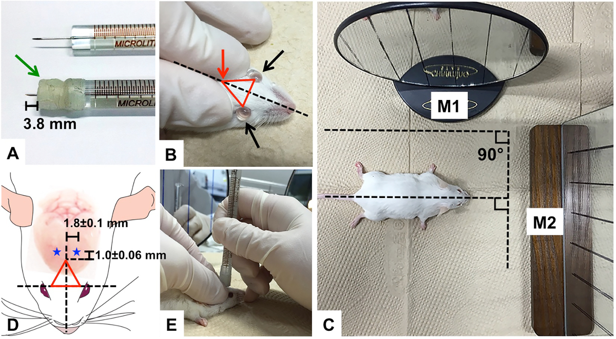
- Wrap the needle of the microsyringe with Parafilm to adjust its length to 3.8 mm to enable maximum accuracy in the depth of the injection into the brain (Figure 1A). (For B6 mice, adjust syringe needle to 3.6 mm).
Note: Compression of the Parafilm will lead to the depth of the injection exceeding 3.6 mm ventral. To prevent the Parafilm from being compressed during needle insertion, densely wrap the needle multiple times. - Wash the inner space of the syringe with 70% ethanol, twice.
- Sonicate the syringe and the needle in a water-bath sonicator for 30 min at RT.
- Wash the inner space of the syringe with 70% ethanol, twice.
- Rinse out the syringe with distilled water and dry it completely in a fume hood for longer than 30 min. Leave the clean syringe under a UV light for 20 min before starting the Aβ injection.
4. Aβ-injected Mouse Model Preparation
Note: Clean the operating field with a disinfectant to maintain sterile conditions and sterilize all surgical instruments for ICV injection using 70% ethanol and UV exposure.
- Anesthetize the mouse by intraperitoneal injection of a mixture of xylazine (20 mg/kg) and tiletamine·HCl/zolazepam·HCl (80 mg/kg) before the ICV injection. Place the mouse on a warm mat to maintain its body temperature while anesthetized and confirm adequate anesthesia by assessing the foot-pinch response.
- Apply sterile PBS drops to both eyes to prevent corneal dryness during the procedure (Figure 1B).
- Prepare two mirrors by drawing multiple straight vertical lines on their surfaces in case of difficulty injecting the syringe perpendicularly.
- Place one mirror (M1) right next to the body of the mouse and the other (M2) in front of the head. Keep M1 parallel to the imaginary midline between the two eyes of the mouse and arrange M2 perpendicular to M1, so that the two planes form a 90° angle (Figure 1C). Place M1 on the left side of the mouse if the researcher is right-handed. Place M1 on the right side of the mouse if the researcher is left-handed.
- Spray 70% ethanol onto the middle of the forehead of the mouse and rub it with dry cotton swabs. Do not wet too large of an area of the forehead to avoid decreasing the body temperature due to evaporation of the alcohol.
- Wipe the same area on the forehead with 2% chlorhexidine solution using a clean cotton swabs. Repeat scrubbings with alcohol and chlorohexidine for total of three times each.
Note: If necessary, shave hair on the forehead of the mouse in order to minimize the possibility of contamination - Locate bregma.
- Using the thumb and index finger, tightly hold down the skin of the forehead, directly above the two eyes to the degree that both eyes protrude. Drag the skin back to make the forehead taut and minimize skull movements under skin.
- Form an invisible triangle by assigning three vertices: the two eyes of the mouse and the point where the thumb and index finger meet, the bregma. (Figures 1B, Red arrow: bregma)
- Locate the injection point with measuring tape: -1.0 ± 0.06 mm posterior to bregma, 1.8 ± 0.1 mm lateral to the sagittal suture, and 2.4 mm in depth (Figure 1D, blue stars: injection points in each hemisphere). (For B6 mice: -0.9 mm posterior, 1.7 mm lateral, and 2.2 mm in depth)
- Fill the syringe with 10 µl Aβ solution or vehicle with excess solution in order to avoid accidental air injection.
- Place the syringe at the injection point (described in protocol 4.7). Make the syringe perpendicular to the plane of injection point. Align reflected images of the syringe in both mirrors with the drawn lines on the mirrors from a fixed perspective (Figure 1C).
- Begin inserting the needle until the parafilm wrapping reaches the skin.
- Keep the hand holding syringe steady and use other hand to inject 5 µl of the Aβ solution or vehicle, slowly over 5 sec without pausing. Make sure the syringe remains perpendicular throughout. After completing the injection, wait 3 to 5 sec before removing the syringe for diffusion.
- Assuming the original triangle position, exert moderate pressure to protect the skull from any unnecessary movements. Remove the syringe without tilting.
- Place the Aβ-injected mouse on warm pad for recovery and maintain its sternal recumbency. Do not leave the mouse unattended until it regains consciousness and do not return the mouse to a cage containing non-surgical mice until it has fully recovered.
- Wash the syringe according to steps 3.3-3.6.
- Postoperatively monitor the mobility of the mice and check for any sign of infection or illness in the mice for 5-7 days.

Figure 1. Aβ Injection into the ICV region. (A) Parafilm-wrapped syringe needle compared to an unmodified syringe needle; (B) PBS drops onto eyes to prevent dryness; the triangle formed on the forehead of mouse with thumb, index finger, and the two eyes of mouse as well as the imaginary midline equidistant from each eye; (C) two mirrors (M1 and M2) with multiple vertical lines drawn on the surfaces to assist perpendicular injection of the syringe needle; (D) the triangle with the injection points, indicated as blue stars (-1.0 ± 0.06 mm from bregma and 1.8 ± 0.1 mm sagittally) on each hemisphere of mouse; Green arrow: parafilm, Red arrow: bregma Please click here to view a larger version of this figure.
5. Confirmation of Aβ Injection by Y-maze and Brain Analyses
- Assess the working memory of Aβ-injected mice by Y-maze tests3,7,8
- Wait 3 days after the injection for the onset of memory deficits.
- Perform the Y-maze tests using a black plastic maze with three identical arms labeled A, B, and C, each of which branches out at a 120° angle from the center of the maze (width x length x height, 10 cm x 40 cm x 12 cm).
- Place a mouse at the start arm (arm A) and allow the subject to freely explore the maze for 8 min.
- Measure the number of entries into each arm and calculate the alternation rate of each subject3,9.
- Directly examine postmortem brains to check if the injection targeted the ICV region appropriately.
- Anesthetize the mouse by the same procedure as outlined in section 4.1. Then sacrifice the mouse by cervical dislocation.
- Decapitate the mouse and remove the skin to expose the skull using surgical scissors. Remove tissues and muscles from the cranium.
- Make cuts on the lambdoid suture (between the parietal and interparietal bones) without damaging the brain. Remove the occipital and interparietal bones, then the parietal and frontal bones. Remove the cranial bone using forceps and isolate the brain from the skull by lifting up the meninges.
- Wash the brain in chilled sterile saline and cut the ICV region of the brain in the coronal direction with a surgical knife (Figure 2).
- Confirm the injection region based on the trace of the needle insertion on the brain tissue (Figure 2). Check if the needle trace has reached one of ventricles. If the needle trace does not meet or pass through the ventricles, exclude that subject's data from analysis of the experimental results.
Access restricted. Please log in or start a trial to view this content.
Results
This section illustrates examples of the results that can be obtained by confirmation of Aβ aggregation and Y-maze assessment of memory deficits. Using the full-length Aβ(1-42) peptide of 42 amino acids, mixture of Aβ monomers, oligomers, and fibrils (Figure 3) was produced. Through the HFIP-induced monomerization step, relatively homogeneous monomers (Lane B) were obtained. After the 7 day incubation, diverse sizes of Aβ aggregates (Lane C) developed. Trimers and tet...
Access restricted. Please log in or start a trial to view this content.
Discussion
The most important step in this protocol is the ICV injection of Aβ. This protocol is designed to inject Aβ into the ICV region of mice without stereotactic instruments11,12. Before starting an experiment, a preliminary period of practice injections with a blue dye instead of Aβ should take place to achieve sufficient dexterity. Immediately after the injection, it is necessary to check whether the pigment injection was performed properly. Carefully take out the brain and check whether the approp...
Access restricted. Please log in or start a trial to view this content.
Disclosures
The authors have nothing to disclose.
Acknowledgements
This research was supported by a grant of the Korea Health Technology R&D Project through the Korea Health Industry Development Institute (KHIDI), funded by the Ministry of Health & Welfare, Republic of Korea (grant number: H14C04660000).
Access restricted. Please log in or start a trial to view this content.
Materials
| Name | Company | Catalog Number | Comments |
| ICR mouse | Orientbio | male, 6-8 weeks, 27-29 g of body weight | |
| C57BL/6 mouse | Orientbio | male, 6-8 weeks, 21-23 g of body weight | |
| Amyloid-beta1-42 | in house synthesis | stock concentration: 1 mM/DMSO, injected concentration: 100 μM/10% DMSO and 90% PBS | |
| ICV injection syringe (26s gauge) | Hamilton | 80308 | |
| Evans blue dye (EBD) | abcamBIochemicals | ab120869 | 1% EBD in PBS |
| DMSO | Sigma | D2650 | |
| PBS | gibco | 10010-023 | |
| Gradi-GelTM II Gradient PAGE Analysis Kit | ELPiS Biotech | EBS-1056 | 15% Gel |
| Precision Plus ProteinTM Dual Xtra Standards | Bio-Rad | 161-0377 | |
| Silver-Staining Kit | GE-Healthcare | 17-1150-01 | |
| LabDiet | Orientbio | 5053 |
References
- Kam, T. I., Gwon, Y., Jung, Y. K. Amyloid beta receptors responsible for neurotoxicity and cellular defects in Alzheimer's disease. Cell Mol Life Sci. 71, 4803-4813 (2014).
- Elder, G. A., Gama Sosa, M. A., De Gasperi, R. Transgenic mouse models of Alzheimer's disease. Mt Sinai J Med. 77, 69-81 (2010).
- Bryan, K. J., Lee, H., Perry, G., Smith, M. A., Casadesus, G. Methods of Behavior Analysis in Neuroscience Frontiers in Neuroscience. Buccafusco, J. J. , (2009).
- Van Dam, D., De Deyn, P. P. Animal models in the drug discovery pipeline for Alzheimer's disease. Br J Pharmacol. 164, 1285-1300 (2011).
- Choi, J. W., Kim, H. Y., Jeon, M., Kim, D. J., Kim, Y. Efficient access to highly pure beta-amyloid peptide by optimized solid-phase synthesis. Amyloid. 19, 133-137 (2012).
- Rahimi, F., Maiti, P., Bitan, G. Photo-induced cross-linking of unmodified proteins (PICUP) applied to amyloidogenic peptides. J Vis Exp. , (2009).
- Kim, H. V., et al. Amelioration of Alzheimer's disease by neuroprotective effect of sulforaphane in animal model. Amyloid. 20, 7-12 (2013).
- Jackson, L. L. VTE on an elevated T-maze. J Comp Psychol. 36, 9(1943).
- Kim, H. Y., et al. Taurine in drinking water recovers learning and memory in the adult APP/PS1 mouse model of Alzheimer's disease. Sci Rep. 4, 7467(2014).
- Paxinos, G., Franklin, B. J. The Mouse Brain in Stereotaxic Coordinates. 2nd edn. , Academic Press. (2001).
- Ledo, J. H., et al. Amyloid-beta oligomers link depressive-like behavior and cognitive deficits in mice. Mol Psychiatry. 18, 1053-1054 (2013).
- Laursen, S. E., Belknap, J. K. Intracerebroventricular injections in mice. Some methodological refinements. J Pharmacol Methods. 16, 355-357 (1986).
- Bromley-Brits, K., Deng, Y., Song, W. Morris water maze test for learning and memory deficits in Alzheimer's disease model mice. J Vis Exp. , (2011).
- Figueiredo, C. P., et al. Memantine rescues transient cognitive impairment caused by high-molecular-weight abeta oligomers but not the persistent impairment induced by low-molecular-weight oligomers. J Neurosci. 33, 9626-9634 (2013).
- Haley, T. J., McCormick, W. G. Pharmacological effects produced by intracerebral injection of drugs in the conscious mouse. Br J Pharmacol Chemother. 12, 12-15 (1957).
- Harkany, T., et al. Neuroprotective approaches in experimental models of beta-amyloid neurotoxicity: relevance to Alzheimer's disease. Prog Neuropsychopharmacol Biol Psychiatry. 23, 963-1008 (1999).
- Wang, D., et al. The allosteric potentiation of nicotinic acetylcholine receptors by galantamine ameliorates the cognitive dysfunction in beta amyloid25-35 i.c.v.-injected mice: involvement of dopaminergic systems. Neuropsychopharmacology. 32, 1261-1271 (2007).
- Yamada, K., Nabeshima, T. Animal models of Alzheimer's disease and evaluation of anti-dementia drugs. Pharmacol Ther. 88, 93-113 (2000).
- Cho, S. M., et al. Correlations of amyloid-beta concentrations between CSF and plasma in acute Alzheimer mouse model. Sci Rep. 4, 6777(2014).
Access restricted. Please log in or start a trial to view this content.
Reprints and Permissions
Request permission to reuse the text or figures of this JoVE article
Request PermissionExplore More Articles
This article has been published
Video Coming Soon
Copyright © 2025 MyJoVE Corporation. All rights reserved