Method Article
Imaging the Human Immunological Synapse
* These authors contributed equally
In This Article
Summary
This protocol images both the immunological synapse formation and the subsequent polarized secretory traffic towards the immunological synapse. Cellular conjugates were formed between a superantigen-pulsed Raji cell (acting as an antigen-presenting cell) and a Jurkat clone (acting as an effector helper T lymphocyte).
Abstract
The purpose of the method is to generate an immunological synapse (IS), an example of cell-to-cell conjugation formed by an antigen-presenting cell (APC) and an effector helper T lymphocyte (Th) cell, and to record the images corresponding to the first stages of the IS formation and the subsequent trafficking events (occurring both in the APC and in the Th cell). These events will eventually lead to polarized secretion at the IS. In this protocol, Jurkat cells challenged with Staphylococcus enterotoxin E (SEE)-pulsed Raji cells as a cell synapse model was used, because of the closeness of this experimental system to the biological reality (Th cell-APC synaptic conjugates). The approach presented here involves cell-to-cell conjugation, time-lapse acquisition, wide-field fluorescence microscopy (WFFM) followed by image processing (post-acquisition deconvolution). This improves the signal-to-noise ratio (SNR) of the images, enhances the temporal resolution, allows the synchronized acquisition of several fluorochromes in emerging synaptic conjugates and decreases fluorescence bleaching. In addition, the protocol is well matched with the end point cell fixation protocols (paraformaldehyde, acetone or methanol), which would allow further immunofluorescence staining and analyses. This protocol is also compatible with laser scanning confocal microscopy (LSCM) and other state-of-the-art microscopy techniques. As a main caveat, only those T cell-APC boundaries (called IS interfaces) that were at the right 90° angle to the focus plane along the Z-axis could be properly imaged and analyzed. Other experimental models exist that simplify imaging in the Z dimension and the following image analyses, but these approaches do not emulate the complex, irregular surface of an APC, and may promote non-physiological interactions in the IS. Thus, the experimental approach used here is suitable to reproduce and to confront some biological complexities occurring at the IS.
Introduction
The main goal of the method is to generate immunological synapse (IS) cell-to-cell conjugates formed by an antigen-presenting cell (APC) pulsed with SEE superantigens and an effector Th cell, and to register the images corresponding to the first stages of immunological synapse formation and the subsequent trafficking events (occurring both in the APC and the Th cell), that eventually will lead to polarized secretion at the IS. The establishment of the IS by T lymphocytes upon binding of their cell receptor (TCR) to antigens bound to MHC-II on the APC organizes an extremely dynamic, malleable and critical instance involved in antigen-specific, humoral and cellular immune responses1,2. The IS is defined by the formation of a special supramolecular activation complex (SMAC) pattern characterized by an actin reorganization process3. Upon IS construction by T lymphocytes with an APC, the polarization of the secretory vesicles towards the IS appears to be implicated in polarized secretion at the synaptic gap. This focused machinery appears to unambiguously supply the immune system with a superbly regulated stratagem to enhance the efficacy of critical secretory effector roles of T lymphocytes, while reducing nonspecific, cytokine-arbitrated stimulation of bystander cells, killing of irrelevant target cells and apoptotic suicide via activation-induced cell death (AICD)4.
The consequence of the IS varies on the nature of both the T lymphocyte and the APC. The synaptic contact of Th cells (typically CD4+ cells) with the APC displaying antigen-associated to MHC-II produces the activation of the T cell (cytokine secretion, proliferation, etc.) and, in some instances, apoptosis via AICD4. For cytotoxic T lymphocytes (CTLs) (principally CD8+ cells) interacting with APC presenting antigen associated to MHC-I, the outcome differs on the pre-stimulation or not of the CTLs with antigen. Thus, naïve CTLs identifying antigen-MHC-I complexes on the APC are "primed" to destroy target cells and divide. Primed CTLs also establish synapses with target cells (i.e., cells infected by viruses or tumor cells) producing antigen-specific cell extermination5,6.
The polarized secretion of exosomes at the immunological synapse is a developing and challenging area of research involved in relevant immune responses7. It has been demonstrated that multivesicular bodies (MVB) carrying intraluminal vesicles (ILVs) experience polarized transportation towards the IS8,9 (Video 1) upon TCR stimulation with antigen. The fusion of these MVB at the synaptic membrane induces their degranulation and the release of the ILVs as exosomes to the synaptic cleft8,10. This occurs in IS formed by Th-type Jurkat cells that were challenged with SEE superantigen-coated Raji cells acting as APC11, TCR-stimulated CD4+ lymphoblasts, and primed CTLs. Thus, synapses made by Jurkat cells constitute a valuable model to study polarized secretory traffic of exosomes. In addition, several decades of investigation have shown that many fundamental insights into TCR signaling came from studies with transformed T-cell lines, and indeed the best known of these model systems is the Jurkat leukemic T-cell line12.
The formation of a fully developed IS produces several crucial biological outcomes, including activation of Th cells, activation of naïve CTLs or target cell killing by primed CTLs, apart from anergy or AICD5. Therefore, there are two major types of secretory IS established by T lymphocytes that result in very diverse, but similarly critical, immune effector functions1,6,13. On one hand, the IS from primed cytotoxic T lymphocytes (CTLs) induces rapid polarization (ranging from seconds to few minutes) of lytic granules (called "secretory lysosomes") towards the IS. The degranulation of the lytic granules induces the secretion perforin and granzymes to the synaptic cleft14, which are pro-apoptotic molecules. The secreted perforin and granzymes subsequently induce killing of the target cells15,16. CTLs develop temporary synapses, lasting only few minutes, as the target cells are murdered3,17. This is probably due to the circumstance that the optimal CTL task requires a rapid and temporary contact in order to distribute as many lethal strikes as possible to numerous target cells3,17. In contrast, Th lymphocytes such as Jurkat cells generate stable, long-standing IS (from 10-30 min up to hours), since this appears to be necessary for both directional and incessant secretion of stimulating cytokines3,17. The cytokines are also enclosed in secretory vesicles and some of them (i.e., IL-2, IFN-γ) experience polarized transport to the IS17 and secretion. One essential characteristic of the IS is the formation of exploratory, weak and transient contacts between the T cell and the APC (Video 1) that may produce a stronger interaction and the establishment of a mature IS, providing that the TCR identifies the cognate antigen-MHC complexes and that appropriate co-stimulatory connections are established5. Both the beginning of the initial contacts and the founding of a mature, totally productive IS, are inherently stochastic, fast and asynchronous processes5,18. Furthermore, there is a scarce frequency in the creation of cell-to-cell conjugates19, which may constitute a challenge for imaging techniques (please refer to Results and Discussion sections).
Another main challenge in examining polarization of the microtubule-organizing center (MTOC) and secretory granules in T lymphocytes is that the entire process is fast (seconds to few minutes), particularly in CTLs. Considering these facts, most early approaches confronted an end point strategy in which APC/target cells and T lymphocytes are mixed jointly and converged by low speed centrifugation, to favor cell-to-cell conjugate creation, incubated for several minutes, fixed and subsequently evaluated for the relocation of MTOC and/or secretory vesicles towards the IS20. This approach has two significant limitations: no lively trafficking data was achieved and high levels of background MTOC/secretory granules polarization were obtained, probably due to the stochastic character of IS establishment18. Furthermore, any correlation between TCR-stimulated, initial signaling events (i.e., intracellular calcium rises, actin reorganization) and secretory vesicle polarization is problematic to investigate. Thus, imperative provisions for appropriate imaging of the IS in living cells combine to enhance the cell-to-cell conjugate materialization, to synchronize the generation of the IS and, when possible, to guarantee the establishment of cellular conjugates at defined microscope XY fields and Z positions. Several strategies have been developed in order to avoid all these problems. It is out of the scope of this paper to explain these methods, their benefits and weaknesses. Please refer to the previously published reviews tackling these important points1,4,5,21.
The fact that IS made by Th lymphocytes are long-lived, and the circumstance that in Th lymphocytes the MTOC, lymphokine-containing secretory granules and MVB take from several minutes up to hours to move and dock to the IS22 makes the Th-APC IS an ideal candidate for imaging using the protocol described here.
Protocol
1. Preparation of Slides to Adhere Raji Cells
- Add 150 µL of fibronectin (100 µg/mL) per well to an 8 microwell chamber slide (plastic-bottom chamber slide) and incubate it for 30 min to 1 h at 37 °C. This adhesion substrate will allow the binding of Raji cells to the well bottom (Step 2), the formation of living conjugates with Jurkat cells (Step 4) cells, and time-lapse microscopy capture (step 6), and is also compatible afterwards with optional paraformaldehyde (PFA) fixation (step 7).
NOTE: For acetone fixation, use glass bottom chamber slides and poly-L-lysine (20 µg/mL) instead of fibronectin, since acetone dissolve plastic. The 8 microwell chamber slide (1 cm2 well, 300 µL maximum volume) or equivalent is a flexible and appropriate format. - Aspirate fibronectin using a 200 μL automatic pipette and wash each well with 200 µL of PBS for 2 min with gentle shaking. Repeat this wash one more time. The chamber slide can be stored at this stage with PBS for 1-2 weeks at 4 °C.
2. Adhesion of Raji Cells to the Chamber Slides and 7-amino-4-chloromethylcoumarin (CMAC) Labeling
- Transfer 10 mL of a confluent (1-2 x 10⁶ cells/mL) pre-culture of Raji cells to a 15 mL, V-bottom tube. Mix well and use 10 µL to count the cells on Neubauer chamber or equivalent.
- Centrifuge the remaining cells at 300 x g for 5 min at room temperature. Aspirate and discard the supernatant.
- Gently resuspend the cell pellet in warm complete culture medium (RPMI 1640 supplemented with 10% FCS, 2 mM glutamine, 10 mM HEPES, 100 U/mL penicillin, and 100 µg/mL streptomycin) at a concentration of 106 cells/mL. Use the equation given below:
([ ]initial x Vinitial = [ ]final x Vfinal), where [ ]initial represents initial cell concentration, Vinitial = initial volume of the cell suspension, [ ]final = final cell concentration, Vfinal = final volume of the cell suspension.
NOTE: Depending on the cell concentration of the starting culture, it is possible to collect more cells than needed but it is important to maintain the remaining cells in culture (37 °C) till the end of the experiment in order to prevent potential problems (see Step 2.6). - Label Raji cells to allow for their identification during the synaptic conjugate formation. In this experiment, 7-amino-4-chloromethylcoumarin (CMAC) labeling is performed in step 2.4.2.
- Transfer the required number of Raji cells in the culture medium to a 2 mL tube. For the 8 microwell chamber slide, a total of 1.6 mL of the cell suspension is needed (200 µL per well).
- Add CMAC to a final concentration of 10 µM. Keep the cells in the dark by covering the tube with aluminum foil since CMAC is light-sensitive. 200 µL containing 2 x 10⁵ Raji cells are needed per 1 cm2 well. Thus, if 8 wells need to be prepared, 1.6 x 10⁶ Raji cells are required.
NOTE: Labeling Raji cells with cell tracker blue (CMAC, UV excitation and blue emission) distinguishes them from Th cells when the synaptic conjugates are formed. This dye is compatible with PFA and acetone fixatives and allows further immunofluorescence procedures. Try to avoid light exposition. The labeling of Raji cells in a pool with CMAC followed by resuspension before the adhesion of the Raji cells to the fibronectin-coated chamber slides ensures the homogeneous labeling of Raji cells with CMAC among different wells.
- Resuspend CMAC-stained cells and, after aspiration of the PBS in the chamber slide from step 1.2., transfer 200 µL of the cell suspension to each well of the fibronectin-coated chamber slides prepared in step 1.1-1.2. Incubate the chamber slide at 37 °C, 5% CO2 for 30 min-1 h.
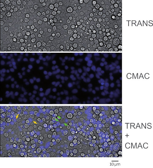
NOTE: The adhesion and CMAC labeling will simultaneously occur at this step and this saves time. Please be aware that Raji cells will sediment quickly and caution needs to be taken to maintain a homogeneous concentration in the cell suspension before seeding. It is not necessary to wash CMAC at this stage by centrifugation, since CMAC washing will be more easily done at step 2.7 (when the labeled Raji cells are already adhered to the chamber slides). Since CMAC is present in the cell suspension in large excess, the blue fluorescence background is too high to distinguish the blue-stained cells. Check cell CMAC fluorescence in step 2.7 after CMAC washing. - Ensure that Raji cells are adhered to the bottom of the wells by gentle shaking of the chamber slides on the microscope. Ensure that the cells display gaps among each other and are not confluent (Figure 1, middle panel). 50-60% of cell confluence is appropriate.
- If most of the cells efficiently adhere to the chamber slide and no cell gaps are observed, wash each well with warm complete medium and resuspend the medium with a 200 μL automatic pipet to detach cell excess. Check confluence after each resuspension step.
- If the cells do not adhere, repeat the adhesion step again and increase adhesion time and/or cell number.
NOTE: It is possible to stop here, incubate the chamber slide at 37 °C, 5% CO2 overnight (O/N), and continue with the protocol the next day. Please confirm the next day that Raji cells remain adhered and CMAC-labeled by using fluorescence microscopy.
- Wash each well again carefully with warm supplemented RPMI to eliminate excess CMAC and check for blue emission with the fluorescence microscope (Figure 1).
NOTE: To avoid the use of immersion oil (sticky and viscous) and high numerical aperture oil objectives, extra-long distance (i.e., 20x or 40x) objectives can be used to quickly check CMAC labeling using the fluorescence microscope.
3. Pulse of CMAC — Labeled Raji Cells with Staphylococcal Enterotoxin E
- Add Staphylococcal Enterotoxin E (SEE, 1 µg/mL) to each well. SEE can be conveniently diluted in cell culture medium (working solution at 100 µg/mL) from the SEE frozen stocks (1 mg/mL in PBS). Use 2 μL of the 100x working solution per 200 μL microwell.
CAUTION: Use gloves for this step and dispose of the used tip into the biohazard box. - Incubate the chamber slide at 37 °C, 5% CO2 for at least 30 min. The SEE effect lasts for at least 3-4 h.
NOTE: SEE can be added to the wells at different time points when required, if distinct time-lapse setups are planned (step 5) and/or depending of the start time point for the end point experiments (step 6).
4. Preparation of Jurkat Cells
- Use a previously growing culture of Jurkat cells (1-2 x 10⁶ cells/mL) for this experiment. Use cells from a standard culture flask or from a previous transfection following standard electroporation protocols, as previously described23. The transfection of Jurkat cells will allow time lapse visualization of the traffic of secretory granules in living cells. For instance, when GFP-CD63 (a marker of MVB) is expressed the movement of GFP-CD63-decorated vesicles can be recorded (Video 1).
- Observe the cells under a phase contrast microscope. If an excess of dead cells (>20-30%) are observed, perform Ficoll density gradient centrifugation using standard protocols24, to eliminate the excess of dead cells (dead cells exhibit higher density than living cells) prior to use (see Discussion).
- Transfer the cells to a 15 mL tube, V-bottom tube, and use 10 µL for counting using hemocytometer.
- Centrifuge the remaining cells as described in step 2.2. Discard the supernatant and resuspend the cells at the same concentration as Raji cells (1 x 10⁶/mL) using fresh, warm culture medium. Follow steps 2.2-2.3.
- Maintain the Jurkat cells in culture (37 °C, 5% CO2) while waiting for the Step 4.
NOTE: In the second option (transfection), the number of living cells is going to be much lower than in the first one. Thus, consider using a higher starting cell culture volume in order to have enough cells for the experiment. From 10 x 106 Jurkat cells per electroporation cuvette and transfection, only 2-4 x 10⁶ Jurkat cells will survive after 48 h of transfection and some of these cells will be lost during the Ficoll step. Thus, one electroporation cuvette is generally sufficient to challenge the adhered SEE-pulsed Raji cells from 8 micro wells (1.6 x 10⁶ transfected Jurkat cells needed).
5. Co-seeding of Raji and Jurkat Cells
- Take the chamber slides containing the CMAC-labeled, SEE-pulsed, adhered Raji cells out of the incubator from step 3.2. It is not necessary to wash the CMAC at this stage since this was previously done in Step 2.7.
- Aspirate carefully the culture medium of each well, one by one, from one corner of the well using an automatic 200 μL pipette. Do not let the medium in the well dry out completely.
- Immediately replace the medium with 200 µL of resuspended Jurkat cells in cell culture medium (1 x 10⁶/mL) prepared in step 4.5. If time lapse imaging is performed, go to step 6 immediately after this step, since Jurkat cells tend to sediment and form synaptic conjugates very quickly. For convenience, the microwells containing SEE-pulsed, adhered Raji cells that do not receive seeding with Jurkat cells at this stage should be maintained with cell culture medium until subsequent challenge with Jurkat cells. This will flexibly allow subsequent challenge with Jurkat cells for additional time lapse or reverse kinetic, end point experimental approaches.
- If a time lapse is going to be performed, quickly proceed to step 6. This involves the co-culture for 1-2 h on the microscope stage incubator or equivalent at 37 °C, 5% CO2 to allow the synaptic conjugate formation and simultaneous image acquisition. If end point analysis, but no time lapse, is foreseen check conjugate formation after the co-culture period using the microscope (as in Figure 1) before fixing the cells (step 7).
6. Time Lapse Imaging of Emerging Synaptic Conjugates
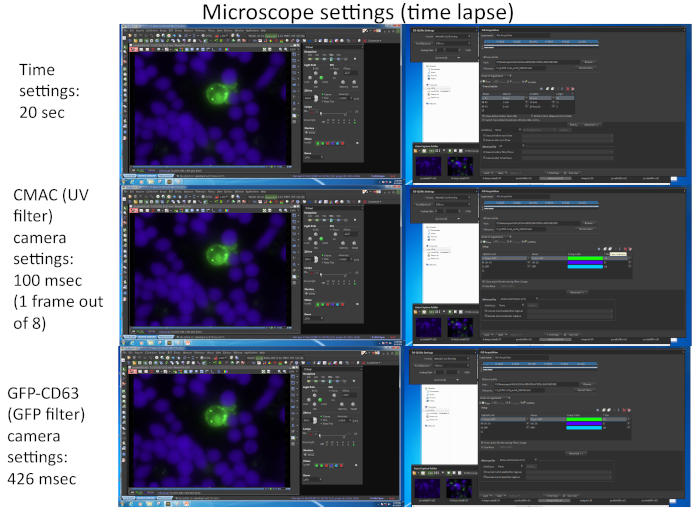
- Prepare the microscope and incubation chamber prior to imaging. For the example shown in Video 1, the detailed microscopy settings are shown in Figure 2.
NOTE: If a time lapse experiment is planned, all the microscope settings and complements (ambient cell culture chamber, etc.) should be prepared before adding the Jurkat suspension to the chamber slide with adhered Raji cells. The following steps are described for a commercial microscope (Table of Materials). However, any inverted fluorescence microscope equipped with a cell culture incubator can be used.- Use a microscope with a 60x oil-immersion, high numerical aperture when imaging polarized traffic.
- Ensure that the automatic focus system is switched on and adjust the offset to focus the Raji cells bound to the bottom surface. Please refer to Figure 1, Video 1 and Video 2.
- After Jurkat addition to each well containing the adhered Raji cells in step 5.3, quickly locate the microwell chamber slide on the pre-heated (1-2 h) microscope stage incubator (i.e., OKOlab) and select some XY positions with the microscope, fields in which it is likely to record an emerging IS formation made by, for instance, a Jurkat-transfected cell falling into the microscope focus.
- Use a pre-heated microscope stage incubator since it was observed that a temperature-stabilized stage maintains stable X,Y,Z positions. Criteria for a convenient XY field are: well-focused and non-confluent Raji cells (i.e., displaying gaps among cells) and the presence of transfected Jurkat cells (this can be checked by combining transmittance and UV or GFP channels). Jurkat cells will sediment very quickly (few minutes) on the chamber slide, and the chances to image emerging synapses will decrease with time (Figure 1). It is possible either to finish the experiment after the defined time lapse or proceed to the Step 7 and fix the conjugates for subsequent immunofluorescence and analyses.
NOTE: It is possible to select up to 16 different microscope fields from up to 4 different microwells for simultaneous, multi-well time-lapse acquisition with the proper temporal resolution (1-2 min per frame). The limitation relies on both the number and intensity (affecting camera exposition) of the diverse fluorochromes to be imaged (dependent on the number of expressed fluorescent proteins, apart from CMAC). One way to increase the frame rate is recording for the CMAC channel in only one out of each "n" time frames for GFP (i.e., n = 8, as shown in Figure 2), since Raji cells are adhered to the well bottom and do not easily move as Jurkat cells. In addition, this benefits the cell viability, since frequent UV light exposure may damage the cells. Try to adjust the time frame rate to 1 frame every 1 minute or less (i.e., 20 s per frame in Video 1, Figure 2) since the polarization of MVB takes a few min to hours to complete. A microscope equipped with a motorized epi-fluorescence turret and appropriate band-pass fluorescence filters or equivalents are necessary to perform this multichannel capture.
7. End Point Formation of Synaptic Conjugates and Fixation
- If only an endpoint experiment is planned (1-2 hours incubation to allow synaptic conjugate formation is appropriate), incubate the chamber slide at 37 °C, 5% CO2 for 1-2 h. Check conjugate formation after the culture period (as in Figure 1) and, subsequently, fix the conjugates with acetone or PFA (fixation will depend on the antigens and antibodies to be used in the subsequent immunofluorescence). In this case the incubation does not require a microscope stage incubator. Please refer to Video 3 for an example.
- To fix the cells, wash the well by gentle shaking with warm RPMI (37 °C) medium without FCS (albumin from serum may precipitate with acetone fixation). Aspirate and add 200 µL of PFA or pre-chilled acetone to each well. Incubate the chamber slide at room temperature (RT) or on ice, respectively, for 20 min.
NOTE: For the acetone fixation, pre-chill the acetone at -20 °C and pre-chill the chamber slide at 4 °C. Remove plastic lids when acetone is used to fix the cells cultured in the 8 well glass-bottom chamber slides. - Wash each well twice with PBS and add 200 µL of quenching solution (PBS, 50 mM NH4Cl). Incubate the chamber slide at 4 °C.
NOTE: At this stage the chamber slide can stay for at least one month at 4 °C before performing the immunofluorescence protocol, as described8. The lid prevents evaporation.
8. Image Processing
- Perform post-acquisition image deconvolution (i.e. Huygens deconvolution or equivalent, Table of Materials) of time-lapse series and/or still photos of fixed cells. Deconvolute by employing an appropriate software (i.e., using the "wide field" optical option in Huygens) and the correct optical parameters. Deconvolution necessitates for the image processing of the measured point spread function (PSF) of the microscope4.
- Alternatively, use the software to calculate the idealized PSF by automatic loading of the optical parameters included among the metadata from the microscope files. These optical parameters comprise fluorochrome wavelength, refraction index, numerical aperture of the objective, and the imaging technique (confocal, wide-field, etc.)4.
- Subsequently, the imaging software uses the PSF and diverse deconvolution algorithms (i.e., QMLE and CMLE in Huygens software) in a step-by-step accumulative, calculation process, whose results than can be continuously visualized and stopped (or resumed) when required by the user. At this stage the user can change the number of convolutions and/or the signal to noise ratio and resume deconvolution. The deconvolution software works well with time lapse series (X,Y,T) (Video 2) and Z-stacks (X,Y,Z) (Video 3). The deconvoluted channels were subsequently merged to the CMAC, raw channel, since cytosolic, diffuse fluorochromes do not improve by deconvolution4,9.
Results
We have followed the described protocol in order to generate Jurkat-Raji immune synapse conjugates and to properly image the early stages of IS formation. Our aim was to improve the early approaches20 previously followed to study the polarization of the MTOC and the secretory machinery towards the IS. These approaches were based on an end point strategy that did not allow imaging of the IS formation or the early synaptic events, since in these strategies the IS formation obligatory occurred in the pellet of mixed, centrifuged cells, but not on a microscope. Our protocol was designed to avoid this main caveat, since the approach was based on the use of an appropriate cell concentration (Steps 2 and 3), to favor the formation of the cell-to-cell IS conjugates on the own microscope chamber slides (8 micro well chamber slide),mounted on the pre-heated microscope stage incubator (in Step 4). This strategy induces the IS formation, simultaneously to the time-lapse imaging capture (Video 1).
Figure 1 represents synaptic Jurkat-Raji conjugates that were obtained following the protocol (step 5.2). The image represents the first frame from a representative, time-lapse experiment. 2 x 10⁵ Raji cells and 2 x 10⁵ Jurkat cells were added to a 1 cm2 well. The upper panel shows transmittance channel, the middle panel consists of CMAC (blue) channel (Raji cells), and the lower panel shows both transmittance plus CMAC merged channels. Yellow arrows label some synaptic conjugates, as a reference, whereas green arrows indicate synaptic conjugates made by one Jurkat cell and several Raji cells (complex conjugates). Decreasing cell concentrations (105 or less cells in the 1 cm2 well) will circumvent the formation of complex cellular conjugates, but there may not be enough cell conjugates for subsequent analysis of polarized traffic (see below), that in turn will decrease also the chances to find and to image emerging synaptic conjugates.
Figure 2 represents screenshots corresponding to the imaging parameters used for the simultaneous capture of the two different fluorochromes (CMAC and GFP-CD63) by using appropriate software (i.e., NIKON NIS_AR) in a representative time lapse experiment corresponding to Video 1.
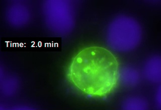
Video 1 (Immunological synapses, raw) represents a Jurkat T lymphocyte expressing GFP-CD63 (a marker of multivesicular bodies-MVB, green vesicles) and the formation of a double synapse between 1 Jurkat cell and 2 Raji cells labelled with CMAC, in blue (one of them undergoes engulfment). The simultaneous movement of MVB inside the Jurkat cell towards synapse areas was recorded (capture frame rate = 1 frame every 20 seconds for GFP-CD63; video reproduction speed = 2 frames per s). The synaptic conjugates were obtained and imaged following the protocol described above (Step 6.2), which allowed imaging of emerging synapses (Video 1). The simultaneous capture of both GFP-CD63 and CMAC fluorescence channels was performed by using an appropriate software (i.e., NIS-AR, Table of Materials). The video represents the raw data from a representative, time-lapse experiment. The automatic focus system was defined with an appropriate offset with respect to the Raji cells bound to the glass bottom, to assure a stable focus along the experiment.
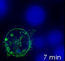
Video 2 (immunological synapses, after deconvolution) corresponds to Video 1, but the deconvolution of the GFP-CD63 fluorescence channel images was performed by employing an appropriate deconvolution software (i.e., Huygens), using the "wide field" optical option and the proper optical parameters (step 8). This deconvoluted channel was subsequently merged to the CMAC, raw channel. The improvement of both signal-to-noise ratio and sharpness of the image is evident. The deconvolution was performed post-acquisition, as described above. For specific details regarding the deconvolution software please refer to4.
Video 3 (immunological synapse, after fixing and immunofluorescence staining) represents a Z-stack (z-step size = 0.8 µm, 5 frames) of a fixed IS conjugate after the step 6 of the protocol (acetone fixation). After fixation, immunofluorescence was performed following standard protocols25 by using Phalloidin to visualize the F-actin (green), anti-CD63 to visualize MVB (magenta), anti-γ-tubulin to visualize MTOC (red). CMAC (blue) labels the Raji cell. Subsequently the conjugate was imaged by epifluorescence and several channels deconvoluted using the "wide field" optical option and the proper optical parameters (step 7). The deconvoluted channels were subsequently merged to the CMAC, raw channel, as indicated in the different panels (CMAC plus Transmittance-TRANS, CMAC plus Phallodin, CMAC plus anti-CD63 and CMAC plus anti-γ-tubulin, respectively). White arrow labels the synapse, whereas green arrow labels the MVB and the yellow arrow labels the MTOC. The detailed quantification of MVB and MTOC polarization has been explained elsewhere25.
The approach presented here involves the formation of cell-to-cell conjugates and, simultaneously, the time-lapse acquisition by wide-field fluorescence microscopy (WFFM) followed by image processing (post-acquisition deconvolution). This tactic improved the signal-to-noise ratio (SNR) of the images, enhanced their temporal resolution, and allowed the synchronized acquisition of several fluorochromes in emerging synaptic conjugates4. In addition, the protocol is well matched with subsequent end point cell fixation methods (paraformaldehyde, acetone or methanol), which would allow further immunofluorescence staining and analyses25 (Video 3). This protocol is also compatible with laser scanning confocal microscopy and other state-of-the-art microscopy techniques.

Figure 1: Representative double-size microscopy field of synaptic conjugates. The image represents the first frame from a representative, time-lapse experiment following the protocol. The upper panel shows the transmittance channel, the middle panel CMAC channel (Raji cells) and the lower panel both merged channels. Yellow arrows label some synaptic conjugates, as a reference. Green arrows indicate complex synaptic conjugates (i.e., one Jurkat cell establishing synapses with more than one Raji cell). Captured with a 40x EWD (0.6 NA) objective. Please click here to view a larger version of this figure.

Figure 2: Time lapse microscope settings. The image corresponds to several screenshots corresponding to the imaging parameters used for the simultaneous capture of the two different fluorochromes (CMAC and GFP-CD63) using appropriate software (i.e., NIKON NIS_AR) in a representative time lapse experiment corresponding to Video 1. Each frame for GFP-CD63 channel was captured every 20 s. Only one frame for a UV channel out of eight frames for GFP channel was captured in order to maintain cell viability along the experiment. Please click here to view a larger version of this figure.

Video 1: Immunological synapses made by a Jurkat cell expressing GFP-CD63, raw data. Raji B cells labelled with cell tracker blue (CMAC, blue) were pulsed with SEE for 30 min and synapses with Jurkat cells expressing GFP-CD638 were formed. Time-lapses corresponding to GFP-CD63 and CMAC channels were captured (20 s per frame; video reproduction speed = 2 frames per s) and a representative example is shown. Captured with a 60x PLAN APO (1.4 NA) objective. Please click here to view this video (Right click to download).

Video 2: Immunological synapses made by a Jurkat cell expressing GFP-CD63, after deconvolution. Same as Video 1, but images were deconvoluted using a deconvolution software. The improved signal-to noise ratio and the enhanced sharpness, due to the elimination of contaminant out of focus fluorescence, is evident. Please click here to view this video (Right click to download).

Video 3: Fixed and immunostained immunological synapse. The image shows a representative fixed synaptic conjugate after Step 7 of the Protocol and subsequent immunofluorescence. CMAC (blue) labels raji cells, transmittance (TRANS) to show the synaptic conjugate (white arrow), Phalloidin labels F-actin (green), anti-CD63 labels MVB (magenta, green arrow) and anti-γ-tubulin labels MTOC (red, yellow arrow), respectively. These channels were imaged by epifluorescence, deconvoluted and merged as indicated. The video includes a Z-stack (z-step size = 0.8 μm, 5 frames). White arrow labels the synaptic contact area, whereas the green arrow labels the MVB and the yellow arrow labels the MTOC. Please click here to view this video (Right click to download).
Discussion
A limitation of this protocol is that not all synapses will be ideally oriented perpendicular to the optical axis. Using this technique, there is no way to predict and/or to influence the ideal orientation for immune synapse imaging. To fix this problem, we exclude from the subsequent analyses all the randomly captured synapses that finally do not fulfill the ideal criteria. These synapses, opportunely enough, are not very frequent. However, it is possible to circumvent this limitation using several experimental approaches4.
The polarized CD63 release (degranulation) can be quantified by other complementary techniques such as cell surface staining of CD63 (CD63 relocalization to the cell surface) in living cells (not fixed and not permeabilized), after step 6, and subsequent washing and fixation, as previously shown8. In addition, CD63 release on exosomes8,9 and exosome quantification by nanoparticle tracking analyses9,25 can be performed. These approaches are certainly compatible with our protocol, provided fixation is being performed after the cell surface immunofluorescence of living cells.
We have found the ideal number of cells (for a 1 cm2 well in the 8-well chamber slide) is 4 x 10⁵ cells (2 x 10⁵ Raji cells and 2 x 10⁵ Jurkat cells) since they adhere efficiently to the bottom of the well. Binding efficiency to plastic (using fibronectin), or glass bottom (using poly-L-lysine) microslides is not usually a problem. Higher cell numbers may produce no gaps among adhered cells and, subsequently, complex synaptic conjugates (Figure 1); both situations are not desirable when single cell-to-cell conjugates need to be imaged in, for instance, MTOC or secretory granule polarization experiments. Lower number of cells may decrease the chances to find conjugates, especially when transfected Jurkat cells are challenged with APC to generate synapses. Please note that we have observed that a temperature-stabilized stage incubator in place on the microscope X/Y stage before the onset of the protocol (i.e., 1-2 h in advance to stabilize the stage/microscope setup) maintains stable X,Y,Z parameters, which is crucial for proper imaging. Automatic focus system will eventually compensate small Z variations.
When certain gene transduction techniques such as electroporation are used to express fluorescent chimeric proteins (i.e., GFP-CD63) in the Jurkat clones (Video 1), a considerable fraction of cells may die after electroporation. This can be a problem since although dead cells do not form synapses, when they are in excess over the living, transfected cells, they may interfere with the formation of conjugates made by living Th cells. We have found that careful elimination of dead cells from transfected cultures by using a density gradient medium following standard protocols before the conjugate formation step can indeed increase the chances to properly image conjugates. In addition, low transfection efficiencies (<20%) can be an important caveat since this, combined with a moderate conjugate formation efficiency (around 60%)25, will decrease the probability to find conjugates made by transfected cells. This is not a problem when non-transfected cells are used to obtain conjugates in end point experiments and subsequent fixation. The 8 microwell chamber slides are compatible with conventional immunofluorescence protocols. This increases the flexibility of the above protocol with different purposes. Fixation with acetone can be a problem to consider when using chamber slides with plastic-bottom wells. However, there are commercially available 8 microwell microscope chamber slides containing glass bottoms, which are compatible with acetone fixation. Remove plastic lids when acetone is used to fix the cells cultured in 8 microwell glass-bottom chamber slides.
It is recommended that the microscope is equipped with a motorized XY stage, motorized epi-fluorescence turret and automatic focus system (e.g., Perfect Focus System) or equivalent supplements. When multi-well acquisition is required25, the automatic focus system will ensure a stable focus all along the experiment. Previous experience indicates that by establishing an appropriate focus offset on the Raji cells, both the movement of T cells (Video 1) and the microscope stage/chamber slide movements in XY multi-point experiments, may be compensated. This is indeed convenient for multiwell time-lapse capture.
A black and white, panchromatic and cooled charged coupled device (CDD) camera was used but higher-sensitivity, fluorescence scientific complementary metal-oxide semiconductor (sCMOS) camera is desirable, since this will decrease camera exposure times and will enhance temporal resolution. The short camera exposure times we have used (ranking form 100 ms to 500 ms) combined with the automatic fluorescence shutter allows prolonged time lapse capture (up to 24 h) with an adequate time resolution (1 frame per minute or less, for up to 16 XY positions) without significant fluorescence bleaching and/or loss in cell viability. The motorized stage allows multi-point (XY) capture and increases the chance to find and image the emerging and developing synapses in the ideal orientation, but also permits image acquisition in multiwell chamber slides when different Jurkat clones need to be simultaneously conjugated25. High numerical aperture of the objective (i.e. 60x, 1.4) is necessary in order to obtain the best results when analyzing the traffic of secretory granules.
The RAJI-SEE-Jurkat constitutes a well-established immunological synapse model that has been used by a myriad of researchers since it was originally described11. We have adapted our protocol to this model in order to properly image the early stages of IS formation. Our aim was to improve the early approaches20 previously followed to study the polarization of the MTOC and the secretory machinery towards the IS. It is remarkable that the conjugates made with this protocol produce F-actin reorganization at the synapse, configuring a canonical SMAC, concomitantly to the MVB polarized traffic25. These crucial events have been also analyzed and validated by confocal microscopy25.
Kinetic differences in polarized traffic exist among different types of IS. For example, the polarized transport of lytic granules from CTLs takes place in seconds or very few minutes, whereas several cytokine-containing vesicles from Th lymphocytes take from minutes up to several hours to finish. These temporal dissimilarities must be taken into account in advance, in order to design the best strategy and to select the most appropriate experimental and imaging approach, since for some imaging stratagems (i.e., laser scanning confocal microscopy (LSCM)), time can be a limiting factor since capture time is much higher than the appropriate time resolution (1 min or less)4. This is not a limitation when wide-field fluorescence microscopy (WFFM) is used as described in the protocol above. Since in CTLs, the polarization of MTOC towards the synapse lasts only a few minutes3,6,17, diverse specific state-of the art microscopy approaches different to that described here (but harboring higher spatial and temporal resolution) are necessary in order to properly image these synapses26,27, mainly when several microscope fields (multi-point capture) are imaged. These high-resolution, new approaches can be also utilized for imaging the synapses made by Th lymphocytes, although economical and/or logistic reasons (i.e., the core equipment required for some of these imaging techniques costs 6-7 times more than the one described here) could certainly constitute a limitation for these state of the art imaging methods4. The fact that IS made by Th lymphocytes are long-lived, and the circumstance that in Th lymphocytes the MTOC, the lymphokine-containing secretory vesicles and MVB take from several minutes up to hours to transport and dock to the IS22, makes this protocol an ideal, affordable approach for imaging the Th-APC IS.
WFFM in combination with post-acquisition image deconvolution constitutes an interesting approach and not only economic reasons support this strategy. The intrinsic poor resolution in the Z axis (the most important caveat of the technique) can be improved by using post- acquisition image deconvolution4 (compare Video 1 with Video 2). Deconvolution uses a calculation-based, image processing approach that can improve signal to noise ratio and image resolution and contrast27 up to 2 times, down to 150-100 nm in XY axis and 500 nm in the Z axis4.
The use of higher-sensitivity, high readout speed and wide dynamic range, new fluorescence sCMOS cameras will improve the quality of the images and will reduce fluorescence bleaching. The flexibility offered by the cell-to-cell conjugation protocol described here allows the combination of the described cellular approach with several state of the art microscopy techniques, both in living cells but also in fixed cells, and the expected outcome will indeed improve our knowledge of the immunological synapse.
Although we have implemented and validated the protocol by using easy-to-handle, well established cell lines, the potential the approach may allow visualization of more physiological interactions when primary T cells and different types of antigen-presenting cells (such as dendritic cells of macrophages) are used5. In this context, this protocol has been also extended and validated by using superantigen (SEB)-pulsed mouse EL-4 cell line used as an APC, to challenge primary mouse T lymphoblasts9. Indeed primary T lymphocytes, CTLs in particular, rendered more short-lived and dynamic synaptic contacts (see Supplemental Video 8 in reference9) to those seen with the SEE-Raji and Jurkat model. The variability of synaptic contact modes can best be seen for primary T-cell interactions with dendritic cells or B cells in two-dimensional in vitro tissue equivalents that can be also recorded and analyzed by using this protocol. In addition, apart from superantigens, the technique can be used to image other types of synapses. For instance, it could be used in a TCR transgenic, antigen-specific T cell model, for instance using the ovalbumin specific murine OT1/OT2 system or by transfection of T cells with antigen-specific T cell receptors. This opens a myriad of experimental possibilities for the immediate future.
Disclosures
The authors declare no conflict of interest.
Acknowledgements
We acknowledge all the past and present members of the lab for their generous contribution. This work was supported by grants from the Spanish Ministerio de Economía y Competitividad (MINECO), Plan Nacional de Investigación Científica (SAF2016-77561-R to M.I., which was in part granted with FEDER-EC funding). We acknowledge Facultad de Medicina (UAM) and Departamento de Audiovisuales of the Facultad de Medicina for their support and the facilities provided to produce the video. We acknowledge NIKON-Europe for the continuous and superb technical and theoretical support. Free access to this article is sponsored by Nikon.
Materials
| Name | Company | Catalog Number | Comments |
| Camera Nikon DS-QI1MC | Nikon | MQA11550 | Cooled Camera Head |
| CMAC | ThermoFisher Scientific | C2110 | Cell tracker blue |
| JURKAT cells | ATCC | ATCC TIB-152 | Effector T lymphocytes |
| μ-Slide 8 well ibiTreat, μ-Slide 8 well Glass-Bottom | IBIDI | Cat.No: 80826, 80827 | Cell culture and cell imaging supports |
| Microscope NIKON Eclipse Ti-E | Nikon | NIKON Eclipse Ti-E | Wide-field fluorescence, fully-motorized microscope equipped with Perfect Focus System (PFS) option |
| Microscope Stage Incubator with 3-channel manual gas mixer and gas bubbler/ humidity module | OKOLAB | H201-NIKON-TI-S-ER | Cell culture atmosphere |
| Raji Cells | ATCC | ATCC CCL-86 | APC |
| RPMI medium GIBCO | ThermoFisher Scientific | 21875034 | Culture medium |
| Streptococcus Enterotoxin E (SEE) | Toxin Technology, Inc | EP404 | Bacterial Toxin |
| Software Huygens Essential | SVI | Huygens Essential | Image Deconvolution software |
| Software ImageJ | NIH | Image J | Image software |
| Software Nikon NIS-AR | Nikon | NIS-Elements AR | Image capture and analysis software |
References
- Fooksman, D. R., et al. Functional anatomy of T cell activation and synapse formation. Annual Review of Immunology. 28, 79-105 (2010).
- de la Roche, M., Asano, Y., Griffiths, G. M. Origins of the cytolytic synapse. Nature Reviews. Immunology. 16, 421-432 (2016).
- Griffiths, G. M., Tsun, A., Stinchcombe, J. C. The immunological synapse: a focal point for endocytosis and exocytosis. The Journal of Cell Biology. 189, 399-406 (2010).
- Calvo, V., Izquierdo, M. Imaging Polarized Secretory Traffic at the Immune Synapse in Living T Lymphocytes. Frontiers in Immunology. 9, 684 (2018).
- Friedl, P., den Boer, A. T., Gunzer, M. Tuning immune responses: diversity and adaptation of the immunological synapse. Nature Reviews. Immunology. 5, 532-545 (2005).
- Xie, J., Tato, C. M., Davis, M. M. How the immune system talks to itself: the varied role of synapses. Immunological Reviews. 251, 65-79 (2013).
- Colombo, M., Raposo, G., Théry, C. Biogenesis, Secretion, and Intercellular Interactions of Exosomes and Other Extracellular Vesicles. Annual Review of Cell and Developmental Biology. 30, 255-289 (2014).
- Alonso, R., et al. Diacylglycerol kinase alpha regulates the formation and polarisation of mature multivesicular bodies involved in the secretion of Fas ligand-containing exosomes in T lymphocytes. Cell Death & Differentiation. 18, 1161-1173 (2011).
- Mazzeo, C., Calvo, V., Alonso, R., Merida, I., Izquierdo, M. Protein kinase D1/2 is involved in the maturation of multivesicular bodies and secretion of exosomes in T and B lymphocytes. Cell Death & Differentiation. 23, 99-109 (2016).
- Mittelbrunn, M., et al. Unidirectional transfer of microRNA-loaded exosomes from T cells to antigen-presenting cells. Nature Communications. 2, 282 (2011).
- Montoya, M. C., et al. Role of ICAM-3 in the initial interaction of T lymphocytes and APCs. Nature Immunology. 3, 159-168 (2002).
- Abraham, R. T., Weiss, A. Jurkat T cells and development of the T-cell receptor signalling paradigm. Nature Reviews. Immunology. 4, 301-308 (2004).
- Huse, M., Quann, E. J., Davis, M. M. Shouts, whispers and the kiss of death: directional secretion in T cells. Nature Immunology. 9, 1105-1111 (2008).
- Peters, P. J., et al. Cytotoxic T lymphocyte granules are secretory lysosomes, containing both perforin and granzymes. The Journal of Experimental Medicine. 173, 1099-1109 (1991).
- Vignaux, F., et al. TCR/CD3 coupling to Fas-based cytotoxicity. The Journal of Experimental Medicine. 181, 781-786 (1995).
- de Saint Basile, G., Menasche, G., Fischer, A. Molecular mechanisms of biogenesis and exocytosis of cytotoxic granules. Nature Reviews. Immunology. 10, 568-579 (2010).
- Huse, M. Microtubule-organizing center polarity and the immunological synapse: protein kinase C and beyond. Frontiers in Immunology. 3, 235 (2012).
- Yi, J., et al. Centrosome repositioning in T cells is biphasic and driven by microtubule end-on capture-shrinkage. The Journal of Cell Biology. 202, 779-792 (2013).
- Jang, J. H., et al. Imaging of Cell-Cell Communication in a Vertical Orientation Reveals High-Resolution Structure of Immunological Synapse and Novel PD-1 Dynamics. Journal of Immunology. 195, 1320-1330 (2015).
- Kupfer, A., Singer, S. J. Cell biology of cytotoxic and helper T cell functions: immunofluorescence microscopic studies of single cells and cell couples. Annual Review of Immunology. 7, 309-337 (1989).
- Dustin, M. L., Depoil, D. New insights into the T cell synapse from single molecule techniques. Nature Reviews. Immunology. 11, 672-684 (2011).
- Dustin, M. L. Supported bilayers at the vanguard of immune cell activation studies. Journal of Structural Biology. 168, 152-160 (2009).
- Jambrina, E., et al. Calcium influx through receptor-operated channel induces mitochondria-triggered paraptotic cell death. The Journal of Biological Chemistry. 278, 14134-14145 (2003).
- Fuss, I. J., Kanof, M. E., Smith, P. D., Zola, H. Isolation of whole mononuclear cells from peripheral blood and cord blood. Current Protocols in Immunology. , 1 (2009).
- Herranz, G., et al. Protein Kinase C delta Regulates the Depletion of Actin at the Immunological Synapse Required for Polarized Exosome Secretion by T Cells. Frontiers in Immunology. 10, 851 (2019).
- Ritter, A. T., et al. Actin depletion initiates events leading to granule secretion at the immunological synapse. Immunity. 42, 864-876 (2015).
- Combs, C. A., Shroff, H. Fluorescence Microscopy: A Concise Guide to Current Imaging Methods. Current Protocols in Neuroscience. 79, (2017).
Reprints and Permissions
Request permission to reuse the text or figures of this JoVE article
Request PermissionExplore More Articles
This article has been published
Video Coming Soon
Copyright © 2025 MyJoVE Corporation. All rights reserved