
Visualization of Craniofacial Development in the sox10: kaede Transgenic Zebrafish Line Using Time-lapse Confocal Microscopy
September 30th, 2013
•Visualization of experimental data has become a key element in presenting results to the scientific community. Generation of live time-lapse recording of growing embryos contributes to better presentation and understanding of complex developmental processes. This protocol is a step-by-step guide to cell labeling via photoconversion of kaede protein in zebrafish.
Tags
Related Videos

Monitoring Actin Disassembly with Time-lapse Microscopy

Two-photon axotomy and time-lapse confocal imaging in live zebrafish embryos

Live Imaging of the Zebrafish Embryonic Brain by Confocal Microscopy
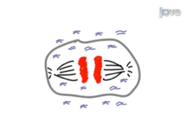
Time-lapse Imaging of Mitosis After siRNA Transfection

Multicolor Time-lapse Imaging of Transgenic Zebrafish: Visualizing Retinal Stem Cells Activated by Targeted Neuronal Cell Ablation
Time-lapse Microscopy of Early Embryogenesis in Caenorhabditis elegans

Electroporation of Craniofacial Mesenchyme
Quantitative Analysis of Random Migration of Cells Using Time-lapse Video Microscopy
Analyzing Craniofacial Morphogenesis in Zebrafish Using 4D Confocal Microscopy
High-resolution Time-lapse Imaging and Automated Analysis of Microtubule Dynamics in Living Human Umbilical Vein Endothelial Cells
ABOUT JoVE
Copyright © 2024 MyJoVE Corporation. All rights reserved