
Full-Field Optical Coherence Microscopy for Histology-Like Analysis of Stromal Features in Corneal Grafts
October 21st, 2022
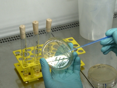
•We describe use of full-field optical coherence microscopy as a method for high quality assessment of corneal donor stroma. This protocol can be used to identify features indicative of health or disease, and is aimed at improving the screening and selection of donor tissues, and hence the results of keratoplasty.
Tags
Related Videos
Neutrophil Extracellular Traps: How to Generate and Visualize Them

Microwave Assisted Rapid Diagnosis of Plant Virus Diseases by Transmission Electron Microscopy
Intravital Microscopy of the Spleen: Quantitative Analysis of Parasite Mobility and Blood Flow
Single-cell Analysis of Bacillus subtilis Biofilms Using Fluorescence Microscopy and Flow Cytometry

Recurrent Herpetic Stromal Keratitis in Mice, a Model for Studying Human HSK
Generation of Lymph Node-fat Pad Chimeras for the Study of Lymph Node Stromal Cell Origin

Isolation of Murine Lymph Node Stromal Cells

Isolation and Transplantation of Different Aged Murine Thymic Grafts.
Analysis of Yersinia enterocolitica Effector Translocation into Host Cells Using Beta-lactamase Effector Fusions

Preparation of CD4+ T Cells for Analysis of GD3 and GD2 Ganglioside Membrane Expression by Microscopy
ABOUT JoVE
Copyright © 2024 MyJoVE Corporation. All rights reserved