需要订阅 JoVE 才能查看此. 登录或开始免费试用。
Method Article
电化学去角质以产生高质量的黑磷
摘要
我们提出了从块状晶体中电化学剥离黑磷 (BP) 的分步程序,黑磷 (BP) 是最有前途的新兴 2D 材料之一,在(光)电子学中的应用,以及通过扫描电子显微镜、原子力显微镜和透射电子显微镜进行形态表征。
摘要
为了从大块晶体中获得高质量的二维 (2D) 材料,在外部控制刺激下的分层至关重要。分层材料的电化学剥离需要简单的仪器,但可提供高质量的剥离 2D 材料,产量高,具有直接的可扩展性;因此,它代表了推进基础研究和工业应用的关键技术。此外,功能化 2D 材料的溶液加工性 使通过喷 墨打印和 3D 打印等不同的打印技术制造(光)电子和能源设备成为可能。本文逐步介绍了从块状晶体中合成黑磷 (BP) 的电化学剥离方案,黑磷 (BP) 是最有前途的新兴 2D 材料之一,即在 N(C4H9)4∙HSO4 在碳酸丙烯酯中存在下对 BP 进行阴极电化学剥离,通过超声处理制备分散体,随后离心分离薄片, 以及通过扫描电子显微镜 (SEM)、原子力显微镜 (AFM) 和透射电子显微镜 (TEM) 进行形态学表征。
引言
由于与分层体类似物相比,2D 材料具有卓越的机械、电气和光学特性,因此在科学界引起了相当大的关注。作为几十年来所有二维材料的前身和研究最多的材料,石墨烯仍然处于前沿发现的聚光灯下,例如膜1、传感器2、催化剂3、能源技术4、拓扑自旋电子器件5 和凝聚态物理学6。受此启发,已经合成和研究了许多其他二维材料,例如金属硫属化物7、层状双氢氧化物8 和氮化硼9。包括 2D 材料(即磷烯10)、MXenes(2D 金属碳化物或氮化物)11 和 2D 聚合物(单层/少层 2D 金属/共价有机框架)12,13 的最新成员,2D 材料系列已发展到由 150 多个成员组成,包括本征绝缘体、半导体、半金属和金属14。
新兴的二维材料,如 BP 15,16,17,18,19,20,21,22、二硫化钼 (MoS 2)23、24、25、26 和硒化铟 (III) (In2Se 3)27,28,29,在科学发现方面显示出相当大的潜力;然而,为了将其优异的理化性质扩展到宏观尺度,迫切需要高效、可重现和低成本的方法。电化学剥离是此类 2D 材料30,31 的大规模生产的一种很有前途的方法,主要是因为它可以在几分钟到几小时内提供克级的高质量和可分散的剥离材料,因为离子在电力下的有效嵌入。
随附的视频演示了使用电化学剥离逐步生产 BP 分散体,BP 是最有前途的新兴 2D 材料之一,应用于(光)电子学,然后是超声处理和离心分离薄片与未剥离颗粒,在各种溶剂中制备剥离 BP 薄片的分散体,以及通过 SEM 进行形态学表征, AFM 和 TEM。
研究方案
注:有关本协议中使用的材料和设备的详细信息,请参阅 材料表 。
1. 通过电化学剥离法合成黑磷 (BP)
- 将大块 BP 晶体切成 ~1-2 mm (≤5 mg) 的小块,并将它们限制在铂纱布内作为阴极。
- 切一块尺寸为 ~2 cm2 的铂箔作为阳极,并将其固定为面向阴极且距离阴极 2 cm。
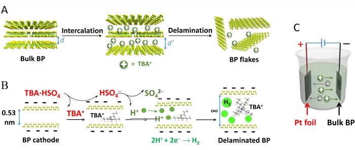
- 为了制备电解液,制备 0.1 M 四正丁基硫酸氢铵 (TBA·HSO4) 在碳酸丙烯酯中(使用无水和脱气溶剂)。填充电化学池,直到溶剂液位达到电极顶部上方至少 5 mm(图 1C)。
- 施加 −8.0 V 的直流电位以开始分层。为防止剥离过程中的降解,请在惰性气氛下进行整个过程(例如,在氩气或氮气下的手套箱内)。
注意:无需用惰性气体连续吹扫电解液。 - 分层完成后(确保不再可见的膨胀 BP 产生),将整个电解质(包括剥离的 BP 片和较大的未剥离颗粒)转移到 50 mL 离心管中。
- 在 15 °C 下以 5,292 × g 离心 10 分钟。弃去电解液,向沉淀物中加入 35 mL 新鲜的碳酸丙烯酯,轻轻摇晃。用碳酸丙烯酯重复离心和洗涤过程 2 次,用最终选择的溶剂(例如,2-丙醇、 N,N-二甲基甲酰胺 [DMF] 或 N-甲基-2-吡咯烷酮 [NMP])重复 2 次。
- 向沉淀产物中加入 5-50 mL 所需溶剂(取决于所需的浓度范围),并在冰冷超声处理浴中对所得产物混合物进行超声处理 10 分钟。
- 将混合物在 15 °C 下以 5,292 × g 离心 10 分钟,以去除厚层 BP 片。
- 将含有单层和少层 (<10 层) BP 薄片的上清液倒入干净的容器中,并将其保存在手套箱中,以便进一步表征或制造器件。
- 使用等式 (1) 表示的重量屈服公式计算分层产量 (η):
η = m1/m2 × 100% (1)
其中 m2 表示起始 BP 晶体的质量,m1 表示分散的 BP 薄片的重量。 - 为了确定 m1,测量从一定体积的 BP 分散体(例如,0.5 mL)的真空过滤中收集的 BP 的重量。使用聚四氟乙烯 (PTFE) 膜(孔径 0.2 μm)进行真空过滤。
- 如需长期储存,请盖上容器瓶盖并将其保存在手套箱内(最多 3 个月)。
2. 用于 SEM、SEM-EDS、AFM 和 TEM 表征的样品制备
注:为了探索合成的 BP 薄片的质量和形态方面,有必要进行表征,例如 SEM32(用于研究 BP 薄片的表面形态)、SEM-EDS33(用于薄片的元素分析)、AFM34,35(用于分析薄片的厚度和横向尺寸)和 TEM36,37(用于检测 BP 薄片的结构缺陷、形状和大小)。上述表征技术的样品制备方案如下所述(第 2.1-2.4 节)。有关上述表征技术的作程序,请参阅引用的参考文献 32,33,34,35,36,37。
- 用于 SEM 表征的样品制备
- 切出一小块硅晶片(尺寸约为 5-7 mm)。
- 先后使用丙酮、甲醇和水清洁硅晶片。然后,通过向基材吹入压缩空气或氮气来干燥基材。
- 通过添加无水 异丙醇,制备第 1 部分制备的 BP 分散液的 0.5 mL 稀释分散液,使其浓度达到 ~0.01 mg/mL。
- 将 1-2 滴制备好的稀释分散体旋涂在手套箱中干净干燥的基材上,并在 100 °C 的热板上干燥 6 小时(在手套箱中)。冷却至室温后,使用样品进行表征。
- 用于 SEM-EDS 表征的样品制备
- 请遵循第 2.1 节中提到的程序;但是,制备 ~0.1 mg/mL 的更浓分散液,而不是步骤 2.1.3 中提到的分散液。
- 用于 AFM 表征的样品制备
- 请遵循第 2.1 节中提到的程序;但是,请使用氧化硅晶片,而不是步骤 2.1.1 中提到的硅晶片。
- 用于 TEM 表征的样品制备
- 如第 2.1.3 节所述制备稀释的 BP 分散体。
- 将一滴分散体滴落在碳微网格上。在手套箱氩气气氛(无需额外加热)中干燥 24 小时后,使用样品进行 TEM 表征。
结果
图 1 展示了 BP 晶体的电化学剥离、TBA·HSO4 和随后的分层,以及反应池设置。

图 1:黑磷晶体电化学剥离机理和反应设置的示意图。 (A) BP 晶体的电化学剥离,(B
讨论
BP 具有 3s2 3p3 的价壳构型,每个磷原子都具有一个孤电子对,这使得磷原子在氧、水和光存在下易快速氧化降解41。为防止降解,建议使用脱气和无水溶剂和试剂,并在惰性气氛下进行生产过程。
在 BP 晶体剥离过程中,产生的 H + 的一部分(方程 1 和方程 2)在 BP 界面处减少,形成 ...
披露声明
作者声明没有利益冲突。
致谢
作者感谢 ERC 对 T2DCP、M-ERA-NET 项目 HYSUCAP、德国教育和研究部 (BMBF) 在 ForMikro(ForMikro)计划下资助的 SPES3 项目、石墨烯旗舰核心 3 881603和新兴印刷电子研究基础设施 (EMERGE) 的资助。EMERGE 项目已根据第 101008701 号赠款协议获得了欧盟 Horizon 2020 研究和创新计划的资助。作者感谢 Markus Löffler 博士的有益讨论和表征,并感谢德累斯顿先进电子中心 (cfaed) 和德累斯顿纳米分析中心 (DCN)。
材料
| Name | Company | Catalog Number | Comments |
| 2-Propanol | Sigma Aldrich | 278475 | anhydrous, 99.5% |
| Atomic force microscopy (AFM) | Bruker Multimode 8 system | ||
| Black phosphorus | Smart Elements | 4504 | Black Phosphorus 5.0 g sealed under Argon in ampoule |
| Centrifuge | Sigma 4-16KS | ||
| Propylene carbonate | Sigma Aldrich | 310328 | anhydrous, 99.7% |
| Scanning electron microscope (SEM) | Zeiss Gemini 500 | ||
| Tetra-n-butylammonium hydrogen sulfate | Sigma Aldrich | 791784 | anhydrous, free-flowing, Redi-Dri, 97% |
| Transmission electron microscopy (TEM) | Zeiss Libra 120 kV |
参考文献
- Yang, Q., et al. Ultrathin graphene-based membrane with precise molecular sieving and ultrafast solvent permeation. Nature Materials. 16 (12), 1198-1202 (2017).
- Goossens, S., et al. Broadband image sensor array based on graphene-CMOS integration. Nature Photonics. 11 (6), 366-371 (2017).
- Yan, H., et al. Single-atom Pd1/graphene catalyst achieved by atomic layer deposition: Remarkable performance in selective hydrogenation of 1,3-butadiene. Journal of the American Chemical Society. 137 (33), 10484-10487 (2015).
- Qu, G., et al. A fiber supercapacitor with high energy density based on hollow graphene/conducting polymer fiber electrode. Advanced Materials. 28 (19), 3646-3652 (2016).
- Calleja, F., et al. Spatial variation of a giant spin-orbit effect induces electron confinement in graphene on Pb islands. Nature Physics. 11 (1), 43-47 (2014).
- Cao, Y., et al. Correlated insulator behaviour at half-filling in magic-angle graphene superlattices. Nature. 556 (7699), 80-84 (2018).
- Manzeli, S., Ovchinnikov, D., Pasquier, D., Yazyev, O. V., Kis, A. 2D transition metal dichalcogenides. Nature Reviews Materials. 2 (8), 1-15 (2017).
- Carrasco, J. A., et al. Liquid phase exfoliation of carbonate-intercalated layered double hydroxides. Chemical Communications. 55 (23), 3315-3318 (2019).
- Lei, W., et al. Boron nitride colloidal solutions, ultralight aerogels and freestanding membranes through one-step exfoliation and functionalization. Nature Communications. 6 (1), 1-8 (2015).
- Kang, J., et al. Solvent exfoliation of electronic-grade, two-dimensional black phosphorus. ACS Nano. 9 (4), 3596-3604 (2015).
- Ding, L., et al. A two-dimensional lamellar membrane: MXene nanosheet stacks. Angewandte Chemie International Edition. 56 (7), 1825-1829 (2017).
- Dong, R., et al. High-mobility band-like charge transport in a semiconducting two-dimensional metal-organic framework. Nature Materials. 17 (11), 1027-1032 (2018).
- Liu, K., et al. On-water surface synthesis of crystalline, few-layer two-dimensional polymers assisted by surfactant monolayers. Nature Chemistry. 11 (11), 994-1000 (2019).
- Choi, W., et al. Recent development of two-dimensional transition metal dichalcogenides and their applications. Materials Today. 20 (3), 116-130 (2017).
- Yang, S., et al. A delamination strategy for thinly layered defect-free high-mobility black phosphorus flakes. Angewandte Chemie International Edition. 57 (17), 4677-4681 (2018).
- Shi, H., et al. Molecularly engineered black phosphorus heterostructures with improved ambient stability and enhanced charge carrier mobility. Advanced Materials. 33 (48), 2105694 (2021).
- Woomer, A. H., et al. Phosphorene: Synthesis, scale-up, and quantitative optical spectroscopy. ACS Nano. 9 (9), 8869-8884 (2015).
- Youngblood, N., Chen, C., Koester, S. J., Li, M. Waveguide-integrated black phosphorus photodetector with high responsivity and low dark current. Nature Photonics. 9 (4), 247-252 (2015).
- Li, L., et al. Black phosphorus field-effect transistors. Nature Nanotechnology. 9 (5), 372-377 (2014).
- Perello, D. J., Chae, S. H., Song, S., Lee, Y. H. High-performance n-type black phosphorus transistors with type control via thickness and contact-metal engineering. Nature Communications. 6 (1), 1-10 (2015).
- Yuan, H., et al. Polarization-sensitive broadband photodetector using a black phosphorus vertical p-n junction. Nature Nanotechnology. 10 (8), 707-713 (2015).
- Huang, Z., et al. Layer-tunable phosphorene modulated by the cation insertion rate as a sodium-storage anode. Advanced Materials. 29 (34), 1702372 (2017).
- Desai, S. B., et al. MoS2 transistors with 1-nanometer gate lengths. Science. 354 (6308), 99-102 (2016).
- Wang, Q. H., Kalantar-Zadeh, K., Kis, A., Coleman, J. N., Strano, M. S. Electronics and optoelectronics of two-dimensional transition metal dichalcogenides. Nature Nanotechnology. 7 (11), 699-712 (2012).
- Xu, X., Yao, W., Xiao, D., Heinz, T. F. Spin and pseudospins in layered transition metal dichalcogenides. Nature Physics. 10 (5), 343-350 (2014).
- Deng, Z., et al. 3D Ordered macroporous MoS2@C nanostructure for flexible Li-ion batteries. Advanced Materials. 29 (10), 1603020 (2017).
- Shi, H., et al. Ultrafast electrochemical synthesis of defect-free In2Se3 flakes for large-Area optoelectronics. Advanced Materials. 32 (8), 1907244 (2020).
- Ding, W., et al. Prediction of intrinsic two-dimensional ferroelectrics in In2Se3 and other III2-VI3 van der Waals materials. Nature Communications. 8 (1), 1-8 (2017).
- Island, J. O., Blanter, S. I., Buscema, M., Van Der Zant, H. S. J., Castellanos-Gomez, A. Gate controlled photocurrent generation mechanisms in high-gain In2Se3 phototransistors. Nano Letters. 15 (12), 7853-7858 (2015).
- Yang, S., Zhang, P., Nia, A. S., Feng, X. Emerging 2D materials produced via electrochemistry. Advanced Materials. 32 (10), 1907857 (2020).
- Li, J., et al. Electrochemically captured Zintl cluster-induced bismuthene for sodium-ion storage. Chemical Communications. 57 (19), 2396-2399 (2021).
- SEM Scanning Electron Microscope A To Z. Basic Knowledge for Using the SEM. JEOL Available from: https://jeol.co.jp/en/applications/pdf/sm/sem_atoz_all.pdf (2006)
- Lang, C., Hiscock, M., Larsen, K., Moffat, J., Sundaram, R. Characterization of two-dimensional transition metal dichalcogenides in the scanning electron microscope using energy dispersive X-ray spectrometry, electron backscatter diffraction, and atomic force microscopy. Applied Microscopy. 45 (3), 131-134 (2015).
- Zhang, H., et al. Atomic force microscopy for two-dimensional materials: A tutorial review. Optics Communications. 406, 3-17 (2018).
- Zahl, P., Zhang, Y. Guide for atomic force microscopy image analysis to discriminate heteroatoms in aromatic molecules. Energy and Fuels. 33 (6), 4775-4780 (2019).
- Backes, C., et al. Guidelines for exfoliation, characterization and processing of layered materials produced by liquid exfoliation. Chemistry of Materials. 29 (1), 243-255 (2017).
- Chang, Y. Y., Han, H. N., Kim, M. Analyzing the microstructure and related properties of 2D materials by transmission electron microscopy. Applied Microscopy. 49 (1), 1-7 (2019).
- Cooper, A. J., Velický, M., Kinloch, I. A., Dryfe, R. A. W. On the controlled electrochemical preparation of R4N+ graphite intercalation compounds and their host structural deformation effects. Journal of Electroanalytical Chemistry. 730, 34-40 (2014).
- Kang, J., et al. Stable aqueous dispersions of optically and electronically active phosphorene. Proceedings of the National Academy of Sciences of the United States of America. 113 (42), 11688-11693 (2016).
- Hanlon, D., et al. Liquid exfoliation of solvent-stabilized few-layer black phosphorus for applications beyond electronics. Nature Communications. 6 (1), 1-11 (2015).
- Favron, A., et al. Photooxidation and quantum confinement effects in exfoliated black phosphorus. Nature Materials. 14 (8), 826-832 (2015).
- Sirisaksoontorn, W., Adenuga, A. A., Remcho, V. T., Lerner, M. M. Preparation and characterization of a tetrabutylammonium graphite intercalation compound. Journal of the American Chemical Society. 133 (32), 12436-12438 (2011).
- You, X., et al. Interfaces of propylene carbonate. The Journal of Chemical Physics. 138 (11), 114708 (2013).
- Hu, G., et al. Black phosphorus ink formulation for inkjet printing of optoelectronics and photonics. Nature Communications. 8 (1), 1-10 (2017).
转载和许可
请求许可使用此 JoVE 文章的文本或图形
请求许可探索更多文章
This article has been published
Video Coming Soon
版权所属 © 2025 MyJoVE 公司版权所有,本公司不涉及任何医疗业务和医疗服务。