需要订阅 JoVE 才能查看此. 登录或开始免费试用。
Method Article
一种幼狨猴的人工饲养方法
摘要
在这里,我们描述了一种在动物孵化器中饲养幼年狨猴的人工饲养方法。该方法大大提高了狨猴幼崽的存活率,为研究在不同产后环境中饲养的具有相似遗传背景的狨猴幼崽的发育提供了机会。
摘要
普通狨猴(Callithrix jacchus)是一种小型且高度社会化的新世界猴子,具有很高的繁殖率,已被证明是生物医学和神经科学研究的引人注目的非人类灵长类动物模型。一些女性生下三胞胎;但是,父母不能全部抚养。为了拯救这些幼崽,我们开发了一种人工饲养方法,用于饲养新生狨猴。在该协议中,我们描述了食物的配方,喂养时间,温度和湿度的配置,以及人工饲养的婴儿对殖民地环境的适应。这种人工饲养方法显著提高了狨猴幼崽的存活率(不人工饲养:45%;人工饲养:86%),并为研究在不同产后环境中饲养的具有相似遗传背景的狨猴幼崽的发育提供了机会。由于该方法实用且易于使用,我们预计它也可以应用于其他使用普通狨猴的实验室。
引言
普通狨猴(Callithrix jacchus)是一种起源于南美洲和中美洲的小型树栖新世界猴子。在过去的几十年里,狨猴在生物医学研究中的使用迅速增长,因为与其他非人类灵长类动物 (NHP) 相比,狨猴的几个关键优势,包括它们的体型更小、更容易在圈养中处理和繁殖、妊娠时间更短、性成熟更早和人畜共患风险较低 1,2,3,4,5,6 .普通狨猴具有与人类相似的大脑结构和大脑功能,并表现出丰富的发声和具有丰富情感的高度社会行为。它是不同类型的神经科学研究的引人注目的 NHP 模型,例如感觉处理研究7、8、9、10、11、12、13、14、声音交流 15、16、17、18、19、脊髓损伤模型 20,21,22,23,帕金森病24,25,26,27,28和年龄相关疾病29。与其他非人灵长类相比,普通狨猴具有较高的繁殖率,可用于转基因修饰30,31,32。这种灵长类动物也广泛用于药理学、血管造影、病原体和免疫研究33、34、35、36、37、38、39。然而,狨猴的供应仍然非常有限,尤其是在中国,无法满足快速增长的科研需求。
在狨猴群落中,成年动物每天喂食一到两次,一些机构改变了幼年狨猴的饮食 40.一般来说,幼年狨猴通常会牢牢抓住父亲或哥哥姐姐的身体进行日常护理,每天多次交给母亲喝奶。一些雌性狨猴会生下三胞胎,在这种情况下,一两个婴儿由于缺乏乳汁而无法存活;此外,有些父母因为缺乏护理经验或其他未知原因而不照顾婴儿。这对许多实验室来说是一个巨大的损失。一些研究报告了圈养环境中成年狨猴的营养管理方法 40,41,42 利用具有不同常量营养素成分、维生素和矿物质的食物和配方,以及不同的富集喂养方案(捣碎、凝胶、纯化或罐装)2,41。之前的一项研究报道了一种针对狨猴三胞胎43 的协作饲养方法,其中照顾者每天带走一个婴儿,全天用手喂养它,并在第二天将其换成另一个三胞胎。虽然这种方法可以让婴儿得到父母的照顾,但它需要有经验的照顾者每天将婴儿从父母的身体中抓起,并且是劳动密集型的。到目前为止,还没有研究报告过对新生狨猴进行详细的、循序渐进的人工饲养方法。
本研究的目标是为那些对狨猴发育感兴趣但资源有限的人提供一种人工饲养方法。与以前的协作抚养方法43相比,现在的方法是一种对婴儿家庭造成较少干扰且易于学习的替代方案。本文基于母乳喂养的基本规则和5年的实践,描述了一种人工饲养幼狨猴的方法,包括食物的准备、喂养时间表、动物孵化器温度和湿度的配置,以及幼年动物对群体环境的适应。
研究方案
所有实验程序均经浙江大学动物使用与护理委员会批准,并遵循美国国立卫生研究院(NIH)指南。
1. 住房和畜牧业44
- 将菌落室设置为12小时:12小时昼夜循环,温度为26-28°C,相对湿度为45%-55%。
- 将 2-6 岁的雄性和雌性狨猴配对,并将它们放在笼子 (850 mm x 800 mm x 800 mm) 中,具有足够的空间和新鲜空气,并配备 24 小时通风系统。
- 在笼子里提供休息板、秋千、栖木和吊床。
- 每天两次用淡水和 30-40 克食物喂养这对狨猴,包括谷物、鸡蛋、红薯、蜂蜜、水果、蔬菜和黄粉虫。
注意:兽医和实验人员必须每天至少检查一次动物设施,以确保立即诊断和治疗任何病人。
2. 狨猴幼崽出生前的准备工作
- 照顾怀孕的狨猴
注意:受孕时间是通过触诊(通常在胚胎期开始后 10-20 天)诊断的,我们还参考了动物受试者的生殖史。- 为繁殖对提供更大的空间和最小的人为干扰。
- 用黄粉虫、鸡蛋、酸奶和干果等额外食物喂养繁殖对,以保证雌性的营养。
- 照顾并经常检查怀孕的狨猴,为它们的分娩做准备。
注意:狨猴的妊娠期估计为 148 天± 4.3 天45。
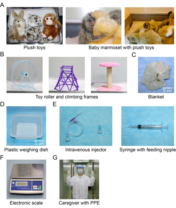
- 准备以下物品:动物培养箱(855 mm [W] x 470 mm [L] x 440 mm [H])、一次性尿布垫(M / L尺寸)、婴儿湿巾、毛绒玩具(图1A)、玩具滚轮(图1B)、攀爬架(图1B)、毯子(10厘米х10厘米,图1C)和电子秤(精度为0.2克,图1F)。
注意: 为避免动物悬挂,毛绒玩具不应具有环形结构。 - 食品和喂食器具的制备
- 准备以下物品:婴儿配方奶粉(适合0-12个月)、婴儿米糊(适合0-6个月)、电水壶、烧杯(100毫升)、加热垫、塑料称量盘(80毫米×80毫米×22毫米,图 1D)、无菌离心管(50毫升)、一次性无菌注射器(1-5毫升)、静脉注射器(用于定制喂养奶嘴)(图1E), 和拭子(80-100厘米)。
注意:为了制作用于喂养的,从连接到注射器的静脉注射器末端切开1厘米(图1E)。
- 准备以下物品:婴儿配方奶粉(适合0-12个月)、婴儿米糊(适合0-6个月)、电水壶、烧杯(100毫升)、加热垫、塑料称量盘(80毫米×80毫米×22毫米,图 1D)、无菌离心管(50毫升)、一次性无菌注射器(1-5毫升)、静脉注射器(用于定制喂养奶嘴)(图1E), 和拭子(80-100厘米)。
- 记录表格准备
- 为每只幼崽狨猴准备一份表格,通常长达数页,记录基本信息,如姓名、出生日期、出生体重、父母、其他感兴趣的基本信息,如头围和尾巴长度、繁殖信息,如繁殖日期和时间、食物摄入量(毫升)、排便状态(是/否, 硬/松)以及培养箱的温度和湿度。
注意:通常,每天测量和记录体重两次,一次在第一餐前,一次在最后一餐前。
- 为每只幼崽狨猴准备一份表格,通常长达数页,记录基本信息,如姓名、出生日期、出生体重、父母、其他感兴趣的基本信息,如头围和尾巴长度、繁殖信息,如繁殖日期和时间、食物摄入量(毫升)、排便状态(是/否, 硬/松)以及培养箱的温度和湿度。

图 1:培养箱中的物品以及喂食工具和配件的照片。 (A) 毛绒玩具;(二)玩具滚轮和爬架;(三)毛毯;(四)塑料称量盘;(五)静脉注射器和注射器配有定制的喂食奶嘴;(六)电子秤;(七)护理人员配备个人防护用品。 请点击这里查看此图的较大版本.
3. 人工饲养程序
- 在预产期之前对房间进行清洁和消毒。
- 将次氯酸或75%乙醇喷洒在地板和桌子上,静置30秒,然后擦拭桌子,拖地。
- 将培养箱的温度设置为35°C,将湿度设置为40%。通常,为了模拟婴儿狨猴的基本温度要求,如 表1 所示,在出生后第14天(P14)之前,将培养箱温度保持在35°C,从P15开始,每3天降低0.5°C。保育箱内保持湿度在40%-45%,接近菌落湿度,保持婴儿皮毛干燥。
- 铺上一次性尿布垫以覆盖培养箱的底盘。
- 为了尽量减少婴儿的压力,在引入婴儿狨猴之前,在保育箱中放几条毯子和毛绒玩具,这些狨猴往往会模仿成年狨猴。
注意:毯子和毛绒玩具在使用前将放在繁殖对的家庭笼子中 1 天。 - 将幼年狨猴放入保育箱中,并在家庭笼子中与父母分开后将它们放在毛绒玩具上。
注意:为了避免社会孤立并符合动物福利,通常选择两个婴儿一起进行人工抚养。- 喂食前穿戴消毒的个人防护装备(PPE, 图1G)。
- 将几条毯子加热至35°C。
- 用温暖的毯子轻轻地抱住幼年狨猴,并获得动物的体重作为出生体重。
- 用温暖的毯子将婴儿狨猴转移到保育箱中。
- 记录,如步骤 2.4 中所述。
- 混合食物成分并喂养幼年狨猴。
- 将5g婴儿配方奶粉溶解在50mL无菌离心管中的30mL 50°C开水中。
注意:不同出生后年龄的婴儿狨猴需要不同的食物食谱。 表2 包括从P1到P60的出生后年龄婴儿配方奶粉、米糊和水的不同剂量。剂量通常足够1天。在第一餐前混合配料,并将其余食物存放在4°C冰箱中。每餐将食物加热至30-35°C。 - 用 1 mL 注射器取 1 mL 食物,并用定制的喂食奶嘴盖住注射器。
注意: 选择合适尺寸的注射器,请参阅 表 2。 - 用加热垫将食物温度保持在 30-35 °C。
- 喂食前加热双手,在保育箱中用一只手用温暖的毯子轻轻握住幼咪狨猴。
- 将喂食奶嘴放入婴儿狨猴的嘴里,同时用照顾者握手的拇指和食指轻轻握住婴儿狨猴的头部,然后以恒定的速度缓慢地将食物从注射器中推出。
注意: 切勿将食物推得比小狨猴吞咽的速度快。快速推动会导致窒息,食物从鼻子通过喉咙流出。这可能导致肺炎等疾病,甚至可能导致死亡。如果食物溢出,幼年狨猴会挣扎。每当发生这种情况时,请停止喂食并小心地擦拭动物脸上的食物。狨猴开始正常表现后继续喂食。
- 将5g婴儿配方奶粉溶解在50mL无菌离心管中的30mL 50°C开水中。
- 幼狨猴进食适量食物后,用温水棉签擦拭肛门,既能清洁肛门,又能促进排便。
- 观察动物几分钟,并检查动物的运动和排便。
- 记录喂食时间、进食量(mL)、排便状态(是/否、硬/松)以及培养箱温度和湿度。
注意: 称重时用温暖的毯子包裹婴儿狨猴,以免婴儿着凉或受伤。 - 通过捡起固体粪便或更换新的一次性尿布垫来保持培养箱的底盘清洁。
- 在P50之前,按照步骤3.3-3.7喂养幼妞狨猴,并使用 表2所示的食物配料剂量和喂养时间和频率。
- 从 P50 开始,幼年狨猴通常可以自愿进食。
- 使用塑料称量盘代替注射器。将食物成分直接混合在盘子中来准备食物;金额见 表2 。
- 将食物盘放入培养箱中,并固定底部以防翻转。观察几分钟,确保幼狨猴吃掉食物。在最初的几次中,通过将动物引诱到食物盘中并引导其嘴多次触摸食物来引导动物自愿进食。
注意: 切勿让动物的鼻子接触食物。通常,动物会在 1 天内学会自愿进食。
4. 幼狨猴返回殖民地前的适应
注意:通常,当幼年狨猴学会自己进食时,人工饲养就完成了。在将它们放回狨猴群落的家庭笼子之前,需要执行一些适应程序。
- 将幼咋猴从动物培养箱移至类似于鸟笼的小笼子(45 cm x 45 cm x 40 cm)中。在每个小笼子上挂一个水瓶(50 mL)。
- 将装有幼狨猴的小笼子转移到殖民地,并将它们放置在靠近家庭笼子的位置。
- 用塑料称重盘分别喂养幼咪狨猴1周,根据为整个群体准备的每日食谱混合食物。
- 每天记录一次体重和排便情况。
5. 幼年狨猴返回家庭笼子
注意:在小笼子里生活7-10天后,幼年狨猴通常能很好地适应群体环境,不再表现出焦虑。
- 早上将幼崽狨猴放回家庭笼子里。
- 观察动物至少 15 分钟,以确保家庭成员和新进入者之间没有咬人、打架或追逐。
注意: 如果发生咬人、打架或追逐,请尽快将婴儿与其他婴儿分开;并尝试在另一天再次将婴儿送回家人身边。如果失败再次发生,请选择另一个家庭进行寄养。育儿经验丰富的家庭群体是寄养的首选。如果没有家人接受,就把毛绒玩具放在一个新的笼子里,陪伴小猴子。 - 停止单独喂养幼咪狨猴,并开始使用蜂群的饮食喂养它们。
- 在幼咬狨回到家庭笼子后 1 周内密切关注它们的活动。
- 每 2 天测量一次幼咪狨猴的体重,并做好记录。如果他们体重减轻,在家庭笼子里用注射器喂他们额外的营养食物。
- 为幼崽狨猴提供日常护理,就像殖民地中的狨猴一样。
结果
体重是动物身体发育的关键指标,在本协议中用作狨猴健康状况的指标。在这项工作中,人工饲养的动物的体重随着年龄的增长而逐渐增加(图2A,n = 16),类似于先前研究中新生婴儿的体重46。为了尽量减少对殖民地繁殖家庭的干扰,我们没有每天对殖民地中的幼年狨猴进行称重。我们获得了父母饲养的动物在出生后1个月及以后的体重,并将其与同龄人?...
讨论
普通狨猴是生物医学和神经科学研究非常有用的非人灵长类模型。然而,狨猴资源太有限,无法满足快速增长的需求。在这项工作中,我们开发了一种人工饲养方法,不仅可以提高狨猴幼崽的存活率,还可以为研究它们的产后发育提供机会。这种人工饲养方法实用且易于学习,因此很容易适用于其他使用普通狨猴的实验室。
一些先天性缺陷通常在出生后的前 2 周内出现。到?...
披露声明
作者没有要披露的利益冲突。
致谢
作者要感谢李明轩对本手稿早期版本的语法和润色。这项工作得到了中国浙江省自然科学基金(LD22H090003)的支持;国家自然科学基金(32170991 32071097),STI2030重大专项2021ZD0204100(2021ZD0204101)和2022ZD0205000(2022ZD0205003);以及浙江大学教育部脑科学与脑机集成前沿科学中心。
材料
| Name | Company | Catalog Number | Comments |
| animal incubator | RCOM, Korea | MX - BL600N, 855 mm (W) x 470 mm (L) x 440 mm (H) | |
| baby milk powder | Meadjohnson, America | suitable for 0-12 months of age, executive standard - GB25596 | |
| baby rice paste | HEINZ, China | suitable for 0-6 months of age, executive standard - GB10769 | |
| baby wipes | babycare, China | soft | |
| beaker | ShuNiu, China | 100 mL | |
| blankets | Grace, China | 10 cm × 10 cm, soft | |
| climbing frame | WowWee, China | firm and no small circular structures | |
| disposable diaper pads | Hi Health Pet, China | either M or L size | |
| disposable sterile syringe | Cofoe, China | 1 mL, 2.5 mL, 3 mL, 5 mL, 10 mL | |
| electronic scale | YouSheng, China | measuring range from 0 to 6,000 g with precision of 0.2 g | |
| intravenous injector | HD, China | 0.55 mm x 20 mm needle | |
| kettle | FGA, China | warm-keeping kettle 1,500 mL | |
| lactulose | BELCOL, China | to solve constipation | |
| plastic weighing dish | SKSLAB, China | 80 mm x 80 mm x 22 mm, used as a bowl | |
| plush toy | Lebiyou, China | soft | |
| probiotic powder | G-Pet, China | to regulate gastrointestinal environment | |
| sterile centrifuge tube | NEST, China | 50 mL | |
| swab | OYEAH, China | 80 - 100 mm | |
| toy roller | WowWee, China | firm and no small circular structures |
参考文献
- Miller, C. T., et al. Marmosets: A neuroscientific model of human social behavior. Neuron. 90 (2), 219-233 (2016).
- Ross, C. N., Colman, R., Power, M., Tardif, S. Marmoset metabolism, nutrition, and obesity. ILAR Journal. 61 (2-3), 179-187 (2020).
- Kishi, N., Sato, K., Sasaki, E., Okano, H. Common marmoset as a new model animal for neuroscience research and genome editing technology. Development, Growth & Differentiation. 56 (1), 53-62 (2014).
- Prins, N. W., et al. Common marmoset (Callithrix jacchus) as a primate model for behavioral neuroscience studies. Journal of Neuroscience Methods. 284, 35-46 (2017).
- Tokuno, H., Watson, C., Roberts, A., Sasaki, E., Okano, H. Marmoset neuroscience. Neuroscience Research. 93, 1-2 (2015).
- Hodges, J. K., Henderson, C., Hearn, J. P. Relationship between ovarian and placental steroid production during early pregnancy in the marmoset monkey (Callithrix jacchus). Journal of Reproduction and Fertility. 69 (2), 613-621 (1983).
- Troilo, D., Judge, S. J. Ocular development and visual deprivation myopia in the common marmoset (Callithrix jacchus). Vision Research. 33 (10), 1311-1324 (1993).
- Mitchell, J. F., Leopold, D. A. The marmoset monkey as a model for visual neuroscience. Neuroscience Research. 93, 20-46 (2015).
- Hung, C. C., et al. Functional MRI of visual responses in the awake, behaving marmoset. NeuroImage. 120, 1-11 (2015).
- Gao, L., Kostlan, K., Wang, Y., Wang, X. Distinct subthreshold mechanisms underlying rate-coding principles in primate auditory cortex. Neuron. 91 (4), 905-919 (2016).
- Gao, L., Wang, X. Subthreshold activity underlying the diversity and selectivity of the primary auditory cortex studied by intracellular recordings in awake marmosets. Cerebral Cortex. 29 (3), 994-1005 (2019).
- Gao, L., Wang, X. Intracellular neuronal recording in awake nonhuman primates. Nature Protocols. 15, 3615-3631 (2020).
- Wang, X., et al. Corticofugal modulation of temporal and rate representations in the inferior colliculus of the awake marmoset. Cerebral Cortex. 32 (18), 4080-4097 (2022).
- Wang, X., et al. Selective corticofugal modulation on sound processing in auditory thalamus of awake marmosets. Cerebral Cortex. 33 (7), 3372-3386 (2022).
- Kajikawa, Y., et al. Coding of FM sweep trains and twitter calls in area CM of marmoset auditory cortex. Hearing Research. 239 (1-2), 107-125 (2008).
- Choi, D., Bruderer, A. G., Werker, J. F., et al. Sensorimotor influences on speech perception in pre-babbling infants: Replication and extension of Bruderer et al. Psychonomic Bulletin & Review. 26 (4), 1388-1399 (2019).
- Eliades, S. J., Miller, C. T. Marmoset vocal communication: Behavior and neurobiology. Developmental Neurobiology. 77 (3), 286-299 (2017).
- Roy, S., Zhao, L., Wang, X. Distinct neural activities in premotor cortex during natural vocal behaviors in a New World primate, the common marmoset (Callithrix jacchus). Journal of Neuroscience. 36 (48), 12168-12179 (2016).
- Simões, C. S., et al. Activation of frontal neocortical areas by vocal production in marmosets. Frontiers in Integrative Neuroscience. 4, 123 (2010).
- Iwanami, A., et al. Transplantation of human neural stem cells for spinal cord injury in primates. Journal of Neuroscience Research. 80 (2), 182-190 (2005).
- Schorscher-Petcu, A., Dupré, A., Tribollet, E. Distribution of vasopressin and oxytocin binding sites in the brain and upper spinal cord of the common marmoset. Neuroscience Letters. 461 (3), 217-222 (2009).
- Bowes, C., Burish, M., Cerkevich, C., Kaas, J. Patterns of cortical reorganization in the adult marmoset after a cervical spinal cord injury. Journal of Comparative Neurology. 521 (15), 3451-3463 (2013).
- Kondo, T., et al. Histological and electrophysiological analysis of the corticospinal pathway to forelimb motoneurons in common marmosets. Neuroscience Research. 98, 35-44 (2015).
- Nash, J. E., et al. Antiparkinsonian actions of ifenprodil in the MPTP-lesioned marmoset model of Parkinson's disease. Experimental Neurology. 165 (1), 136-142 (2000).
- van Vliet, S. A., et al. Neuroprotective effects of modafinil in a marmoset Parkinson model: Behavioral and neurochemical aspects. Behavioural Pharmacology. 17 (5-6), 453-462 (2006).
- van Vliet, S. A., Vanwersch, R. A., Jongsma, M. J., Olivier, B., Philippens, I. H. Therapeutic effects of Delta9-THC and modafinil in a marmoset Parkinson model. European Neuropsychopharmacology. 18 (5), 383-389 (2008).
- Philippens, I. H., t Hart, B. A., Torres, G. The MPTP marmoset model of parkinsonism: a multi-purpose non-human primate model for neurodegenerative diseases. Drug Discovery Today. 15 (23-24), 985-990 (2010).
- Santana, M. B., et al. Spinal cord stimulation alleviates motor deficits in a primate model of Parkinson disease. Neuron. 84 (4), 716-722 (2014).
- Tardif, S. D., Mansfield, K. G., Ratnam, R., Ross, C. N., Ziegler, T. E. The marmoset as a model of aging and age-related diseases. ILAR Journal. 52 (1), 54-65 (2011).
- Sasaki, E., et al. Generation of transgenic non-human primates with germline transmission. Nature. 459, 523-527 (2009).
- Sasaki, E. Prospects for genetically modified non-human primate models, including the common marmoset. Neuroscience Research. 93, 110-115 (2015).
- Park, J. E., Sasaki, E. Assisted reproductive techniques and genetic manipulation in the common marmoset. ILAR Journal. 61 (2-3), 286-303 (2020).
- Smith, D., Trennery, P., Farningham, D., Klapwijk, J. The selection of marmoset monkeys (Callithrix jacchus) in pharmaceutical toxicology. Laboratory Animals. 35 (2), 117-130 (2001).
- Smith, T. E., Tomlinson, A. J., Mlotkiewicz, J. A., Abbott, D. H. Female marmoset monkeys (Callithrix jacchus) can be identified from the chemical composition of their scent marks. Chemical Senses. 26 (5), 449-458 (2001).
- Jagessar, S. A., et al. Induction of progressive demyelinating autoimmune encephalomyelitis in common marmoset monkeys using MOG34-56 peptide in incomplete freund adjuvant. Journal of Neuropathology and Experimental Neurology. 69 (4), 372-385 (2010).
- Kap, Y. S., Laman, J. D., 't Hart, B. A. Experimental autoimmune encephalomyelitis in the common marmoset, a bridge between rodent EAE and multiple sclerosis for immunotherapy development. Journal of Neuroimmune Pharmacology. 5 (2), 220-230 (2010).
- Carrion, R., Patterson, J. L. An animal model that reflects human disease: The common marmoset (Callithrix jacchus). Current Opinion in Virology. 2 (3), 357-362 (2012).
- Jagessar, S. A., et al. Overview of models, methods, and reagents developed for translational autoimmunity research in the common marmoset (Callithrix jacchus). Experimental Animals. 62 (3), 159-171 (2013).
- Feng, Z., et al. Biologically excretable aggregation-induced emission dots for visualizing through the marmosets intravitally: Horizons in future clinical nanomedicine. Advanced Materials. 33 (17), 2008123 (2021).
- Goodroe, A., et al. Current practices in nutrition management and disease incidence of common marmosets (Callithrix jacchus). Journal of Medical Primatology. 50 (3), 164-175 (2021).
- Power, M. L., Koutsos, L., Marini, R., Wachtman, L., Tardif, S., Mansfield, K., Fox, J. Chapter 4 - Marmoset nutrition and dietary husbandry. The Common Marmoset in Captivity and Biomedical Research. , 63-76 (2019).
- Gore, M. A., et al. Callitrichid nutrition and food sensitivity. Journal of Medical Primatology. 30 (3), 179-184 (2001).
- Hearn, J. P., Burden, F. J. Collaborative' rearing of marmoset triplets. Laboratory Animals. 13 (2), 131-133 (1979).
- Cao, X., et al. Effect of a high estrogen level in early pregnancy on the development and behavior of marmoset offspring. ACS Omega. 7 (41), 36175-36183 (2022).
- Watakabe, A., et al. Application of viral vectors to the study of neural connectivities and neural circuits in the marmoset brain. Developmental Neurobiology. 77 (3), 354-372 (2017).
- Takahashi, D. Y., et al. The developmental dynamics of marmoset monkey vocal production. Science. 349 (6249), 734-738 (2015).
- Malukiewicz, J., et al. The gut microbiome of exudivorous marmosets in the wild and captivity. Scientific Reports. 12 (1), 5049 (2022).
- Shigeno, Y., et al. Comparison of gut microbiota composition between laboratory-bred marmosets (Callithrix jacchus) with chronic diarrhea and healthy animals using terminal restriction fragment length polymorphism analysis. Microbiology and Immunology. 62 (11), 702-710 (2018).
- Baxter, V. K., et al. Serum albumin and body weight as biomarkers for the antemortem identification of bone and gastrointestinal disease in the common marmoset. PLoS One. 8 (12), e82747 (2013).
- Tardif, S. D., et al. Characterization of obese phenotypes in a small nonhuman primate, the common marmoset (Callithrix jacchus). Obesity. 17 (8), 1499-1505 (2009).
- Wachtman, L. M., et al. Differential contribution of dietary fat and monosaccharide to metabolic syndrome in the common marmoset (Callithrix jacchus). Obesity. 19 (6), 1145-1156 (2011).
- Power, M. L., Ross, C. N., Schulkin, J., Ziegler, T. E., Tardif, S. D. Metabolic consequences of the early onset of obesity in common marmoset monkeys. Obesity. 21 (12), E592-E598 (2013).
- Shimizu, K., et al. Peer-social response in 4 juvenile marmosets represented the emotional development traits depending on family structure. Neuroscience Research. 65, S244 (2009).
- Schultz-Darken, N., Braun, K. M., Emborg, M. E. Neurobehavioral development of common marmoset monkeys. Developmental Psychobiology. 58 (2), 141-158 (2016).
- Gultekin, Y. B., Hage, S. R. Limiting parental feedback disrupts vocal development in marmoset monkeys. Nature Communications. 8, 14046 (2017).
转载和许可
请求许可使用此 JoVE 文章的文本或图形
请求许可探索更多文章
This article has been published
Video Coming Soon
版权所属 © 2025 MyJoVE 公司版权所有,本公司不涉及任何医疗业务和医疗服务。