Method Article
El cultivo 3D de organoides a partir de criptas intestinales murinas y una sola célula madre para la investigación de organoides
En este artículo
Resumen
Describimos un protocolo para aislar criptas murinas del intestino delgado y cultivar organoides 3D intestinales de las criptas. Además, describimos un método para generar organoides a partir de una sola célula madre intestinal en ausencia de un nicho celular subepitelial.
Resumen
En la actualidad, el cultivo de organoides representa una herramienta importante para los estudios in vitro de diferentes aspectos biológicos y enfermedades en diferentes órganos. Las criptas murinas del intestino delgado pueden formar organoides que imitan el epitelio intestinal cuando se cultivan en una matriz extracelular 3D. Los organoides están compuestos por todos los tipos de células que cumplen diversas funciones homeostáticas intestinales. Estos incluyen células de Paneth, células enteroendocrinas, enterocitos, células caliciformes y células de penacho. Se agregan moléculas bien caracterizadas al medio de cultivo para enriquecer las células madre intestinales (ISC) marcadas con repeticiones ricas en leucina que contienen el receptor 5 acoplado a proteína G y se utilizan para impulsar la diferenciación en linajes específicos; estas moléculas incluyen el factor de crecimiento epidérmico, Noggin (una proteína morfogenética ósea) y R-spondin 1. Además, también se detalla un protocolo para generar organoides a partir de un único ISC positivo para el receptor hepatocelular B2 (EphB2) productor de eritropoyetina. En este artículo de métodos, se describen técnicas para aislar criptas del intestino delgado y un solo ISC de los tejidos y garantizar el establecimiento eficiente de organoides.
Introducción
Los organoides intestinales, que se establecieron por primera vez en 2009, se han convertido en una poderosa herramienta in vitro para estudiar la biología intestinal dada su similitud morfológica y funcional con los tejidos maduros. Recientemente, los avances tecnológicos en organoides cultivados derivados de células madre de tejido adulto han permitido el cultivo a largo plazo de células madre intestinales (ISC) con potencial de autorrenovación y diferenciación. Estos organoides han sido ampliamente utilizados para estudios de investigación básica y traslacional sobre fisiología gastrointestinal y fisiopatología 1,2,3,4,5,6. Los organoides 3D desarrollados por el grupo Clevers proporcionan una poderosa herramienta para estudiar el epitelio intestinal con mayor relevancia fisiológica7. Dado que los organoides intestinales se derivan de células madre tisulares y están compuestos de múltiples tipos de células, recapitulan la funcionalidad del epitelio intestinal. Cabe destacar que una célula madre 5-positiva (Lgr5+) del receptor acoplado a proteínas G que contiene repeticiones ricas en leucina también puede generar organoides 3D sin células de Paneth o un nicho ISC como el nicho epitelial o el nicho estromal7. Sin embargo, la capacidad de formación de organoides de las células Lgr5+ de un solo ordenamiento es baja en comparación con las de las criptas y los dobletes de células ISC-Paneth8.
Un número creciente de estudios han demostrado que los métodos de incubación del ácido etilendiaminotetraacético (EDTA) o la disociación de la colagenasa causan aflojamiento en el epitelio y la liberación de criptas. Como la disociación enzimática puede tener un efecto sobre el estado celular de las criptas, generalmente se usa un método de aislamiento mecánico para disociar el tejido. Aunque la digestión mecánica es una técnica rápida, este método puede estar asociado con rendimientos de criptas inconsistentes o mala viabilidad celular9. Por lo tanto, el tratamiento con EDTA y la disociación mecánica se pueden combinar para generar mejores rendimientos de criptas. Una característica de la metodología mostrada en este artículo es el uso de agitación vigorosa de los fragmentos de tejido después de la quelación con EDTA10. La agitación vigorosa permite el aislamiento eficiente de las criptas de los complejos cripta-vellosidades en el intestino delgado. El grado de agitación manual determina la separación. Por lo tanto, la obtención de criptas de complejos es importante para los experimentadores en este campo. Además, la habilidad adecuada puede reducir la contaminación de las vellosidades al mínimo y aumentar el número de criptas.
Por lo tanto, este protocolo experimental, que emplea organoides del intestino delgado derivados de murina, puede aislar mejor las criptas con fuerza física después del tratamiento con EDTA para la disociación. Se sabe que el patrón de expresión del receptor hepatocelular productor de eritropoyetina B2 (EphB2) refleja en parte el entorno de la cripta. Por ejemplo, las células positivas para EphB2 se organizan de abajo hacia arriba11. La clasificación celular activada por fluorescencia (FACS) se llevó a cabo en función de la expresión de EphB2, y las células obtenidas se dividieron en cuatro grupos: EphB2alta, EphB2med, EphB2baja y EphB2neg. Luego, se demostró el crecimiento de organoides de célulasaltas EphB2 de una sola clasificación en ratones de tipo salvaje (WT).
Protocolo
Todos los experimentos con ratones fueron aprobados por el Comité de Ética Animal de Suntory (APRV000561), y todos los animales se mantuvieron de acuerdo con las directrices del comité para el cuidado y uso de animales de laboratorio. Se utilizó una cepa WT estándar de Mus musculus (C57BL6/J). Se utilizaron ratones machos y hembras de 10 semanas a 20 semanas de edad. Los ratones fueron sacrificados con asfixia porCO2 .
1. Aislamiento del intestino delgado
- Extirpar el intestino delgado, incluyendo el duodeno y la mitad proximal del yeyuno, con tijeras de laboratorio.
- Transfiera el tejido a una placa de Petri y enjuague el intestino delgado con 5 ml de PBS-ABx frío (PBS + penicilina-estreptomicina [1%] + gentamicina [0,5%]) en una jeringa de 5 ml para eliminar el contenido luminal.
- Corte el tejido abierto a lo largo con tijeras de laboratorio y lávelo manualmente con PBS-ABx frío mientras agita.
NOTA: Al raspar las vellosidades con un portaobjetos, se puede reducir la contaminación de las vellosidades12. - Recoja aproximadamente piezas de 5 mm x 5 mm del segmento intestinal con tijeras de laboratorio. Transfiera los fragmentos a un tubo de 50 ml con pinzas y agregue 25 ml de PBS-ABx frío.
- Lave los fragmentos agitando hacia adelante y hacia atrás 10 veces con 25 ml de PBS-ABx frío para eliminar el contenido intestinal en el tubo de 50 ml.
2. Aislamiento de criptas
- Incubar las piezas en PBS-ABx que contiene 2 mM EDTA durante 30 min en hielo sin agitación.
- Para facilitar la solidificación de la matriz extracelular (MEC), incubar previamente una placa de 24 pocillos en una incubadora de cultivo de tejidos a 37 °C.
- Aspire la solución de EDTA del sistema de cultivo celular con una bomba de vacío, agregue 25 ml de PBS-ABx fresco y frío, y luego agite las piezas hacia arriba y hacia abajo vigorosamente a mano 30x-40x para liberar los complejos cripta-vellosidad.
NOTA: Las criptas y vellosidades separadas se pueden verificar mediante la observación microscópica de una gota de 25 μL de la suspensión con un aumento de 4x. - A continuación, filtre la suspensión a través de un colador de 70 μm una vez.
- Centrifugar la suspensión a 390 × g durante 3 min a 4 °C.
- Resuspender el pellet de cripta en 20 mL de DMEM de sorbitol (DMEM avanzado/F12 + penicilina-estreptomicina [1%] + gentamicina [0,5%] + suero bovino fetal [1%] + sorbitol [2%]) con pipeteo, y transferir la suspensión crypt a dos nuevos tubos de 15 mL para dividirlos en dos soluciones de 10 mL para centrifugar a baja velocidad.
NOTA: La masa celular grande y las células/residuos se pueden separar mediante centrifugación a baja velocidad. La masa celular grande está en el pellet, y las células / desechos están en el sobrenadante. - Centrifugar las dos suspensiones de cripta a 80 × g durante 3 min a 4 °C, y luego aspirar suavemente el sobrenadante.
NOTA: Como la formación de pellets es débil, no aspire demasiado. Deje 2 ml de sobrenadante en cada tubo. - Agregue 10 ml de sorbitol DMEM a cada tubo nuevamente. Centrifugar la suspensión a 80 × g durante 3 min a 4 °C.
- Después de aspirar el sobrenadante, dejando 2 ml de sobrenadante en cada tubo, añadir 10 ml de sorbitol DMEM para la resuspensión, y centrifugar la suspensión de la cripta a 80 × g durante un último 3 min a 4 °C.
- Después de aspirar el sobrenadante, dejando 2 ml de sobrenadante en cada tubo, añadir 10 ml de DMEM completo (DMEM avanzado/F12 + penicilina-estreptomicina [1%] + gentamicina [0,5%] + suero fetal bovino [1%]) para la resuspensión del pellet mediante pipeteo hacia arriba y hacia abajo, y dejar durante 1 min.
NOTA: Espere 1 minuto para obtener las criptas flotantes de manera eficiente. - Después de 1 min, recoja cada suspensión de 10 ml para un total de 20 ml y filtre una vez con un colador de células de 70 μm para purificar las criptas.
- Antes de sembrar criptas esencialmente puras, cuente el número de criptas en el DMEM completo filtrado y luego centrifugar a 290 × g durante 3 minutos a 4 ° C.
- Gotea gotas de 25 μL en un plato de 6 cm en tres puntos. Cuente el número de criptas bajo un microscopio con un aumento de 4x y calcule la concentración de criptas por gota de 25 μL.
- Suspender 100 criptas con 40 μL de ECM por pocillo. Pipeta arriba y abajo 5x-10x para obtener una suspensión homogénea de criptas en el ECM, y luego sembrar en una placa de 24 pocillos precalentada a 37 °C.
NOTA: Siempre mantenga el ECM en hielo para evitar la polimerización. Pipetear con cuidado para evitar que se formen burbujas de aire en el ECM. - Incubar la placa de 24 pocillos durante 15 min en una incubadora deCO2 al 5% a 37 °C para la polimerización de la ECM.
- Finalmente, cubra la ECM con 500 μL de medio de cultivo que contenga factor de crecimiento epidérmico (EGF) de ratón, R-espondina 1 de ratón recombinante y Noggin de ratón recombinante a temperatura ambiente. La concentración final de materiales por pocillo es la siguiente: penicilina-estreptomicina (1%), 50 U/mL cada uno; gentamicina (0,5%), 25 μg/ml; EGF: 20 ng/ml; Noggin, 100 ng/mL; R-espondina 1, 500 ng/mL; L-glutamina, 2 mM.
- Iniciar el cultivo de criptas a 37 °C en una incubadora deCO2 al 5%.
NOTA: Para el medio de cultivo para organoides en una placa de 24 pocillos, ver Tabla 1. - Realice imágenes en vivo a largo plazo para observar la morfogénesis organoide con un microscopio de imagen de lapso de tiempo de grabación equipado con un objetivo de 20x cada 3 h durante un máximo de 7 días. Obtenga imágenes en serie apiladas en z a pasos z de 1 μm (1 μm x cinco pasos).
- Cambie el medio cada dos días.
3. Clasificación celular activada por fluorescencia (FACS)
- Aislar las criptas de los ratones (ver sección 2).
- Tratar las criptas aisladas con 2 mL de tripsina durante 30 min a 37 °C.
- Detenga la reacción con 10 ml de PBS y luego pase a través de un filtro celular de 20 μm.
- Centrifugar la solución a 390 × g durante 3 min a 4 °C y volver a suspender con 100 μL de DMEM completo.
- Agregue el anticuerpo anti-EphB2 conjugado con APC (1/50) e incube durante 30 minutos en hielo.
- Lave las células 3 veces con PBS y finalmente agregue 7-amino-actinomicina D (7-AAD) (1/100).
- Ordene las células teñidas a través de FACS.
- Ajuste el factor de escala de área y ordene según el tamaño de la celda (dispersión directa, FSC-A) frente a la granularidad (dispersión lateral, SSC-A).
- Clasifique las celdas negativas y positivas de 7-AAD para su viabilidad con el láser configurado a una longitud de onda de 488 nm y una potencia de 50 mV.
- Delimite las puertas para clasificar las celdas EphB2-high (EphB2high), EphB2-medium (EphB2med), EphB2-low (EphB2low) y EphB2-negative (EphB2neg) con el láser ajustado a una longitud de onda de 640 nm y 100 mV de potencia.
- Iniciar el cultivocelular alto de EphB2 a 37 °C en una incubadora deCO2 al 5%.
4. Organoides cultivados unicelulares
- Realizar el método de aislamiento celular según los niveles superficiales graduados de EphB211, y luego obtener cuatro poblaciones distintas (alta, media, baja y negativa).
- Recolectar, pellet con centrifugación a 390 × g durante 3 min a 4 °C, e incrustar las célulasaltas EphB2 clasificadas en el ECM mediante pipeteo, seguido de siembra en una placa de 24 pocillos (100 singlets/40 μL de ECM/pocillo).
- Al igual que en el paso 2.14, permita que la ECM polimerice y cubra la ECM con un medio de cultivo que contenga un inhibidor de la quinasa asociada a Rho (ROCK) (10 μM) durante los primeros 2 días para mantener las célulasaltas de EphB2.
NOTA: El inhibidor de ROCK es eficaz contra la anoikis. - Inspeccione manualmente las células con un microscopio invertido con un aumento de 40x y observe organoides viables con formación de esferoides y protuberancia de cripta.
Resultados
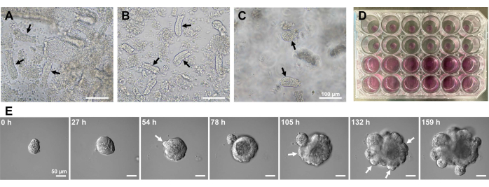
Para generar organoides del intestino delgado del ratón, se puede utilizar una combinación de tratamiento con EDTA y un método de aislamiento mecánico para aislar eficientemente las criptas10,13. Los resultados de este estudio mostraron que casi todas las criptas aisladas se sellaron inmediatamente y aparecieron en forma de cono después de que fueron exprimidas de los nichos epiteliales (Figura 1A). Para minimizar la contaminación de las vellosidades, la suspensión resultante se pasó a través de un filtro celular de 70 μm, y luego el filtrado se centrifugaba. Como algunas criptas se interrumpen durante la filtración y la suspensión, estos pasos deben llevarse a cabo con cuidado. Los resultados mostraron que casi todas las criptas en la fracción final estaban integradas y eran adecuadas para su uso en cultivo (Figura 1B). Para visualizar todas las criptas plateadas individualmente, se colocaron 100 criptas por pocillo (Figura 1C). Después de agregar el medio específico de cultivo de criptas (Figura 1D), el desarrollo de organoides se monitoreó diariamente con un microscopio. Además, el crecimiento de organoides de las criptas se observó mediante imágenes de lapso de tiempo para monitorear su desarrollo (Figura 1E y Video Suplementario S1). Las criptas cultas se comportaron de una manera estereotipada. La luz interna del organoide estaba llena de una masa de células apoptóticas. La proliferación activa y la diferenciación de ISC ocurrieron en la región de la cripta con gemación (Figura 1E y Video Suplementario S1). La gemación se combinó con la migración y proliferación de ISC y la diferenciación de células de Paneth. Las células de Paneth diferenciadas siempre se localizaron en el sitio de gemación (Figura suplementaria S1). Como se confirmó que los organoides eran estables en cultivo utilizando un microscopio invertido a un aumento de 10x, la técnica podría usarse para examinar la formación de criptas en el intestino delgado en desarrollo y para determinar la capacidad de regeneración tisular y la supervivencia a largo plazo de ISC para la producción de nuevas células epitelialesintestinales 14,15,16.
Lgr5 se define como un marcador ISC, y las células murinas Lgr5+ forman organoides 3D7. Sin embargo, como la abundancia de la superficie celular de la proteína LGR5 es baja y hay una falta de anticuerpos anti-LGR5 de alta afinidad, es difícil aislar eficientemente las ISC murinas mediante FACS. EphB2 ha sido previamente identificado como un marcador de superficie para la purificación de ISCs murinos y humanos a partir de tejidos intestinales17,18. El patrón de expresión de EphB2 aumenta la complejidad involucrada en los marcadores ISC. Las células positivas para EphB2 están organizadas en todo el compartimiento proliferativo, alcanzando su punto máximo en la parte inferior de las criptas, mientras que disminuyen en un gradiente hacia la parte superior de las criptas11. Las células de Paneth y las células progenitoras también se localizan en la cripta. Las células de Paneth expresan principalmente EphB3, que es necesario para su posicionamiento, y las células progenitoras sobre ellas en la cripta expresan principalmente EphB2. Por lo tanto, la contaminación de ambos tipos de células puede ocurrir durante el curso de la purificación de ISC utilizando el anticuerpo anti-EphB2. En consecuencia, debe evaluarse su expresión génica marcador y la capacidad de formación de organoides de las células aisladas utilizando EphB2 por el FACS.
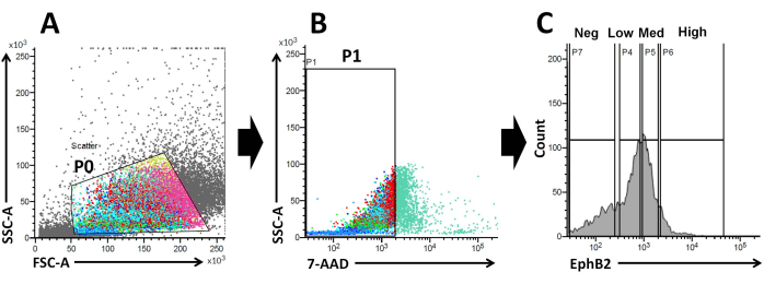
Sobre la base de estos hechos, utilizando el análisis FACS, las células marcadas en superficie EphB2 se pueden aislar de las criptas WT19. Se ha investigado si la expresión de EphB2 puede distinguir entre cuatro grupos con la expresión de marcadores específicos, como los genes marcadores específicos de ISC (Lgr5, Ascl2 y Olfm4) y los genes marcadores específicos de células progenitoras (Ki67, Myc y FoxM1). Este experimento demostró que las célulasaltas de EphB2 eran predominantemente ISC, a diferencia de las célulasmédicas de EphB220,21. Finalmente, con base en el método de aislamiento celular, las células obtenidas se dividieron en cuatro grupos (EphB2alta, EphB2med, EphB2baja y EphB2neg células) (Figura 2). Luego, se cultivaron células individuales que expresaban altos niveles de EphB2 ordenadas por FACS para el crecimiento de organoides. Una sola célulaalta EphB2 se puede aplicar de forma independiente para el tratamiento localizado y recrear estructuras cripto-vellosas autoorganizadas que recuerdan al intestino delgado normal (Figura 3). Sin embargo, las células derivadas de otros grupos (EphB2med, EphB2baja y EphB2neg) no generan organoides20.
En un estudio anterior, ~ 6% de las células Lgr5-GFPhi de una sola clasificación pudieron iniciar organoides cripto-vellosos7. Sin embargo, las células restantes no pudieron generar organoides y murieron dentro de las primeras 12 h7. Los autores presumieron que esto fue el resultado del estrés físico y/o biológico inherente al procedimiento de aislamiento7. También se obtuvo menos del 6% de crecimiento de organoides a partir de célulasaltas de EphB2 de una sola clasificación en ratones WT. Para el día 5 del cultivo, se formaron estructuras similares a esferoides (Figura 3). Del día 7 al día 9, se produjo la evaginación de las manchas para formar criptas (Figura 3). Es importante destacar que la aplicación de un inhibidor de ROCK seleccionado a las célulasaltas de EphB2 de un solo tipo disminuyó la apoptosis inducida por disociación y aumentó la eficiencia del crecimiento de organoides.

Figura 1: Generación de organoides del intestino delgado del ratón. (A) Criptas preparadas por una combinación de quelación de EDTA y disociación mecánica. (B) Criptas purificadas resultantes. (C) Criptas incrustadas en la matriz extracelular. (A-C) Las flechas negras indican criptas. (D) Cultura tridimensional de criptas y organoides. (E) Imágenes representativas de un organoide en crecimiento derivado de una cripta. Las flechas blancas indican la gemación de la cripta. Barras de escala = (A-C) 100 μm y (E) 50 μm. Haga clic aquí para ver una versión más grande de esta figura.

Figura 2: Estrategia de activación por citometría de flujo para obtener una población de células EphB2 positivas (EphB2+) en ratones de tipo salvaje. (A) Los diagramas de dispersión hacia adelante y hacia los lados se utilizan para separar las células de acuerdo con su tamaño y granularidad, respectivamente. (B) La dispersión de fluorescencia se utiliza para separar células viables de acuerdo con la intensidad de fluorescencia 7-AAD (PerCP) de las células. Se eligió la puerta para la población de células 7-AAD negativas. (C) Se eligieron las puertas para las poblaciones celulares EphB2-alta (EphB2alta), EphB2-media (EphB2med), EphB2-baja (EphB2baja) y EphB2-negativa (EphB2neg). Abreviaturas: FSC-A = área de pico de dispersión hacia adelante; SSC-A = área de pico de dispersión lateral; 7-AAD = 7-amino-actinomicina D. Haga clic aquí para ver una versión más grande de esta figura.

Figura 3: Curso temporal del crecimiento de organoides decélulas altas de EphB2 de una sola clasificación en ratones de tipo salvaje. Haga clic aquí para ver una versión más grande de esta figura.
Tabla 1: Medio de cultivo para una placa de 24 pocillos. Haga clic aquí para descargar esta tabla.
Video complementario S1: Imágenes de lapso de tiempo de un organoide en crecimiento. Barra de escala = 50 μm. Haga clic aquí para descargar este archivo.
Figura complementaria S1: Imagen representativa de la tinción de anticuerpos antilisozima en un organoide. Las flechas blancas indican las celdas de Paneth. Abreviatura: DIC = microscopio de contraste de interferencia diferencial. Barra de escala = 10 μm. Haga clic aquí para descargar este archivo.
Discusión
Este protocolo describe un método para aislar consistentemente criptas del intestino delgado y el posterior cultivo de organoides 3D. Para mejorar la velocidad de liberación de criptas, se estableció un método de aislamiento mecánico que implica agitación vigorosa después del tratamiento con EDTA. La composición del medio es diferente del protocolo original de Sato et al.7. El medio original es relativamente costoso. Por lo tanto, en la Tabla 1 se muestra un medio de cultivo y un medio personalizado para organoides murinos del intestino delgado que contienen inhibidores farmacológicos, factores de crecimiento recombinantes y/o medios acondicionados. Wnt3A y N-acetilcisteína no se incluyen en el medio de cultivo en este protocolo. Como las células de Paneth expresan Wnt3, las células producen Wnt3 y apoyan el mantenimiento de ISC. Además, durante el curso del aislamiento de la cripta, no se utiliza el medio acondicionado. El modelo organoide es dinámico y tiene heterogeneidad celular y estructural (células de Paneth, enterocitos, células caliciformes, células enteroendocrinas, células del mechón e ISC). Por lo tanto, estos organoides se pueden utilizar a escala para estudiar cuestiones fundamentales de la biología de los organoides.
El gradiente EphB2 mantiene el tallo ISC y la proliferación a lo largo del eje cripta-vellosidad en el intestino delgado adulto18. La ventaja de hacer organoides a partir de una sola célula EphB2 en comparación con criptas aisladas se relaciona con la comprensión de la biología de los ISC murinos, ya que los ISC desempeñan un papel clave en diversos trastornos intestinales humanos. Las ISC únicas dealta expresión de EphB2 se pueden cultivar para formar organoides de manera similar al desarrollo de organoides a partir de ISC únicas que expresan Lgr5. El paso más importante es dividir con precisión las células en cuatro grupos (EphB2alto, EphB2med, EphB2bajo y EphB2neg) de acuerdo con la expresión EphB2 en las criptas usando FACS. Los gráficos de dispersión hacia adelante frente a los laterales (FSC vs. SSC) se usan comúnmente para identificar celdas de interés en función de su tamaño y granularidad. FSC indica el tamaño de la celda, y SSC se relaciona con la complejidad o granularidad de la celda en la puerta P0 (Figura 2A). En este trabajo, las células que cayeron dentro de la puerta definida (P0) se analizaron posteriormente para determinar su viabilidad. A continuación, se determinó su viabilidad de acuerdo con las poblaciones negativas y positivas de señales de fluorescencia 7-AAD. La frontera entre las células 7-AAD-negativas y -positivas se decidió estrictamente para obtener las negativas con una contaminación celular positiva mínima. Las puertas EphB2 se establecieron aproximadamente en función de la expresión graduada EphB2.
Para confirmar que los cuatro grupos estaban divididos con precisión, se analizó la expresión de ARNm de genes seleccionados. Los niveles de ARNm de los marcadores ISC son altos en las célulasaltas de EphB220. Además, los niveles de ARNm de los marcadores específicos de células progenitoras son relativamente altos en las célulasmédicas EphB220. Sin embargo, la supresión de EphB2 en célulasEphB2 bajas y EphB2neg es baja o negativa en comparación con la de EphB2alta y EphB2med cells20. Las medidas anteriores deben tomarse para garantizar el enriquecimiento de la alta población celular de EphB2 antes del recubrimiento. Sin embargo, el crecimiento de organoides de menos del 6% de las célulasaltas de EphB2 puede deberse a la muerte de las células madre durante el proceso de cultivo, no al temblor vigoroso durante el aislamiento de la cripta. Se ha demostrado que la aplicación de un inhibidor selectivo de la quinasa asociada a Rho (ROCK) a las células madre embrionarias humanas disminuye notablemente la apoptosis inducida por disociación22. Por lo tanto, como cambio técnico, vale la pena intentar agregar el inhibidor de ROCK a una concentración más alta y con una incubación más larga para mejorar la viabilidad.
Las células de Paneth secretoras de Wnt3A junto a los ISC proporcionan un apoyo esencial a los ISC8. De hecho, los dobletes de células ISC-Paneth muestran una capacidad de formación de organoides fuertemente aumentada en comparación con las ISC individuales8. Además, se ha demostrado que la adición de Wnt3A a la concentración de 100 ng/mL durante los primeros 3 días de cultivo aumenta la capacidad formadora de organoides8. Por lo tanto, como otro cambio técnico, la adición de Wnt3A exógeno podría mejorar la capacidad de formación de organoides de ISC únicos dealta expresión de EphB2.
En comparación con los enfoques in vivo, los organoides pueden ser fácilmente utilizados para la manipulación genética, el análisis de fenotipos de malignidad y el cribado de fármacos20,23. Una combinación de quelación con EDTA y un método de aislamiento mecánico es eficaz, reproducible y eficiente en el tiempo para crear organoides del intestino delgado a partir de criptas y puede ser fácilmente seguida por el personal del laboratorio sin ninguna experiencia avanzada. Por lo tanto, la adición del aislamiento mecánico con agitación vigorosa después del tratamiento con EDTA puede establecer eficientemente organoides murinos del intestino delgado ex vivo y proporcionar una herramienta potencial para el cultivo de organoides y el modelado de enfermedades de otros tejidos epiteliales adultos.
Las células epiteliales intestinales están polarizadas y orientadas con el lado apical dirigido hacia la luz. Sin embargo, el lado apical que mira hacia la luz de los organoides 3D está en su interior. Por lo tanto, esta organización impide el acceso al lado apical, que es un problema cuando se estudian los efectos de los componentes luminales, como nutrientes, microbios y metabolitos en las células epiteliales. Para sortear esta desventaja, se ha desarrollado un cultivo de células organoides como monocapas2D 24. En términos de aplicaciones futuras, se utilizará el cultivo de monocapas de células organoides, ya que representa el sistema más eficiente y manejable.
Divulgaciones
Los autores no tienen conflictos de intereses que declarar.
Agradecimientos
Este trabajo fue apoyado por Grants-in-Aid for Scientific Research (C) a T.T. (números de subvención JP17K07495 y JP20K06751). Agradecemos al Prof. Mineko Kengaku por el uso de equipos para la obtención de imágenes de lapso de tiempo a largo plazo (LCV100; Olimpo).
Materiales
| Name | Company | Catalog Number | Comments |
| 1.5 mL Eppendorf tube | Eppendorf | 0030 125.215 | |
| 5 mL syringe | TERUMO | SS-05SZ | |
| 15 mL Falcon tube | Iwaki | 2325-015 | |
| 20 μm cell strainer | Sysmex | 04-004-2325 | |
| 24-well plate | Iwaki | 3820-024 | |
| 50 mL Falcon tube | Iwaki | 2345-050 | |
| 60 mm tissue culture dish | FALCON | 353002 | |
| 70 μm cell strainer | Falcon | 352350 | |
| 100 mm Petri dish | Iwaki | 3020-100 | |
| 7-AAD | BD Biosciences | 559925 | |
| Advanced DMEM/F12 | Gibco | 12634-010 | |
| Alexa Fluor 568 Goat Anti-Mouse IgG (H+L) | Invitrogen | A-11004 | |
| Anti-EphB2 APC-conjugated antibody | BD Biosciences | 564699 | |
| C57BL6/J mice | Japan SLC, Inc. | ||
| Clean bench | HITACHI | CCV-1306E | |
| Confocal laser scanning microscope | Olympus | FV3000 | |
| EDTA (0.5 mol/L) | Nacalai Tesque | 06894-14 | 2 mM |
| FACSMelody | BD Life Sciences-Biosciences | 661762 | |
| Fetal bovine serum | Sigma | 173012 | 1% (v/v) |
| Fiji (software) | https://fiji.sc/ | ||
| Gentamicin (10 mg/mL) | Nacalai Tesque | 16672-04 | 25 μg/mL |
| Hammacher laboratory scissor | SANSYO | 91-1538 | |
| Incubator | Panasonic | MCO-170-PJ | |
| Laboratory tweezer | AS-ONE | 7-164-04 | |
| L-Glutamine 200 mM | Gibco | 25030081 | 2 mM |
| Matrigel | BD Biosciences | 354230 | ECM for 3D organoids |
| Mouse Anti-Human Lysozyme | LSBio | LS-B8704-100 | |
| Murine EGF (20 μg/mL stock solution) | PeproTech | 315-09 | 20 ng/mL |
| PBS 1x | Gibco | 10010-023 | |
| Penicillin-Streptomycin (10,000 U/mL) | Gibco | 15140-122 | 50 U/mL |
| Pipetman (10 μL, 20 μL, 200 μL, and 1,000 μL) | GILSON | 1-6855-12, -13, -15, and -16 | |
| Recombinant murine Noggin (20 μg/mL stock solution | R&D Systems | 1967-NG-025 | 100 ng/mL |
| Recombinant murine R-Spondin 1 (250 μg/mL stock solution) | R&D Systems | 3474-RS-050 | 500 ng/mL |
| Sorbitol | Nacalai Tesque | 32021-95 | 2% (w/v) |
| TE2000-S (inverted microscope) | Nikon | 24131 | |
| Time-lapse image microscope | Olympus | LCV100 | |
| TrypLE Express 1x | Gibco | 12605-010 | |
| ULVAC | ULVAC KIKO Inc. | 100073 | |
| Y-27632 | Fujifilm | 331752-47-7 | 10 μM |
Referencias
- Clevers, H. Modeling development and disease with organoids. Cell. 165 (7), 1586-1597 (2016).
- Seidlitz, T., et al. Human gastric cancer modelling using organoids. Gut. 68 (2), 207-217 (2019).
- Nikolaev, M., et al. Homeostatic mini-intestines through scaffold-guided organoid morphogenesis. Nature. 585 (7826), 574-578 (2020).
- Artegiani, B., Clevers, H. Use and application of 3D-organoid technology. Human Molecular Genetics. 27, R99-R107 (2018).
- Lancaster, M. A., Knoblich, J. A. Organogenesis in a dish: modeling development and disease using organoid technologies. Science. 345 (6194), 1247125 (2014).
- Dedhia, P. H., Bertaux-Skeirik, N., Zavros, Y., Spence, J. R. Organoid models of human gastrointestinal development and disease. Gastroenterology. 150 (5), 1098-1112 (2016).
- Sato, T., et al. Single Lgr5 stem cells build crypt-villus structures in vitro without a mesenchymal niche. Nature. 459 (7244), 262-265 (2009).
- Sato, T., et al. Paneth cells constitute the niche for Lgr5 stem cells in intestinal crypts. Nature. 469 (7330), 415-418 (2011).
- Aronowitz, J. A., Lockhart, R. A., Hakakian, C. S. Mechanical versus enzymatic isolation of stromal vascular fraction cells from adipose tissue. Springerplus. 4 (1), 713 (2015).
- Takahashi, T. New trends and perspectives in the function of non-neuronal acetylcholine in crypt-villus organoids in mice. Methods in Molecular Biology. 1576, 145-155 (2019).
- Batlle, E., et al. β-catenin and TCF mediate cell positioning in the intestinal epithelium by controlling the expression of EphB/ephrinB. Cell. 111 (2), 251-263 (2002).
- Baghdadi, M. B., Kim, T. -. H. Analysis of mouse intestinal organoid culture with conditioned media isolated from mucosal enteric glial cells. STAR Protocols. 3 (2), 101351 (2022).
- Takahashi, T., et al. Non-neuronal acetylcholine as an endogenous regulator of proliferation and differentiation of Lgr5-positive stem cells in mice. FEBS Journal. 281 (20), 4672-4690 (2014).
- Barker, N. Adult intestinal stem cells: Critical drivers of epithelial homeostasis and regeneration. Nature Reviews Molecular Cell Biology. 15 (1), 19-33 (2014).
- Fordham, R. P., et al. Transplantation of expanded fetal intestinal progenitors contributes to colon regeneration after injury. Cell Stem Cell. 13 (6), 734-744 (2013).
- Miyoshi, H., et al. Wnt5a potentiates TGF-β signaling to promote colonic crypt regeneration after tissue injury. Science. 338 (6103), 108-113 (2012).
- Jung, P., et al. Isolation and in vitro expansion of human colonic stem cells. NatureMedicine. 17 (10), 1225-1227 (2011).
- Merlos-Suárez, A., et al. The intestinal stem cell signature identifies colorectal cancer stem cells and predicts disease relapse. Cell Stem Cell. 8 (5), 511-524 (2011).
- Mao, W., et al. EphB2 as a therapeutic antibody drug target for the treatment of colorectal cancer. Cancer Research. 64 (3), 781-788 (2004).
- Takahashi, T., et al. Muscarinic receptor M3 contributes to intestinal stem cell maintenance via EphB/ephrin-B signaling. Life Science Alliance. 4 (9), e202000962 (2021).
- Jung, P., et al. Isolation of human colon stem cells using surface expression of PTK7. Stem Cell Reports. 5 (6), 979-987 (2015).
- Watanabe, K., et al. A ROCK inhibitor permits survival of dissociated human embryonic stem cells. Nature Biotechnology. 25 (6), 681-686 (2007).
- Schulte, L., Hohwieler, M., Müller, M., Klaus, J. Intestinal organoids as a novel complementary model to dissect inflammatory bowel disease. Stem Cells International. 2019, 8010645 (2019).
- Puzan, M., Hosic, S., Ghio, C., Koppes, A. Enteric nervous system regulation of intestinal stem cell differentiation and epithelial monolayer function. Scientific Reports. 8 (1), 6313 (2018).
Reimpresiones y Permisos
Solicitar permiso para reutilizar el texto o las figuras de este JoVE artículos
Solicitar permisoExplorar más artículos
This article has been published
Video Coming Soon
ACERCA DE JoVE
Copyright © 2025 MyJoVE Corporation. Todos los derechos reservados