Method Article
Patrones dinámicos de proteínas inducidos por la luz en membranas modelo
En este artículo
Resumen
Aquí, se describe un protocolo para generar patrones de proteínas regulados por luz y reversibles con alta precisión espacio-temporal en membranas lipídicas artificiales. El método consiste en la fotoactivación localizada de la proteína iLID (dímero mejorado inducible por la luz) inmovilizada en membranas modelo que, bajo luz azul, se une a su proteína asociada Nano (SspB de tipo salvaje).
Resumen
La localización y activación precisas de las proteínas en la membrana celular en un momento determinado da lugar a muchos procesos celulares, incluida la polarización, migración y división celular. Por lo tanto, los métodos para reclutar proteínas para modelar membranas con resolución subcelular y alto control temporal son esenciales para reproducir y controlar dichos procesos en células sintéticas. Aquí, se describe un método para fabricar patrones de proteínas reversibles regulados por luz en membranas lipídicas con alta precisión espacio-temporal. Para ello, inmovilizamos la proteína fotoconmutable iLID (dímero mejorado inducible por la luz) en bicapas lipídicas soportadas (SLB) y en la membrana externa de vesículas unilaminares gigantes (GUV). Tras la iluminación de luz azul local, iLID se une a su socio Nano (SspB de tipo salvaje) y permite el reclutamiento de cualquier proteína de interés (POI) fusionada con Nano desde la solución hasta el área iluminada en la membrana. Este enlace es reversible en la oscuridad, lo que proporciona un enlace dinámico y la liberación del POI. En general, se trata de un método flexible y versátil para regular la localización de proteínas con alta precisión en el espacio y el tiempo utilizando luz azul.
Introducción
La formación de patrones proteicos en las membranas celulares dentro de las regiones subcelulares da lugar a numerosos procesos biológicos, incluyendo la migración, la división y la comunicación localizada de célula a célula 1,2. Estos patrones de proteínas están regulados en el espacio y el tiempo, y son altamente dinámicos. La replicación de estos patrones de proteínas en células sintéticas es esencial para imitar los procesos celulares que surgen de ellas y para comprender mejor cómo funciona dicha regulación a nivel molecular. De manera análoga a lo que se observa para las membranas en células vivas, los métodos para generar patrones de proteínas en membranas artificiales deben capturar su dinámica y proporcionar un control espacio-temporal preciso.
Entre varios estímulos, la luz se destaca por proporcionar el mayor control espacio-temporal y varias ventajas adicionales3. A través de la regulación con luz, es sencillo iluminar un área deseada en cualquier momento deseado con una precisión inigualable. Además, la luz proporciona una alta capacidad de ajuste, ya que se pueden ajustar tanto la intensidad de la luz como la duración del pulso. Además, la luz visible es inofensiva para las biomoléculas, incluidas las proteínas, e incluso es posible abordar múltiples funcionalidades con diferentes longitudes de onda. Por lo tanto, los enfoques sensibles a la luz basados en la luz visible emergen como vías prometedoras para la regulación controlada y biortogonal de los patrones de proteínas en el espacio y el tiempo 4,5,6. El uso de pares de proteínas fotoconmutables de la optogenética, que actúan como dimerizadores inducibles, proporciona un método sencillo para reclutar proteínas específicas en las membranas. En particular, se han formado con éxito patrones de proteínas en membranas artificiales utilizando la interacción desencadenada por la luz azul entre iLID (dímero inducible por luz mejorado, basado en el dominio LOV2 fotoconmutable de Avena sativa) y Nano (SspB de tipo salvaje)7,8, el sistema SpyTag inducible por luz azul (BLISS)9, la proteína tetrámera CarH10 sensible a la luz verde y la interacción inducible por luz roja entre PhyB y PIF611.
Se ha demostrado que la interacción fotoconmutable entre iLID y Nano5 se puede utilizar para foto-modelar proteínas en membranas modelo utilizando luz azul7. La interacción iLID/Nano es reversible en la oscuridad, altamente específica y opera en condiciones fisiológicas. El anclaje de iLID en modelos de membranas lipídicas, como vesículas unilaminares gigantes (GUV) o bicapas lipídicas soportadas (SLB), permite el reclutamiento regulado por luz de Nano a estas membranas, que es reversible en la oscuridad. En particular, observamos que la introducción de un dominio desordenado en el N-terminal de iLID (lo que resulta en una proteína llamada disiLID) como un lazo a una membrana lipídica modelo mejora la eficiencia del reclutamiento de Nano y la dinámica de reversión8.
Mediante el empleo de la interacción disiLID/Nano, hemos desarrollado un método para generar patrones de alto contraste de proteínas de interés (POI) nanofusionadas en SLBs y las membranas externas de los GUVs. Este método permite la creación de patrones de proteínas con una notable resolución espacial y temporal y una alta reversibilidad en cuestión de minutos. El protocolo detallado describe el proceso para el reclutamiento local de proteínas en membranas artificiales. En concreto, esto se consigue mediante la inmovilización de una versión biotinilada de disiLID en SLBs y GUVs a través de la interacción biotina-estreptavidina (SAv). Posteriormente, se recluta Nano marcado con fluorescencia (mOrange-Nano) para estas membranas funcionalizadas disiLID bajo iluminación de luz azul. Nuestro protocolo experimental ofrece un enfoque sencillo y adaptable para lograr el reclutamiento localizado de proteínas en las membranas. Es importante destacar que esta metodología no se limita a las interfaces SLB y GUV reportadas o mOrange-Nano; puede extenderse a otros materiales funcionalizados con disiLID y proteínas fusionadas con Nano.
Protocolo
1. Preparación experimental
- Exprima y purifique biotinilado-disiLID (b-disiLID) y mOrange-Nano (ver Tabla de Materiales) siguiendo los procedimientos previamente informados 7,8.
- Prepare mezclas de lípidos en viales de vidrio con la composición y concentraciones de lípidos seleccionadas. En primer lugar, disuelva los lípidos en cloroformo para obtener una solución lipídica final con una concentración de 1 mg/mL.
- Mezclar lípidos para obtener una composición de 94,9 % mol de 2-dioleoil-sn-glicero-3-fosfocolina (DOPC), 5 % mol de 1,2-dioleoil-sn-glicero-3-fosfoetanolaminaN-(cap biotinil) sal de sodio (DOPE-biotina) y 0,1 % mol de 1,1'-Dioctadecil-3,3,3',3'-Tetrametilindodicarbocianina (DiD) (ver Tabla de Materiales).
NOTA: La mezcla de lípidos se puede ajustar con diferentes proporciones y concentración de DOPE-biotina y/o diferentes colorantes de membrana. La concentración recomendada de DOPE-biotina (5 mol%) permite la formación de una capa de estreptavidina (SAv) de alta densidad en los siguientes pasos.
- Mezclar lípidos para obtener una composición de 94,9 % mol de 2-dioleoil-sn-glicero-3-fosfocolina (DOPC), 5 % mol de 1,2-dioleoil-sn-glicero-3-fosfoetanolaminaN-(cap biotinil) sal de sodio (DOPE-biotina) y 0,1 % mol de 1,1'-Dioctadecil-3,3,3',3'-Tetrametilindodicarbocianina (DiD) (ver Tabla de Materiales).
- Prepare pequeñas vesículas unilaminares (VSU) siguiendo los métodos previamente descritos 7,8,12. Para este paso, se recomienda preparar SUV con un diámetro ≤100 nm.
- Para este estudio, se emplea el método de sonicación. Primero, evapore la solución de cloroformo en el vial de vidrio con un chorro de nitrógeno mientras gira los viales para formar una película lipídica delgada. A continuación, retire el cloroformo residual durante al menos 1 h al vacío.
- Rehidratar la película seca en agua ultrapura con una concentración final de 1 mg/mL de lípidos por vórtice. Por último, sonicar la solución obtenida durante 10 minutos hasta que la solución opaca se vuelva transparente.
NOTA: Guarde la mezcla de lípidos en un tubo de microcentrífuga en el refrigerador durante un máximo de 2 semanas. También se pueden utilizar diferentes métodos de preparación de SUV (por ejemplo, método de extrusión) siempre que el tamaño de los SUV finales sea de ≤ 100 nm.
- Rehidratar la película seca en agua ultrapura con una concentración final de 1 mg/mL de lípidos por vórtice. Por último, sonicar la solución obtenida durante 10 minutos hasta que la solución opaca se vuelva transparente.
2. Reclutamiento de mOrange-Nano para SLBs funcionalizados disiLID
- Añadir 150 μL de NaOH 2 M en cada pocillo de la cámara de fondo de vidrio de 18 pocillos de μ portaobjetos (véase la Tabla de Materiales) e incubar durante 1 h a temperatura ambiente. Posteriormente, retire el NaOH y lave los pocillos de 3 a 5 veces, primero con 150 μL de agua ultrapura, y luego 3 veces con 150 μL de tampón (10 mM Tris pH 7.4, 100 mM de NaCl) que contiene 10 mM de CaCl2.
- Añadir 15 μL de SUV recién preparados (concentración de stock de 1 mg/mL en agua) en los pocillos que contienen 150 μL de tampón con 10 mM de CaCl2 para tener aproximadamente una dilución de factor 10 de SUV en el tampón. Deje que los SUV se incuben durante 30 minutos a temperatura ambiente. Después del tiempo de incubación, se formarán SLB biotinilados.
- Lave los SLB al menos 7 veces con tampón (10 mM Tris pH 7.4, 100 mM NaCl) sin CaCl2 eliminando primero la solución y luego agregando tampón nuevo en cada paso. Se recomienda utilizar 80 μL de tampón para cada paso de lavado.
NOTA: Un lavado óptimo de los SLB se obtiene pipeteando la solución fresca hacia arriba y hacia abajo varias veces sin tocar la superficie. El pipeteo de la solución en los pocillos que contienen los SLB recién formados debe ser suave para reducir la formación de pequeñas burbujas de aire que dañarían los SLB formados. A partir de este momento, los pozos deben contener un volumen suficiente de tampón para evitar que los SLB se sequen. - Para una mayor funcionalización de los SLB biotinilados con SAv, añadir una solución de SAv hasta una concentración final de 250 nM e incubar durante 30 min a temperatura ambiente. A continuación, elimine el exceso de SAv lavándolo con un tampón (10 mM Tris pH 7,4, 100 mM NaCl) al menos 5 veces.
- A partir de este momento, mantenga las muestras bajo una luz roja protectora para evitar la fotoactivación no deseada de las proteínas fotoconmutables. Añadir b-disiILD (ver Tabla de Materiales) hasta una concentración final de 1 μM en el pocillo. Después de 30 minutos de incubación a temperatura ambiente, retire el exceso de proteína lavando con tampón al menos 5 veces.
- Agregue mOrange-Nano (ver Tabla de Materiales) a una concentración final de 200 nM y mantenga la muestra en la oscuridad cubriéndola con papel de aluminio.
- Coloque el portaobjetos de μ bajo el microscopio de fluorescencia y ajuste la configuración de la imagen. Ajuste el láser de 552 nm para la excitación de mOrange-Nano. Ajuste el rango de emisión para optimizar la señal mOrange. La fotoactivación de disiLID se consigue con un láser de 488 nm, utilizando pulsos de luz con intervalos de 2,58 s.
3. Preparación de GUV
- Preparar una solución al 5% (p/v) de alcohol polivinílico (PVA, ver Tabla de Materiales) (MW: 145 000 g/mol) con 100 mM de sacarosa en agua ultrapura, mezclando durante la noche a 80 °C a 400 rpm.
- Prepare una solución lipídica en cloroformo con la composición deseada (concentración final de 10 mg/mL). Para este método, se recomienda una composición compuesta por 10 mg/mL de POPC, 10% mol% de 1-palmitoil-2-oleoil-sn-glicero-3-fosfo-(1'-rac-glicerol) (POPG), 2% mol de DOPE-biotina y 1 mol% de DiD (ver Tabla de Materiales).
- Preparar GUVs con la técnica de hidratación 7,8. En primer lugar, extienda 40 μL de la solución de PVA preparada como una capa fina homogénea sobre un portaobjetos de vidrio de 60 mm x 24 mm, preferiblemente con una punta de pipeta. A continuación, seca la fina capa a 50 °C durante 30 min.
- Extienda 5 μL de la solución lipídica con una aguja sobre la capa de PVA y deje secar a 30 °C durante 1 h.
- Ensamble una cámara en el portaobjetos de vidrio funcionalizado utilizando un espaciador (~40 mm × 24 mm × 2 mm, consulte la Tabla de materiales) y un segundo portaobjetos de vidrio.
- Agregue 1 mL de tampón de rehidratación (10 mM Tris pH 7.4, 100 mM NaCl) en la cámara durante 1 h a temperatura ambiente para formar GUV. Después de 1 hora, invierta la cámara y golpee suavemente las superficies de vidrio con la punta de una pipeta.
- Retire con cuidado el portaobjetos de vidrio de un lado para abrir la cámara construida y coseche los GUV con una pipeta.
- Coloque la solución en un tubo de plástico y deje que los GUV se asienten durante 2 h.
4. Reclutamiento de mOrange-Nano para GUV funcionalizados disiLID
- Agregue una solución de SAv a los GUV recién cosechados y déjelos reposar durante 30 minutos a temperatura ambiente.
NOTA: Los siguientes pasos deben realizarse con luz roja protectora para evitar la fotoactivación de disiLID. - Añada 1 μM de b-disiLID a la solución de GUVs y coloque la muestra durante 30 min en la oscuridad, cubriéndola con papel de aluminio.
- Trate previamente una cámara de fondo de vidrio de 18 pocillos de μ portaobjetos con 150 μL de solución de BSA (3% p/v en agua) durante 10 min. A continuación, retire la solución BSA y lave los pocillos con 150 μL de agua ultrapura 3 veces.
- Añada 145 μL de 200 nM mOrange-Nano en tampón (10 mM Tris pH 7,4, 100 mM NaCl) al pocillo.
- A continuación, añada 5 μL de GUVs decorados con b-disiLID a la solución y espere ~15 min para que los GUVs se asienten.
- Coloque el portaobjetos de μ bajo el microscopio confocal. Excite la muestra a 552 nm para visualizar la fluorescencia mOrange (λex = 557 nm; λem = 576 nm) y a 638 nm para visualizar DiD (λex = 644 nm; λem = 665 nm) en las membranas GUV. El reclutamiento de mOrange-Nano se activa con pulsos de luz azul (488 nm, intensidad del 1%) cada 5,3 s para minimizar los efectos de fotoblanqueo no deseados.
NOTA: Las longitudes de onda de excitación se pueden adaptar en función del tipo de microscopio utilizado. Otras longitudes de onda de excitación comunes disponibles para microscopios son también los láseres de 532 nm o 561 nm y de 633 nm, 647 nm, 639 nm o 640 nm.
Resultados
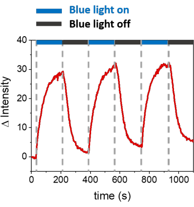
Los procedimientos descritos permiten la formación de SLBs para reclutar mOrange-Nano en las membranas sintéticas. En la Figura 1A se muestra la formación de mOrange-Nano definidos con el patrón de los SLB funcionalizados con b-disiLID. A medida que una región de interés (ROI) cuadrada (24 μm × 24 μm) en el SLB se ilumina con luz azul de 488 nm, se observa un rápido aumento de la señal de fluorescencia en el canal mOrange (mostrado en rojo) en el ROI dentro de 200 s. El patrón muestra bordes muy definidos y afilados (Figura 1B), lo que indica un alto control espacial sobre el área fotoactivada. La interacción es rápida y totalmente reversible ya que se interrumpe la iluminación con luz azul. Este método también permite la formación de patrones a lo largo de varios ciclos de iluminación (Figura 2). Ciclos alternos de ~200 s de luz azul y 200 s de oscuridad conducen a un reclutamiento reversible de mOrange-Nano en el área seleccionada durante múltiples tiempos con valores comparables de intensidad de fluorescencia Δ en los patrones.
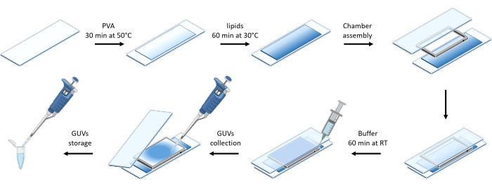
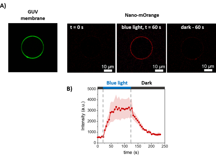
La Figura 3 muestra la representación esquemática de la preparación de los GUV. El reclutamiento de mOrange-Nano también se observa en los GUV. Se muestra que los GUV colocados en la oscuridad no exhiben fluorescencia mOrange (Figura 4A). A medida que los GUV se iluminan globalmente con luz azul, se observa fluorescencia mOrange, que se colocaliza con el colorante de membrana GUV (DiD). La interacción es altamente reversible ya que la iluminación está terminada. La cuantificación de la intensidad de mOrange en la membrana GUV a lo largo del tiempo muestra el reclutamiento rápido y efectivo de proteínas, así como la reversibilidad completa (Figura 4B).

Figura 1: Imágenes de microscopía de fluorescencia de SLBs funcionalizados con b-disiLID. Las imágenes de fluorescencia en presencia de mOrange-Nano antes (A) y durante (B) la iluminación de luz azul local (488 nm) en el ROI. Barra de escala = 20 μm. (C) Intensidad de fluorescencia de mOrange medida en el ROI para SLBs funcionalizados con b-disiLID (a = 200 s). La figura es una adaptación de Di Iorio et al.8. Haga clic aquí para ver una versión más grande de esta figura.

Figura 2: Intensidad de fluorescencia de mOrange reclutada en el ROI en SLBs decorados con b-disiLID durante tres ciclos de reclutamiento. Después de cada paso de fotoactivación, la fluorescencia mOrange-Nano aumentó dentro del ROI. El patrón alcanza la saturación dentro de los 120 s, y la fluorescencia disminuye dentro de los 120 s, casi hasta los niveles de fondo. No se observa pérdida de calidad del patrón en diferentes ciclos de luz/oscuridad azul. La figura es una adaptación de Di Iorio et al.8. Haga clic aquí para ver una versión más grande de esta figura.

Figura 3: Representación esquemática de la preparación de los GUVs utilizando el método de hidratación suave. El esquema ofrece una representación visual de los varios escalones y de la cámara construida con dos correderas de vidrio y un espaciador. Haga clic aquí para ver una versión más grande de esta figura.

Figura 4: Mediciones de microscopía de fluorescencia del reclutamiento dependiente de la luz de mOrange-Nano en las membranas de GUVs. (A) Imágenes de fluorescencia de un GUV funcionalizado con disiLID en presencia de mOrange-Nano. En verde está el tinte de membrana de los GUV, y en rojo está la fluorescencia mOrange antes, durante y después de la iluminación con luz azul. Barras de escala = 10 μm. (B) Intensidad de fluorescencia de mOrange localizada en GUV a lo largo del tiempo. Tras la iluminación, la fluorescencia mOrange (mostrada en rojo) en la membrana lipídica alcanza la intensidad máxima en 60 s, con un aumento de la intensidad de fluorescencia de 5,9 veces. A medida que se detiene la iluminación, la fluorescencia mOrange disminuye a casi valores previos a la iluminación dentro de los 60 segundos (con una recuperación del 90%). La figura es una adaptación de Di Iorio et al.8. Haga clic aquí para ver una versión más grande de esta figura.
Discusión
Hemos descrito un método para el reclutamiento localizado de proteínas mOrange-Nano en membranas modelo, como la bicapa lipídica soportada y las vesículas unilamelares gigantes mediante el uso de la proteína fotoconmutable disiLID8. Los aspectos que contribuyen a la calidad del patrón incluyen la calidad de las proteínas, así como la buena calidad de los SLB y GUV.
Para garantizar una buena calidad de la proteína después de la expresión y la purificación, es importante evaluar primero las propiedades fotoconmutables de disiLID. Para ello, hay que medir la absorción del cofactor FMN en la oscuridad y después de la iluminación con luz azul. Se espera que los espectros UV-Vis de disiLID muestren el característico triple pico del cofactor FMN en la oscuridad, que disminuye significativamente con la iluminación de luz azul y se recupera en la oscuridad13. Este comportamiento fotoconmutable es crucial para obtener un reclutamiento reproducible y reversible en los siguientes pasos. Trabajar con luz roja protectora y exponer disiLID a una iluminación externa mínima durante la preparación de las muestras aumenta el rendimiento de los experimentos.
Otro paso crítico, y quizás el más crucial, es la formación de SLB adecuados. Los defectos en las membranas y/o la formación de SLB no homogéneos (es decir, la presencia de multicapas o SLB parcheados) afectarán la calidad del patrón de proteínas. Por lo tanto, para los usuarios inexpertos, se recomienda reproducir el protocolo marcando los SUV con algunos tintes de membrana, como DiD y DiO, para formar SLB marcados con fluorescencia. De esta manera, las propiedades y la calidad de los SLB se pueden caracterizar bien con microscopía de fluorescencia. Las mediciones de FRAP representan un enfoque típico para evaluar la calidad de un SLB mediante la evaluación de la fluidez de las membranas. Alternativamente, en el caso de los SLB biotinilados como los descritos en este protocolo, se puede utilizar SAv marcado con fluorescencia (por ejemplo, Atto 488-SAv) para visualizar y evaluar la calidad de los SLB.
La primera parte del protocolo describe la formación de patrones en los SLB. Para garantizar un resultado óptimo, es importante añadir mOrange-Nano a los SLB y dejar que la muestra se incube en la oscuridad durante 15 minutos. Durante la fotoactivación, la selección del ROI no se restringe a un tamaño específico. Sin embargo, la intensidad del láser y el tiempo de exposición deben regularse para reducir el fotoblanqueo no deseado de las proteínas fluorescentes.
Este método no se limita a las proteínas biotiniladas, y se pueden utilizar otros enfoques para anclar disiLID a los SLB. Por ejemplo, el disiLID marcado con His puede expresarse y anclarse en SLB que contienen Ni-NTA. Sin embargo, es crucial expresar Nano y disiLID con etiquetas diferentes para evitar el reemplazo de las proteínas en los SLB. Este método también permite la posibilidad de invertir el orden de las proteínas, funcionalizando así las SLB con Nano y reclutando disiLID (o proteínas fusionadas con disiLID) tras la iluminación con luz azul.
Para el control dinámico de la localización de proteínas, la localización reversible de la proteína en la región seleccionada debería ser posible repetidamente. Para lograr esto, la concentración de Nano (200 nM) en la solución es un parámetro crítico para obtener una alta reversibilidad.
Otra preocupación es el reclutamiento de Nano en la superficie GUV funcionalizada con disiLID. Al igual que en el caso del patrón de proteínas en SLBs, este método puede extenderse a diferentes estrategias de funcionalización de membranas. En este protocolo, todo el GUV se iluminó con luz azul para reclutar mOrange-Nano en toda la superficie del GUV. Sin embargo, la selección de pequeños ROIs localizados en la membrana GUV debería conducir a la localización precisa de proteínas en un área más restringida.
Este método presenta solo una limitación relacionada con la elección del fluoróforo empleado para la obtención de imágenes del Nano reclutamiento en la membrana de los SUV o GUV. En particular, deben evitarse los fluoróforos con un espectro de excitación en el rango de luz azul, ya que su uso interferirá con la fotoactivación de (dis)iLID. Por lo tanto, se recomienda la elección de fluoróforos en el rango de luz verde o roja (por ejemplo, mOrange o Cy5) para este tipo de experimento.
El diseño de disiLID ofrece una forma sencilla y adaptable de mejorar el reclutamiento de proteínas locales a las membranas y amplía el rango dinámico de iLID y Nano de la optogenética4. Estos métodos se centran en el reclutamiento de Nano en membranas mímicas como bicapas lipídicas y GUV. Sin embargo, este enfoque es extensible a las numerosas herramientas optogenéticas en las células donde (dis)iLID o Nano están unidos a una membrana.
Divulgaciones
Los autores no tienen nada que revelar.
Agradecimientos
Este trabajo fue financiado por el Consejo Europeo de Investigación ERC Starting Grant ARTIST (# 757593 S.V.W.). DDI agradece a la Fundación Alexander von Humboldt por una beca postdoctoral.
Materiales
| Name | Company | Catalog Number | Comments |
| µ-Slide 18 Well | Ibidi | 81817 | For SLB preparation |
| 25 µL Microliter Syringe | Hamilton | Model 702 N | For the preparation of lipid mixture and spreading the lipid solution on the PVA layer |
| Biotinyl Cap PE (1,2-dioleoyl-sn-glycero-3-phosphoethanolamine-N-(cap biotinyl)) (sodium salt) | Avanti Polar Lipids | 870273C | For SUVs preparation |
| CaCl2 (Calcium chloride) | Sigma-Aldrich | C5670 | For SLB formation |
| Cover Slips 24 mm x 60 mm | Engelbrecht | K12460 | For GUVs formation |
| DiD (1,1'-Dioctadecyl-3,3,3',3'- Tetramethylindodicarbocyanine) | Thermo Fisher Scientific | D7757 | Membrane dye |
| disiLID | Sequence: MGGSGLNDIFEAQKIEWHEGGSH HHHHHGSMAATELRGVVGPGPAA IAALGGGGAGPPVGGGGGRGDA GPGSGAASGTVVAAAAGGPGPG AGGVAAAGPPAPPTGGSGGSGA GGSGSAGEFLATTLERIEKNFVIT DPRLPDNPIIFASDSFLQLTEYSR EEILGRNCRFLQGPETDRATVRK IRDAIDNQTEVTVQLINYTKSGKK FWNVFHLQPMRDYKGDVQYFIG VQLDGTERLHGAAEREAVMLIKK TAFQIAEAANDENYF | ||
| DOPC (1,2-di-(9Z-octadecenoyl)-sn-glycero-3-phosphocholine) | Avanti Polar Lipids | 850375P | For SUVs lipid composition |
| Eppendorf Protein LoBind microcentrifuge tubes | Merk | EP0030108116-100EA | For collecting freshly made GUVs |
| mOrange-Nano | Sequence: MRGSHHHHHHGSKIEEGKLVI WINGDKGYNGLAEVGKKFEKDT GIKVTVEHPDKLEEKFPQVAATG DGPDIIFWAHDRFGGYAQSGLLA EITPDKAFQDKLYPFTWDAVRYN GKLIAYPIAVEALSLIYNKDLLPNP PKTWEEIPALDKELKAKGKSALM FNLQEPYFTWPLIAADGGYAFKY ENGKYDIKDVGVDNAGAKAGLTF LVDLIKNKHMNADTDYSIAEAAFN KGETAMTINGPWAWSNIDTSKVN YGVTVLPTFKGQPSKPFVGVLSA GINAASPNKELAKEFLENYLLTDE GLEAVNKDKPLGAVALKSYEEELA KDPRIAATMENAQKGEIMPNIPQM SAFWYAVRTAVINAASGRQTVDEA LKDAQTNSSSNNNNNNNNNNLGI EGTTENLYFQGSVSKGEENNMAI IKEFMRFKVRMEGSVNGHEFEIE GEGEGRPYEGFQTAKLKVTKGG PLPFAWDILSPQFTYGSKAYVKH PADIPDYFKLSFPEGFKWERVMN FEDGGVVTVTQDSSLQDGEFIYK VKLRGTNFPSDGPVMQKKTMG WEASSERMYPEDGALKGEIKMR LKLKDGGHYTSEVKTTYKAKKPV QLPGAYIVGIKLDITSHNEDYTIVE QYERAEGRHSTGGMDELYKGG SGTSSPKRPKLLREYYDWLVDN SFTPYLVVDATYLGVNVPVEYVK DGQIVLNLSASATGNLQLTNDFIQ FNARFKGVSRELYIPMGAALAIYA RENGDGVMFEPEEIYDELNIG | ||
| NaCl (Sodium chloride) | Sigma-Aldrich | S9888 | For buffer |
| NaOH (Sodium hydroxide) | Sigma-Aldrich | 1064980500 | For surface activation in SLB formation |
| POPC (1-Palmitoyl-2- oleoylphosphatidylcholine) | Avanti Polar Lipids | 850457C | For GUVs lipid composition |
| PVA (Polyvinyl alcohol) fully hydrolyzed | Sigma-Aldrich | 8148940101 | For GUVs formation |
| SP8 confocal laser scanning microscope | Leica | ||
| Streptavidin | TermoFisher | 434301 | |
| Sucrose | Sigma-Aldrich | 84097 | For GUVs formation |
| Tris hydrochloride | Sigma-Aldrich | 10812846001 | For buffer |
Referencias
- Kretschmer, S., Schwille, P. Pattern formation on membranes and its role in bacterial cell division. Curr Opin Cell Biol. 38, 52-59 (2016).
- Yang, H. W., Collins, S. R., Meyer, T. Locally excitable Cdc42 signals steer cells during chemotaxis. Nat Cell Biol. 18 (2), 191-201 (2016).
- Caldwell, R. M., et al. Optochemical control of protein localization and activity within cell-like compartments. Biochem. 57 (18), 2590-2596 (2018).
- Kennedy, M. J., et al. Rapid blue-light-mediated induction of protein interactions in living cells. Nat Methods. 7 (12), 973-975 (2010).
- Guntas, G., et al. Engineering an improved light-induced dimer (iLID) for controlling the localization and activity of signaling proteins. Proc Natl Acad Sci. 112 (1), 112-117 (2015).
- Levskaya, A., Weiner, O. D., Lim, W. A., Voigt, C. A. Spatiotemporal control of cell signalling using a light-switchable protein interaction. Nature. 461 (7266), 997-1001 (2009).
- Bartelt, S. M., et al. Dynamic blue light-switchable protein patterns on giant unilamellar vesicles. Chem Commun. 54 (8), 948-951 (2018).
- Di Iorio, D., Bergmann, J., Higashi, S. L., Hoffmann, A., Wegner, S. V. A disordered tether to iLID improves photoswitchable protein patterning on model membranes. Chem. Commun. 59 (29), 4380-4383 (2023).
- Hartzell, E. J., Terr, J., Chen, W. Engineering a blue light inducible Spytag system (BLISS). J Am Chem Soc. 143 (23), 8572-8577 (2021).
- Xu, D., Bartelt, S. M., Rasoulinejad, S., Chen, F., Wegner, S. V. Green light lithography: a general strategy to create active protein and cell micropatterns. Mater Horiz. 6 (6), 1222-1229 (2019).
- Jia, H., et al. Light-induced printing of protein structures on membranes in vitro. Nano Lett. 18 (11), 7133-7140 (2018).
- Di Iorio, D., Verheijden, M. L., vander Vries, E., Jonkheijm, P., Huskens, J. Weak Multivalent Binding of influenza hemagglutinin nanoparticles at a sialoglycan-functionalized supported lipid bilayer. ACS Nano. 13 (3), 3413-3423 (2019).
- Kasahara, M., Torii, M., Fujita, A., Tainaka, K. FMN binding and photochemical properties of plant putative photoreceptors containing two LOV domains, LOV/LOV proteins. J Biol Chem. 285 (45), 34765-34772 (2010).
Reimpresiones y Permisos
Solicitar permiso para reutilizar el texto o las figuras de este JoVE artículos
Solicitar permisoThis article has been published
Video Coming Soon
ACERCA DE JoVE
Copyright © 2025 MyJoVE Corporation. Todos los derechos reservados