
Esame obiettivo dell'addome I: ispezione e auscultazione
Panoramica
Fonte: Alexander Goldfarb, MD, Assistente Professore di Medicina, Beth Israel Deaconess Medical Center, MA
Le malattie gastrointestinali rappresentano milioni di visite in ufficio e ricoveri ospedalieri ogni anno. L'esame fisico dell'addome è uno strumento cruciale nella diagnosi delle malattie del tratto gastrointestinale; inoltre, può aiutare a identificare i processi patologici nei sistemi cardiovascolari, urinari e di altro livello. Come esame fisico in generale, l'esame della regione addominale è importante per stabilire il contatto medico-paziente, per raggiungere la diagnosi preliminare e selezionare i successivi test di laboratorio e di imaging e determinare l'urgenza delle cure.
Come con le altre parti di un esame fisico, l'ispezione visiva e l'auscultazione dell'addome vengono eseguite in modo sistematico in modo che non vengano persi potenziali risultati. Particolare attenzione dovrebbe essere prestata a potenziali problemi già identificati dalla storia del paziente. Qui assumiamo che il paziente sia già stato identificato e abbia avuto una storia presa, sintomi discussi e aree di potenziale preoccupazione identificate. In questo video non esamineremo la storia del paziente; invece, andremo direttamente all'esame fisico.
Prima di arrivare all'esame, esaminiamo brevemente i punti di riferimento della superficie della regione addominale, dell'anatomia addominale e della topografia. Ecco un elenco di punti di riferimento utili: margini costali, processo xifoide, muscolo addominale retto, linea alba, ombelico, cresta iliaca, legamento inguinale e sinfisi pubica. L'esame addominale copre l'area in basso dai margini xifoidi e costali in modo superiore alla sinfisi pubica inferiormente.
Per scopi diagnostici e descrittivi, l'addome è suddiviso in quattro quadranti: quadrante superiore destro (spesso designato come RUQ), quadrante superiore sinistro (LUQ), quadrante inferiore destro (RLQ) e quadrante inferiore sinistro (LLQ)(Figura 1). La topografia più dettagliata dell'addome lo divide in 9 regioni: ipocondriaca destra e sinistra, lombare destra e sinistra, iliaca destra e sinistra, e anche regioni epigastriche, ombelicali e ipogastriche nel mezzo (Figura 2).
Ricorda quali organi in genere proiettano in ogni regione addominale (Figura 3). È essenziale conoscere bene l'anatomia e la topografia della regione per documentare e interpretare adeguatamente i reclami e i sintomi di un paziente, nonché i risultati fisici durante l'esame.
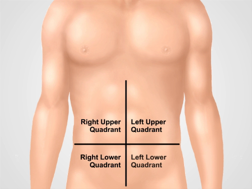
Figura 1. Quattro quadranti addominali. L'addome può essere diviso in quattro regioni da due linee immaginarie che si intersecano all'ombelico: vengono mostrati il quadrante superiore destro (spesso designato come RUQ), il quadrante superiore sinistro (LUQ), il quadrante inferiore destro (RLQ) e il quadrante inferiore sinistro (LLQ).
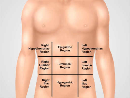
Figura 2. Nove regioni addominali. Le linee midclavicolari e i piani subcostali e intertubercolari separano l'addome in nove regioni: regione epigastrica, regione ipocondriaca destra, regione ipocondriaca sinistra, regione ombelicale, regione lombare destra, regione lombare sinistra, regione ipogastrica, regione inguinale destra e regione inguinale sinistra. I termini per le regioni epigastriche, ombelicali, ipogastriche e sovrapubiche sono i più comunemente usati nella pratica clinica.
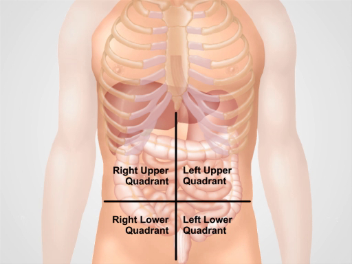
Figura 3. Posizione di diversi organi nelle quattro regioni addominali. Organi nella cavità addominale e la loro posizione rispetto a quattro quadranti addominali.
Procedura
1. Preparazione
- Prima di iniziare l'esame fisico dell'addome, assicurarsi che il paziente sia a suo agio e abbia svuotato la vescica.
- Posizionare comodamente il paziente in posizione supina, possibilmente con la testa del paziente sostenuta da un cuscino e le ginocchia leggermente flesse. Le braccia del paziente devono essere laterali e non piegate dietro la testa, poiché ciò tende la parete addominale.
- Chiedi il permesso di esporre la zona addominale del paziente ("Va bene se sposto il foglio per ispezionare lo stomaco?"). Drappeggiare il paziente in un modo che mantenga la modestia da un lato, ma non comprometta l'esame dall'altro. L'addome è esposto da sopra lo xifoide alla regione sovrapubica; anche l'inguine è esposto.
- Assicurati che ci sia abbastanza luce e che il rumore sia ridotto al minimo (spegnendo la TV o la radio nella stanza).
- Prima di avvicinarsi al paziente, determinare se sono necessarie precauzioni per il contatto.
- Lavarsi le mani (o in alternativa utilizzare preparati antibatterici topici), quindi riscaldare le mani e lo stetoscopio. Come con altre parti di un esame fisico, posizionati sul lato destro del paziente e spiega al paziente ogni fase dell'esame man mano che progredisce.
- Ricorda: l'esame deve essere eseguito in modo strutturato, seguendo una particolare procedura. L'esame completo iniziale è il primo, e una volta identificati alcuni risultati patologici, segue un esame più dettagliato di quei risultati specifici. Le aree di dolore o tenerezza dovrebbero essere esaminate per ultime. Una volta che il paziente è posizionato e pronto per essere esaminato, la sequenza della procedura è la seguente: ispezione, auscultazione, percussione e palpazione. La differenza di sequenza rispetto ad altre parti dell'esame si basa sul fatto che la palpazione può influenzare i suoni intestinali e quindi è fatta per ultima.
2. Ispezione
- Prima di iniziare l'esame, spiega che l'addome del paziente verrà ispezionato. ("Ho bisogno di ispezionare il tuo stomaco ora").
- Ispezionare visivamente la pelle per eruzioni cutanee / ecchimosi, ittero, vene dilatate, strie, lesioni, lividi e cicatrici. Se sono presenti cicatrici, chiedi al paziente di loro e documentale nella storia del paziente.
- Esaminare la forma dell'addome.
- È piatto, scafoide o protuberante? Gli addominali scafoidi possono essere visti nei pazienti cachettici. Le protuberanze addominali globali possono derivare da gas, liquidi o grassi
- L'addome sembra simmetrico o no? L'asimmetria è un segnale di avvertimento e può suggerire masse o organomegalia.
- Le parti dell'addome sono sporgenti? I fianchi sporgenti sono un segno di accumulo di liquido nell'addome (ascite).
- Verificare la visibile ernia e masse addominali
- Prestare attenzione alla presenza di pulsazioni visibili o peristalsi, che di solito rappresentano un problema serio. A volte la peristalsi visibile può essere vista nell'ostruzione intestinale.
- Si noti la presenza di tubi, cateteri e altri dispositivi.
- Prestare particolare attenzione all'area ombelicale. Esaminare per protuberanze, infiammazioni, ernie, spostamenti lungo la linea verticale e scolorimento della pelle violacea. La decolorazione della pelle violacea indica un sanguinamento intraperitoneale sottocutaneo ed è associata a pancreatite emorragica acuta.
3. Auscultazione
I suoni addominali sono tipicamente generati dalla peristalsi e dal flusso sanguigno e talvolta dagli sfregamenti per attrito. L'auscultazione viene eseguita in due o tre cicli, ogni volta ascoltando un particolare suono, piuttosto che cercare di ascoltare tutti i suoni contemporaneamente. Inizialmente ascolta i suoni intestinali, quindi concentrati sui suoni vascolari o sui lividi. Infine, anche se raro, controlla se ci sono sfregamenti di attrito.
- Spiega la procedura al paziente ("Ora ascolterò il tuo stomaco / pancia")
- Preriscaldare lo stetoscopio.
- Usando il diaframma dello stetoscopio, ascolta i suoni intestinali su ciascuno dei quattro quadranti addominali per 30-40 s; notare la loro frequenza e carattere. Dovrebbero essere sentiti suoni gorgoglianti che si verificano a una frequenza di 5-34 al minuto.
- I suoni intestinali sono variabili, quindi in un paziente asintomatico l'assenza di suoni intestinali richiede un ascolto più lungo. L'assenza di suoni intestinali in un paziente con dolore addominale è un segnale di avvertimento e potrebbe indicare ileo paralitico. Se i suoni intestinali sembrano assenti, auscultate per un massimo di 3 minuti nel quadrante inferiore destro. Anche i suoni intestinali iperattivi sono anormali. I suoni intestinali aumentati e acuti possono essere associati alle fasi iniziali dell'ostruzione intestinale.
- Ascolta diverse strutture vascolari in 7 diverse posizioni (sopra l'arteria renale destra, l'aorta, l'arteria renale sinistra, le arterie iliache e le arterie femorali) per almeno 5 s ciascuna.
- I suoni vascolari udibili sono chiamati bruits e sono causati da un flusso turbolento nelle grandi arterie (ad esempio, aorta, iliaca, arterie renali). Durante l'auscultazione i bruit producono un suono "swishing". La loro presenza può indicare stenosi dell'arteria renale, aneurisma dell'aorta addominale e stenosi dell'arteria iliaca e femorale. I suoni vascolari possono essere ascoltati nel 4-20% degli individui sani, ma una bruit sistolico-diastolica sulle arterie renali in un paziente con ipertensione suggerisce fortemente la malattia rinnovoculare.
- Ascolta gli sfregamenti di attrito sul fegato e sulla milza usando la campana dello stetoscopio. Lo sfregamento per attrito è una scoperta rara che indica l'infiammazione della superficie peritoneale dell'organo da infezione, tumore o infarto.
Applicazione e Riepilogo
In questo video abbiamo esaminato l'anatomia dell'addome e imparato come eseguire i primi due passaggi dell'esame addominale: ispezione e auscultazione. Prima di iniziare l'esame, assicurarsi che il paziente sia a suo agio, ben posizionato e adeguatamente drappeggiato. Non esaminare mai un paziente attraverso un camice. Assicurati che le mani siano lavate e calde. Chiedi sempre a un paziente il permesso di eseguire l'esame e spiega ogni fase della procedura. Inizia con un'ispezione visiva dell'addome. Prendi nota del contorno addominale e della simmetria, delle eruzioni cutanee, delle cicatrici di precedenti interventi chirurgici e lesioni, delle vene distese e delle peristalse e pulsazioni visibili. Se si sospetta la presenza di ascite, ernie o masse, confermare questi risultati tramite ulteriori manovre che vengono affrontate più avanti in questa raccolta di video.
Omettere l'ispezione visiva e / o le fasi di auscultazione sono errori comuni durante un esame addominale e possono influire negativamente sulla capacità di un medico di raggiungere una diagnosi corretta. Un'attenta ispezione della zona addominale è particolarmente importante. Spesso un medico esperto può fare una diagnosi preliminare basata solo sulla storia e l'ispezione di un paziente. Una combinazione di diversi segni patologici è di particolare valore diagnostico. Ad esempio, ittero, ascite, angiomi ragno e caput medusae (vene distese che circondano l'ombelico) possono essere contemporaneamente presenti in un paziente con cirrosi epatica.
Una volta completata l'ispezione visiva, segue l'auscultazione. Auscultate separatamente per i suoni intestinali e per i bruits. Eseguire sempre l'auscultazione prima delle percussioni addominali e della palpazione. L'auscultazione addominale è di importanza clinica, specialmente in un paziente sintomatico. I risultati devono essere interpretati nel contesto della storia del paziente: ad esempio, l'assenza di suoni intestinali in un paziente con dolore addominale suggerisce una catastrofe addominale (come la peritonite o le fasi successive di un'ostruzione intestinale), ma è normale nei pazienti postoperatori per alcuni giorni dopo l'intervento chirurgico addominale.
Tags
Vai a...
Video da questa raccolta:

Now Playing
Esame obiettivo dell'addome I: ispezione e auscultazione
Physical Examinations II
202.6K Visualizzazioni

Esame obiettivo dell'occhio
Physical Examinations II
77.2K Visualizzazioni

Esame oftalmoscopico
Physical Examinations II
68.0K Visualizzazioni

Esame obiettivo dell'orecchio
Physical Examinations II
55.1K Visualizzazioni

Esame di naso, seni, cavità orale e faringe
Physical Examinations II
65.8K Visualizzazioni

Esame obiettivo della tiroide
Physical Examinations II
105.0K Visualizzazioni

Esame obiettivo dei linfonodi
Physical Examinations II
387.4K Visualizzazioni

Esame obiettivo dell'addome II: percussione
Physical Examinations II
248.2K Visualizzazioni

Esame obiettivo dell'addome III: palpazione
Physical Examinations II
138.5K Visualizzazioni

Esame obiettivo dell'addome IV: valutazione del dolore addominale acuto
Physical Examinations II
67.3K Visualizzazioni

Esame rettale maschile
Physical Examinations II
114.5K Visualizzazioni

Esame completo del seno
Physical Examinations II
87.6K Visualizzazioni

Esame obiettivo ginecologico I: valutazione dei genitali esterni
Physical Examinations II
307.1K Visualizzazioni

Esame obiettivo ginecologico II: esame con lo speculum
Physical Examinations II
150.4K Visualizzazioni

Esame obiettivo ginecologico III: palpazione rettovaginale e bimanuale
Physical Examinations II
147.7K Visualizzazioni
Personale delle biblioteche
Copyright © 2025 MyJoVE Corporation. Tutti i diritti riservati