Aby wyświetlić tę treść, wymagana jest subskrypcja JoVE. Zaloguj się lub rozpocznij bezpłatny okres próbny.
Method Article
Immunohistological Labeling of Microtubules in Sensory Neuron Dendrites, Tracheae, and Muscles in the Drosophila Larva Body Wall
W tym Artykule
Podsumowanie
To understand how complex cell shapes, such as neuronal dendrites, are achieved during development, it is important to be able to accurately assay microtubule organization. Here we describe a robust immunohistological labeling method to examine microtubule organization of dendritic arborization neuron sensory dendrites, trachea, muscle, and other Drosophila larva body wall tissues.
Streszczenie
To understand how differences in complex cell shapes are achieved, it is important to accurately follow microtubule organization. The Drosophila larval body wall contains several cell types that are models to study cell and tissue morphogenesis. For example tracheae are used to examine tube morphogenesis1, and the dendritic arborization (DA) sensory neurons of the Drosophila larva have become a primary system for the elucidation of general and neuron-class-specific mechanisms of dendritic differentiation2-5 and degeneration6.
The shape of dendrite branches can vary significantly between neuron classes, and even among different branches of a single neuron7,8. Genetic studies in DA neurons suggest that differential cytoskeletal organization can underlie morphological differences in dendritic branch shape4,9-11. We provide a robust immunological labeling method to assay in vivo microtubule organization in DA sensory neuron dendrite arbor (Figures 1, 2, Movie 1). This protocol illustrates the dissection and immunostaining of first instar larva, a stage when active sensory neuron dendrite outgrowth and branching organization is occurring 12,13.
In addition to staining sensory neurons, this method achieves robust labeling of microtubule organization in muscles (Movies 2, 3), trachea (Figure 3, Movie 3), and other body wall tissues. It is valuable for investigators wishing to analyze microtubule organization in situ in the body wall when investigating mechanisms that control tissue and cell shape.
Protokół
1. Preparation of reagents
Notes before beginning: Dissection and immunohistochemical staining are carried out in a magnetic chamber and the larva is pinned down using specially shaped insect pins. Detailed instructions on the construction of a magnetic chamber, and preparation of these pins can be found in related references 14,15. In brief, a 1x1cm square hole is cut into a magnetic sheet and a coverslip affixed to the rear of the sheet to make a small chamber. The sides of the chamber are sealed with epoxy glue; after this glue has set the chamber is washed several times with 70% ethanol before use. Dissection insect pins are prepared by bending to the required shape and then glued onto a metal tab14,15. Alternatively to a metal tab, we have used an inverted flat-head steel drawing pin with a handle made from a cut-off yellow tip. Use of this magnetic chamber arrangement allows close control over pin positioning and tissue stretching during dissection.
To drive reporter gene expression in different subsets of DA neurons investigators may use several different Gal4 lines (summarized by Shimono and colleagues16). Many of these lines are available from public stock centers. In this representative protocol, we carry out immunostaining of a line in which two contrasting classes of DA neuron are co-labeled: the most simple- class I and most complex- class IV (P10-Gal417,18, UAS-mCD8::Kusabira-Orange (KO)).
- Prepare Ca++-free HL3.1 saline
- In mM: 70 NaCl, 5 KCl, 20 MgCl2 , 10 NaHCO3 , 5 HEPES, 115 sucrose, and 5 trehalose; pH 7.219. Filter-sterilize and store at 4°C. Note: Ca++-free solution prevents muscle contraction during dissection.
- Prepare 2x PHEM buffer
- In mM: 130 PIPES, 60 HEPES, 20 EGTA, 4 MgSO4; pH 7.0. Filter-sterilize and store at 4°C.
Note: These materials will not dissolve until the pH approaches 7.0.
- In mM: 130 PIPES, 60 HEPES, 20 EGTA, 4 MgSO4; pH 7.0. Filter-sterilize and store at 4°C.
- Prepare the fixative freshly immediately before the fixation.
- In order to prepare a 25ml solution, first mix 2g paraformaldehyde, 100μl 1M NaOH and 10ml water in a 50ml Falcon tube.
- Shake fixative in a 55°C water bath with shaker until the solution is clear. (Total volume will be about 11.5 ml.)
- Cool fixative on ice.
- Add 12.5ml 2x PHEM buffer.
- Adjust pH to 7.0 with 1M HCl.
- Fill the solution with water until the volume is 25ml.
- Filter the solution using Whatman paper.
2. Larval dissection
Note before beginning: Microtubule networks, and particularly those in sensory dendrites, will breakdown rapidly after the initiation of dissection. Achieving fast dissection in less than five minutes followed by immediate fixation are key factors in the success of this protocol.
- Wash the larva in water and quickly move them into this drop using a loop of hair.
- Orient the larva dorsal side up and the ventral side on the glass. Note: This orientation is to examine the ventral cluster neurons. To examine dorsal cluster neurons, invert the orientation.
- Use the center insect-pin to pin the anterior end near the mouth hooks. Place the pin close to the end for best results.
- Place a drop of HL3.1 saline in the dissection chamber.
- Cut the very posterior tip of the larva off with microscissors. Note: This step opens an aperture at the posterior end of the larva that will allow access for the microscissors (step 2.7).
- Grab with the forceps the region of gut that is now poking out of the aperture at the posterior end of the larva. Gently pull out the whole gut.
- Place the tip of one blade of the microscissors through the aperture and cut along the ventral midline towards the anterior.
- Using the corner pins, pin the now free corners, posterior first, then anterior. Simultaneously gently stretch open the larval fillet.
3. Fixation, blocking, staining, and mounting of larval fillets
Notes before beginning: All fixation and staining steps are carried out in the dissection chamber. During this process, be careful not to knock the insect pins holding the larva as this may lead to tissue damage. To prevent the experiment from drying out, do all staining steps in a small Tupperware container surrounded by moistened tissues.
- Aspirate the Ca++-free HL3.1 buffer using a yellow tip. Immediately add the fixative using another pipetteman.
- Gently pipette up and down to mix the fixative with the remaining traces of Ca++-free HL3.1 buffer in the chamber. Immediately aspirate it and then add fresh fixative into the chamber.
- Incubate at room temperature for 20 minutes.
- Wash 6x 10mins in PBST (0.1% Triton X-100 in PBS).
- Block with 5% goat serum in PBST for 20 mins at RT.
- Replace the blocking solution with primary antibodies diluted in 5% goat serum in PBST. The primary antibodies used are mouse anti-α-Tubulin (DM1A) and rat anti-CD8 (5H10) both diluted 1/1000.
Note: The investigator may wish to substitute mouse anti-α-Tubulin (DM1A) with mouse anti-Futsch (22C10)20,21 diluted 1/1000 in some circumstances (see discussion). - Incubate overnight (for at least 16h) at 4°C.
- Wash 6x 10mins in PBST.
- Add secondary antibody solution diluted in 5% goat serum in PBST. The secondary antibodies used are Alexa Fluor 488 goat anti-mouse IgG and Cy3 donkey anti-rat IgG. Keep the sample covered to prevent fluorophore photo-bleaching22.
- Incubate either at RT for two hours, or overnight at 4°C followed by one hour at room temperature.
- Wash 6x 10mins in PBST.
- Place the larval fillet on the slide cuticle-side down, mount in 80% glycerol, and seal the sides of the coverslip with nail polish for a 'quick' mount.
Note: For improved tissue clearing, and a permanent stable sample, mount in DPX as previously described23.
4. Representative Results:
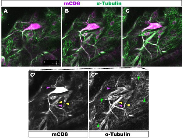
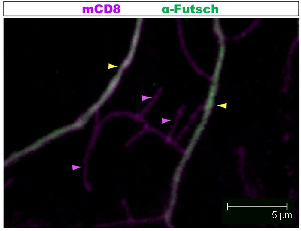
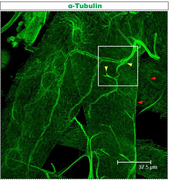
Fluorescent staining was examined under a confocal microscope. In Figures 1-2, different branches within a dendrite arbor have different cytoskeletal organization. Figure 1 shows a region of the arbor of a class IV DA neuron at the 1st instar larval stage. The whole arbor is marked with mCD8::KO and detected using an anti-CD8 antibody and fluorescent secondary (Cy3). Tubulin is detected using anti α-Tubulin antibody and fluorescent secondary (Alexa Fluor 488). The main branches are positive for Tubulin, some thin side branches are Tubulin-negative. Movie 1 is a set of serial sections through a similar staining of a class I DA neuron. Figure 2 shows a region of the arbor of a class IV DA neuron stained with antibodies against Futsch and CD8 at the 1st instar larval stage. The main branches are Futsch-positive, some thin side branches are Futsch-negative. Figure 3. Tracheae in the larval body wall show a complex microtubule organization. Movies 2 and 3 show serial sections of staining through body wall muscles and trachea.

Figure 1. Fig. 1 shows a region of the arbor of a class IV DA neuron at the 1st instar larval stage. The whole arbor is marked with mCD8::KO and detected using anti-CD8 antibody and fluorescent secondary (Cy3). Tubulin is detected using anti-&-Tubulin antibody and fluorescent secondary (Alexa 488). Panels A-C sequential confocal z-sections (0.5μm), C'-C", single antibody staining from panel C. Example of branches with (yellow arrowhead) or without (purple arrowhead) microtubules are highlighted. Red arrowheads highlight microtubules in the underlying epithelial cells.

Figure 2. Fig. 2 shows a similar region of the arbor of a class IV da neuron stained with antibodies against Futsch and CD8 at the 1st instar larval stage. The whole arbor is marked with mCD8::KO and detected using anti-CD8 antibody and fluorescent secondary (Cy3). Futsch is detected using anti-Futsch antibody and fluorescent secondary (Alexa 488). The main branches are Futsch-positive, some thin side branches are Futsch-negative. Example of branches with (yellow arrowhead) or without (purple arrowhead) Futsch are highlighted.

Figure 3. Trachea stained using the anti-Tubulin protocol described above. This larva is third instar and dissected as previously described23,24. Movie 3 shows enlarged serial sections from the field marked by a square.
Movie 1. Serial sections tracing Tubulin staining throughout the dendritic arbor of a class I neuron. The whole arbor is marked with mCD8::KO (Magenta) and detected using anti-CD8 antibody and fluorescent secondary (Cy3). Tubulin (Green) is detected using an anti-α-Tubulin antibody and fluorescent secondary (Alexa Fluor 488). Scale: one side of the video image corresponds to 46.88μm in the section. Click here to view the Movie.
Movie 2. Serial sections tracing Tubulin staining in body wall muscles of a third instar larva. Tubulin is detected using anti-α-Tubulin antibody and fluorescent secondary (Alexa Fluor 488). Scale: one side of the video image corresponds to 46.88μm in the section. Click here to view the Movie.
Movie 3. Serial sections tracing Tubulin staining in a body wall trachea of a third instar larva, marked with a square in Figure.3. Tubulin is detected using anti-α-Tubulin antibody and fluorescent secondary (Alexa Fluor 488). Scale: one side of the video image corresponds to 46.88μm in the section. Click here to view the Movie.
Dyskusje
To understand how complex cell shapes are achieved it is important to be able to accurately assay microtubule organization. Here we describe a robust immunohistological labeling method to assay microtubule organization of dendritic arborization neuron sensory dendrites. In addition to staining sensory neurons, this method achieves robust immunohistological staining of trachea, muscles and other body wall tissues.
We use this protocol to examine microtubule organization in the developing sensor...
Ujawnienia
We have nothing to disclose.
Podziękowania
We thank RIKEN for funding. P10-Gal4 was a kind gift of Alain Vincent (Université Paul Sabatier, Toulouse, France).
Materiały
| Name | Company | Catalog Number | Comments |
| Forceps | Dumont | 11251-20 | |
| Microscissors | Fine Science Tools | 15000-08 | |
| Mouse anti-α-tubulin (Clone: DM1A) | Sigma-Aldrich | T9026 | Dilution 1/1000 |
| Mouse anti-Futsch (Clone: 22C10), supernatant | Developmental Studies Hybridoma Bank | 22C10 | Dilution 1/1000 |
| Rat anti-CD8 (Clone: 5H10) | Caltag | MCD0800 | Dilution 1/1000 |
| Alexa Fluor 488 anti-mouse IgG | Invitrogen | A-11001 | Dilution 1/500 |
| Cy3 anti-Rat IgG | Jackson ImmunoResearch | 712-166-150 | Dilution 1/200 |
Odniesienia
- Schottenfeld, J., Song, Y., Ghabrial, A. S. Tube continued: morphogenesis of the Drosophila tracheal system. Curr. Opin. Cell. Biol. 22, 633-639 (2010).
- Gao, F. B., Brenman, J. E., Jan, L. Y., Jan, Y. N. Genes regulating dendritic outgrowth, branching, and routing in Drosophila. Genes Dev. 13, 2549-2561 (1999).
- Corty, M. M., Matthews, B. J., Grueber, W. B. Molecules and mechanisms of dendrite development in Drosophila. Development. 136, 1049-1061 (2009).
- Moore, A. W. Intrinsic mechanisms to define neuron class-specific dendrite arbor morphology. Cell. Adh. Migr. 2, 81-82 (2008).
- Grueber, W. B., Jan, L. Y., Jan, Y. N. Tiling of the Drosophila epidermis by multidendritic sensory neurons. Development. 129, 2867-2878 (2002).
- Nishimura, Y. Selection of Behaviors and Segmental Coordination During Larval Locomotion Is Disrupted by Nuclear Polyglutamine Inclusions in a New Drosophila Huntington's Disease-Like Model. J. Neurogenet. 24, 194-206 (2010).
- Ramon y Cajal, S. . Histology of the nervous system of man and vertebrates, 1995 translation. , (1911).
- London, M., Hausser, M. Dendritic computation. Annu. Rev. Neurosci. 28, 503-532 (2005).
- Jinushi-Nakao, S. Knot/Collier and cut control different aspects of dendrite cytoskeleton and synergize to define final arbor shape. Neuron. 56, 963-978 (2007).
- Li, W., Gao, F. B. Actin filament-stabilizing protein tropomyosin regulates the size of dendritic fields. J. Neurosci. 23, 6171-6175 (2003).
- Ye, B. Differential regulation of dendritic and axonal development by the novel Kruppel-like factor Dar1. J. Neurosci. 31, 3309-3319 (2011).
- Parrish, J. Z., Xu, P., Kim, C. C., Jan, L. Y., Jan, Y. N. The microRNA bantam functions in epithelial cells to regulate scaling growth of dendrite arbors in drosophila sensory neurons. Neuron. 63, 788-802 (2009).
- Sugimura, K. Distinct developmental modes and lesion-induced reactions of dendrites of two classes of Drosophila sensory neurons. J. Neurosci. 23, 3752-3760 (2003).
- Budnik, V., Gorczyca, M., Prokop, A. Selected methods for the anatomical study of Drosophila embryonic and larval neuromuscular junctions. Int. Rev. Neurobiol. 75, 323-365 (2006).
- Sullivan, W., Ashburner, M., Hawley, R. S. . Drosophila Protocols. , (2000).
- Shimono, K. Multidendritic sensory neurons in the adult Drosophila abdomen: origins, dendritic morphology, and segment- and age-dependent programmed cell death. Neural. Dev. 4, 37-37 (2009).
- Colomb, S., Joly, W., Bonneaud, N., Maschat, F. A concerted action of Engrailed and Gooseberry-Neuro in neuroblast 6-4 is triggering the formation of embryonic posterior commissure bundles. PLoS One. 3, 2197-2197 (2008).
- Dubois, L. Collier transcription in a single Drosophila muscle lineage: the combinatorial control of muscle identity. Development. 134, 4347-4355 (2007).
- Feng, Y., Ueda, A., Wu, C. F. A modified minimal hemolymph-like solution, HL3.1, for physiological recordings at the neuromuscular junctions of normal and mutant Drosophila larvae. J Neurogenet. 18, 377-402 (2004).
- Hummel, T., Krukkert, K., Roos, J., Davis, G., Klambt, C. Drosophila Futsch/22C10 is a MAP1B-like protein required for dendritic and axonal development. Neuron. 26, 357-370 (2000).
- Zipursky, S. L., Venkatesh, T. R., Teplow, D. B., Benzer, S. Neuronal development in the Drosophila retina: monoclonal antibodies as molecular probes. Cell. 36, 15-26 (1984).
- Brent, J., Werner, K., McCabe, B. D. Drosophila Larval NMJ Immunohistochemistry. J. Vis. Exp. (25), e1108-e1108 (2009).
- Karim, M. R., Moore, A. W. Morphological Analysis of Drosophila Larval Peripheral Sensory Neuron Dendrites and Axons Using Genetic Mosaics. J. Vis. Exp. (57), e3111-e3111 (2011).
- Brent, J. R., Werner, K. M., McCabe, B. D. Drosophila Larval NMJ Dissection. J. Vis. Exp. (24), e1107-e1107 (2009).
- Tao, J., Rolls, M. M. Dendrites have a rapid program of injury-induced degeneration that is molecularly distinct from developmental pruning. J. Neurosci. 31, 5398-5405 (2011).
- Yamamoto, M., Ueda, R., Takahashi, K., Saigo, K., Uemura, T. Control of axonal sprouting and dendrite branching by the Nrg-Ank complex at the neuron-glia interface. Curr. Biol. 16, 1678-1683 (2006).
- Mattie, F. J. Directed microtubule growth, +TIPs, and kinesin-2 are required for uniform microtubule polarity in dendrites. Curr. Biol. 20, 2169-2177 (2010).
- Pawson, C., Eaton, B. A., Davis, G. W. Formin-dependent synaptic growth: evidence that Dlar signals via Diaphanous to modulate synaptic actin and dynamic pioneer microtubules. J. Neurosci. 28, 11111-11123 (2008).
- Williams, D. W., Tyrer, M., Shepherd, D. Tau and tau reporters disrupt central projections of sensory neurons in Drosophila. J. Comp. Neurol. 428, 630-640 (2000).
Przedruki i uprawnienia
Zapytaj o uprawnienia na użycie tekstu lub obrazów z tego artykułu JoVE
Zapytaj o uprawnieniaPrzeglądaj więcej artyków
This article has been published
Video Coming Soon
Copyright © 2025 MyJoVE Corporation. Wszelkie prawa zastrzeżone