需要订阅 JoVE 才能查看此. 登录或开始免费试用。
Method Article
对于未螯合钆基于核酸适配体传感器(III)
摘要
The use of polydeoxynucleotide (44-mer aptamer) molecules for sensing unchelated gadolinium(III) ion in an aqueous solution is described. The presence of the ion is detected via an increase in the fluorescence emission of the sensor.
摘要
A method for determining the presence of unchelated trivalent gadolinium ion (Gd3+) in aqueous solution is demonstrated. Gd3+ is often present in samples of gadolinium-based contrast agents as a result of incomplete reactions between the ligand and the ion, or as a dissociation product. Since the ion is toxic, its detection is of critical importance. Herein, the design and usage of an aptamer-based sensor (Gd-sensor) for Gd3+ are described. The sensor produces a fluorescence change in response to increasing concentrations of the ion, and has a limit of detection in the nanomolar range (~100 nM with a signal-to-noise ratio of 3). The assay may be run in an aqueous buffer at ambient pH (~7 - 7.4) in a 384-well microplate. The sensor is relatively unreactive toward other physiologically relevant metal ions such as sodium, potassium, and calcium ions, although it is not specific for Gd3+ over other trivalent lanthanides such as europium(III) and terbium(III). Nevertheless, the lanthanides are not commonly found in contrast agents or the biological systems, and the sensor may therefore be used to selectively determine unchelated Gd3+ in aqueous conditions.
引言
在临床诊断中,这是由该技术的固有敏感性限制磁共振成像(MRI)的重要性日益增加,已导致在研究的快速增长到新的基于钆的造影剂(GBCAs)1的开发。 GBCAs是被给药,以提高图像质量的分子,并且它们通常具有配位的多齿配体的三价钆离子(钆3+)的化学结构。这络合作为未螯合的Gd 3+是有毒至关重要;它已经牵涉于肾源性系统纤维化的一些患者的肾疾病或衰竭2的开发。因此,检测该水性自由离子是确保GBCAs的安全工具。未螯合的Gd 3+的GBCA溶液存在往往是在配体和离子的复合物的解离,或者displacemen之间不完全反应的结果其他生物金属阳离子3吨 。
在目前用于确定的Gd 3+,那些在通用性和适用性4方面依靠色谱法和/或光谱法秩最高的存在下的几种技术。在他们的优势是较高的灵敏度和准确度,分析各种样品基质(包括人血清5,尿液和头发6,废水7和造影剂配方8),和多个钆3+络合物的同时定量(一个列表的能力的研究之前,2013是在全面审查Telgmann 等人 )4所述。唯一的缺点是,一些这些方法需要仪器仪表(如电感耦合等离子体质谱)4,一些实验室可能无法访问。在小说GBCA发现在研究和论证的概念层面,AR上下文elatively更方便,快速和成本有效的基于分光法(如紫外吸收或荧光)可以作为一种有价值的替代。考虑到这些应用中,水的Gd 3+的荧光基于适体的传感器被开发9。
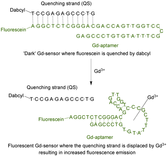
适体(钆适体)是与通过指数富集(SELEX)9的配体的系统进化的过程中分离出的碱基序列特异性的44碱基长的单链DNA分子。适应适体为荧光传感器,荧光团被连接到线料,然后将其与经由13互补碱基( 图1)淬火链(QS)杂交的5'末端。了QS标记有在3'末端的暗淬灭剂分子。在不存在的Gd 3+,所述传感器(钆传感器)的,由1:分别钆适体和QS的2摩尔比,将具有最小的荧光发射是由于吨从荧光基团淬灭Ø能量转移。在加入含水的Gd 3+的将从钆适体置换的QS,从而增加荧光发射。

图 1. 传感器(GD-传感器),由标有荧光素(荧光基团),并标有DABCYL 13个碱基长的淬链(QS)的44个碱基长的适体(GD-适体)(暗淬灭剂)的。在不存在未螯合的Gd 3+的,传感器的荧光是最小的。与另外的Gd 3+的,发生的QS位移和增加的荧光发射是观察。 请点击此处查看该图的放大版本。
有目前,人们常用的基于分光法检测ING水的Gd 3+。该测定使用了分子二甲酚橙,其经历在其最大吸收波长从433至573纳米时螯合离子10的换档。这两个最大吸收的比率可以用来量化未螯合的Gd 3+的量。适体传感器是一种替代(也可以是互补的),以在二甲酚橙法,因为这两种方法具有不同的反应条件(如pH值和所使用的缓冲溶液的组合物),目标的选择性,定量的线性范围,和检测方式9。
研究方案
注:分子生物学级水在所有的缓冲和解决方案的准备工作时使用。所有一次性管(离心及PCR)和枪头是DNase-和无RNA酶的。请咨询使用前的所有化学品的材料安全数据表(MSDS)。强烈推荐合适的个人防护装备(PPE)的使用。
1.适体母液的制备
- 购买2股多脱氧核苷酸的市场。通过高效液相色谱法(HPLC)命令与纯化两条链。
串1(GD-适体):
5' - / 56-FAM / AGGCTCTCGGGACGACCAGTTGGTCCCGCTTTATGTGTCCCGAG-3'
链2(QS):
5'-GTCCCGAGAGCCT / 3Dab / -3' - 溶解每条链在水中使钆适体和的QS100μM的个别储备溶液。
- 存放于-20°C这些解决方案。该溶液是稳定的迄今,为3年。
- 为了尽量减少FREEZ电子解冻周期,存储解决方案,股价在10微升等分。
2. 2倍的Gd-传感器溶液的制备
- 制备测定缓冲液(20mM HEPES,2mM的MgCl 2的,150毫摩尔NaCl,5mM的KCl)中。调整用NaOH和HCl pH值至7.4〜,并通过一次性无菌瓶顶过滤器具有0.2微米PES膜过滤器。存放在无菌瓶中。如果过滤,并妥善保存(在常温下),缓冲稳定到目前为止,2年。
- 稀释钆适体原液(从步骤1.2)在测定缓冲液497微升和QS原液(从步骤1.2)的2微升1微升。拌匀,用旋涡。其在2×钆传感器溶液浓度是200和400nm,分别。
注意:在此步骤中制备的2倍的Gd-传感器溶液的体积为500微升,这是足以测试6 - 7不同浓度的Gd 3+的校准曲线和造影剂溶液(步骤3)。每个样品将在384孔板给复孔。- 根据需要被测试的Gd 3+解的数目调整2倍的Gd-传感器溶液的体积。
- 转移2×钆传感器溶液到9的PCR管,用50微升到每个管中。放置管在热循环仪。
- 在热循环仪设置程序以加热在管至95℃的溶液中,保持5分钟,然后经〜15分钟缓慢地冷却该溶液至25℃(以速率〜0.05 - 0.1℃/ S)。加热和冷却循环是确保钆适体和QS之间最佳杂交。部分杂交结果不完全淬火和传感器的一个更高的背景荧光。如果热循环仪是不可用,进行利用热水浴代替这个过程。
- 一旦冷却到25℃,立即使用该溶液,或保持在热循环仪(最高达约2小时),直到准备好是用过的。当水浴用于加热,离开该管在浴中当水慢慢冷却到室温。
3.构建荧光校准曲线和钆造影剂的溶液检测未螯合GD的存在3+
- 溶解GdCl 3在测定缓冲液固体(相同的缓冲液如在步骤2.1)。
- 通过连续稀释,在最终所需浓度的校准曲线(2×溶液)的双制备各6个不同的Gd 3+在微量离心管溶液100微升。
- 例如,为了构建一个校准为0,50,100,200,400(仅无GdCl 3缓冲液),和钆3+为800nm,制备含有0,100,200,400,800,和1600纳米的解决方案的离子。确保始终包括"空白"0纳米的Gd 3+作为阴性对照。
- 溶解造影剂是测试编在测定缓冲液中。准备2个或3个不同的浓度通过连续稀释的造影剂的解决方案。
注:测试3种不同浓度造影剂溶液的建议。这是为了确保这些浓度的线性范围内。如果测试没有显示线性关系的样品中,减少造影剂的浓度使用。 - 服用含有从步骤2.5 2倍的Gd-传感器解决方案出来的热循环仪的PCR管。
- 添加来自步骤3.2的每个的Gd 3+溶液的50微升到含有2倍的Gd-传感器溶液9 PCR管6。吹打向上和向下混合。每个PCR管现在包含钆3+的所需浓度进行测试,100nM的钆适体,和200nM的QS。
- 到含有2倍的Gd-传感器溶液剩余的PCR管,从步骤3.3添加造影剂溶液的50微升。通过上下吹打几次拌匀。
- 孵育在Solutions在PCR管在室温下约5分钟。它们可以被放置长达30分钟。
- 传送每个管的45微升到384孔板中。每个PCR管会给重复孔中。
- 记录的每个孔中的荧光在平板阅读器。在GD-传感器设计中使用的荧光团(FAM),分别有495和520 nm的激发和发射最大值,对供应商的网站上所列出。选择取决于在板阅读器是否monochromator-或基于过滤器的合适的激发和发射波长或过滤器。
- 绘制荧光任意荧光单位(AFU)对钆3+的浓度曲线图。
- 画出图作为对抗的Gd 3+的荧光浓度倍数变化。由"空白"解决方案的AFU(0纳米的Gd 3+)将每个浓度的AFU计算荧光倍数变化。荧光倍数变化将使n个结果ormalization,应AFU显示一些周期性的(不同天等 )的变化。
- 比较含有造影剂的溶液和"空白",它是含0纳米GdCl 3(仅缓冲)该溶液的荧光发射。
注:GBCA溶液的更高的荧光意味着未螯合的Gd 3+的存在,这可能需要造影剂的进一步纯化。未螯合的Gd 3+的存在量可以利用在步骤3.10或3.11构建的校准曲线来估算。
结果
在未螯合的Gd 3+的存在下的Gd-传感器溶液的典型荧光变化示于图2。发射可被绘制为荧光倍数变化( 图2A)或在原始荧光读数( 图2B)以任意单位(AFU)。两条曲线在> 3μM收率低于1μM和饱和信号的线性范围为钆浓度3+非常相似的校准曲线。检测限为〜100纳米带3的信噪比。
...
讨论
使用基于适体的Gd-传感器,荧光发射的增加正比于观察未螯合的Gd 3+的浓度。为了最大限度地减少样品的使用量,该测定可以在384孔微量板以每孔45微升总样品体积运行。在此设计中,荧光素(FAM)和DABCYL(DAB)的选择主要基于试剂的成本;修改发射波长的荧光团不同的配对和猝灭剂,可以使用11。
要注意的是,以获得与该传感器最好的结果是很重要的,过程的...
披露声明
The authors declare that they have no competing financial interests.
致谢
We would like to gratefully acknowledge Dr. Milan N Stojanovic from Columbia University, New York, NY for valuable scientific input. This work is supported by funding from the California State University East Bay (CSUEB) and the CSUEB Faculty Support Grant-Individual Researcher. O.E., T.C., and A.L. were supported by the CSUEB Center for Student Research (CSR) Fellowship.
材料
| Name | Company | Catalog Number | Comments |
| Gd-aptamer | IDTDNA | Input sequence and fluorophore modification in the order form | A fluorophore with a different emission wavelength may be used. The aptamer may also be ordered from another company. |
| Quenching strand | IDTDNA | Input sequence and quencher modification in the order form | A different quencher for optimal energy transfer from the fluorophore may be used. The aptamer may also be ordered from another company. |
| Molecular biology grade water | No specific manufacturer, both DEPC or non-DEPC treated work equally well | ||
| Gadolinium(III) chloride anhydrous | Strem | 936416 | Toxic |
| HEPES | Fisher Scientific | BP310-500 | |
| Magnesium chloride anhydrous | MP Biomedicals | 0520984480 - 100 g | |
| Sodium Chloride | Acros Organics | 327300025 | |
| Potassium chloride | Fisher Scientific | P333-500 | |
| Sodium hydroxide, pellets | Fisher Scientific | BP359 | Corrosive |
| Hydrochloric acid | Fisher Scientific | SA49 | Toxic and corrosive |
| 384-well low flange black flat bottom polystyrene NBS plates | Corning | 3575 | Plates which are suitable for fluorescence reading are required. |
| Nalgene Rapid-Flow sterile disposable bottle top filter | Thermo Scientific | 5680020 | The bottle top is fitted with 0.2 micron PES membrane |
| Disposable sterile bottles 250 mL | Corning | 430281 | A larger or smaller bottle may be used |
| 1.5 mL microcentrifuge tubes | No specific manufacturer, as long as they are DNAse and RNAse-free | ||
| 0.2 mL PCR tubes | No specific manufacturer, as long as they are DNAse and RNAse-free | ||
| Micropipets | No specific manufacturer | ||
| Pipet tips (non filter) of appropriate sizes | No specific manufacturer, as long as they are DNAse and RNAse-free | ||
| Equipment | |||
| Plate reader | Biotek Synergy H1 | Plate readers from other manufacturers would work equally well |
参考文献
- Shen, C., New, E. J. Promising strategies for Gd-based responsive magnetic resonance imaging contrast agents. Curr. Opin. Chem. Biol. 17 (2), 158-166 (2013).
- Cheong, B. Y. C., Muthupillai, R. Nephrogenic systemic fibrosis: a concise review for cardiologists. Tex. Heart Inst. J. 37 (5), 508-515 (2010).
- Hao, D., Ai, T., Goerner, F., Hu, X., Runge, V. M., Tweedle, M. MRI contrast agents: basic chemistry and safety. J Magn. Reson. Imaging. 36 (5), 1060-1071 (2012).
- Telgmann, L., Sperling, M., Karst, U. Determination of gadolinium-based MRI contrast agents in biological and environmental samples: a review. Anal. Chim. Acta. 764, 1-16 (2013).
- Frenzel, T., Lengsfeld, P., Schirmer, H., Hütter, J., Weinmann, H. -. J. Stability of gadolinium-based magnetic resonance imaging contrast agents in human serum at 37 degrees C. Invest. Radiol. 43 (12), 817-828 (2008).
- Loreti, V., Bettmer, J. Determination of the MRI contrast agent Gd-DTPA by SEC-ICP-MS. Anal. Bioanal. Chem. 379 (7), 1050-1054 (2004).
- Telgmann, L., et al. Speciation and isotope dilution analysis of gadolinium-based contrast agents in wastewater. Environ. Sci. Technol. 46 (21), 11929-11936 (2012).
- Cleveland, D., et al. Chromatographic methods for the quantification of free and chelated gadolinium species in MRI contrast agent formulations. Anal. Bioanal. Chem. 398 (7), 2987-2995 (2010).
- Edogun, O., Nguyen, N. H., Halim, M. Fluorescent single-stranded DNA-based assay for detecting unchelated gadolinium(III) ions in aqueous solution. Anal. Bioanal. Chem. 408 (15), 4121-4131 (2016).
- Barge, A., Cravotto, G., Gianolio, E., Fedeli, F. How to determine free Gd and free ligand in solution of Gd chelates. A technical note. Contrast Med. Mol. Imaging. 1 (5), 184-188 (2006).
- Johansson, M. K. Choosing reporter-quencher pairs for efficient quenching through formation of intramolecular dimers. Methods Mol. Biol. 335, 17-29 (2006).
- Sherry, A. D., Caravan, P., Lenkinski, R. E. A primer on gadolinium chemistry. J. Magn. Reson. Imaging. 30 (6), 1240-1248 (2009).
- Shakhverdov, T. A. A cross-relaxation mechanism of fluorescence quenching in complexes of lanthanide ions with organic ligands. Opt. Spectrosc. 95 (4), 571-580 (2003).
- Brittain, H. G. Submicrogram determination of lanthanides through quenching of calcein blue fluorescence. Anal. Chem. 59 (8), 1122-1125 (1987).
转载和许可
请求许可使用此 JoVE 文章的文本或图形
请求许可探索更多文章
This article has been published
Video Coming Soon
版权所属 © 2025 MyJoVE 公司版权所有,本公司不涉及任何医疗业务和医疗服务。