需要订阅 JoVE 才能查看此. 登录或开始免费试用。
Method Article
黑质神经黑色素敏感磁共振成像的标准化数据采集
摘要
该协议显示了如何获取黑质的神经黑色素敏感磁共振成像数据。
摘要
多巴胺能系统在健康的认知(例如,奖励学习和不确定性)和神经精神疾病(例如,帕金森病和精神分裂症)中起着至关重要的作用。神经黑色素是多巴胺合成的副产物,积聚在黑质的多巴胺能神经元中。神经黑色素敏感磁共振成像(NM-MRI)是一种测量这些多巴胺能神经元中神经黑色素的非侵入性方法,可直接测量黑质中多巴胺能细胞丢失和多巴胺功能的代理测量。尽管NM-MRI已被证明可用于研究各种神经精神疾病,但它受到下-上方向的有限视野的挑战,导致意外排除部分黑质可能导致数据丢失。此外,该领域缺乏用于获取NM-MRI数据的标准化协议,这是促进大规模多站点研究和临床转化的关键步骤。该协议描述了一步一步的NM-MRI体积放置程序和在线质量控制检查,以确保获得覆盖整个黑质的高质量数据。
引言
神经黑色素 (NM) 是一种深色色素,存在于黑质 (SN) 和腔规则位点 (LC) 的去甲肾上腺素能神经元中1,2。NM由胞质多巴胺和去甲肾上腺素的铁依赖性氧化合成,并储存在体细胞3的自噬液泡中。它首先出现在 2-3 岁左右的人类中,并在1,4,5 岁时累积。
在SN和LC神经元的含NM的液泡内,NM与铁形成复合物。这些NM-铁配合物是顺磁性的,允许使用磁共振成像(MRI)对NM进行无创可视化6,7。可以可视化 NM 的 MRI 扫描称为 NM 敏感 MRI (NM-MRI),并使用直接或间接磁化转移效应来提供高 NM 浓度区域(例如 SN)与周围白质之间的对比度8,9。
磁化转移对比是大分子结合的水质子(被磁化转移脉冲饱和)与周围自由水质子之间相互作用的结果。在NM-MRI中,据信NM-铁配合物的顺磁性缩短了周围自由水质子的T1 ,从而减少了磁化转移效应,因此NM浓度较高的区域在NM-MRI扫描中出现高强度10。相反,SN周围的白质具有高大分子含量,导致较大的磁化转移效应,因此这些区域在NM-MRI扫描上出现低信号,从而在SN和周围白质之间提供高对比度。
在SN中,NM-MRI可以提供多巴胺能细胞丢失的标志物11和多巴胺系统功能12。这两个过程与几种神经精神疾病有关,并得到了大量临床和临床前工作的支持。例如,在精神分裂症中广泛观察到多巴胺功能异常;使用正电子发射断层扫描(PET)的体内研究表明纹状体多巴胺释放增加13,14,15,16和多巴胺合成能力增加17,18,19,20,21,22.此外,尸检研究表明,精神分裂症患者的基底神经节23 和 SN24,25 中的酪氨酸羟化酶(参与多巴胺合成的限速酶)水平升高。
一些研究调查了多巴胺能细胞丢失的模式,特别是在帕金森病中。验尸研究表明,SN的色素多巴胺能神经元是帕金森病神经变性的主要部位26,27,虽然帕金森病中的SN细胞丢失与正常衰老中的细胞损失无关28,但它与疾病的持续时间相关29.与大多数研究多巴胺能系统的方法不同,NM-MRI的非侵入性,成本效益和无电离辐射使NM-MRI成为多功能生物标志物30。
本文中描述的NM-MRI方案旨在提高NM-MRI的受试者内和受试者间可重复性。该协议可确保SN的完全覆盖,尽管NM-MRI扫描在下-上方向的覆盖范围有限。该协议使用矢状面、冠状面和轴向三维 (3D) T1 加权 (T1w) 图像,应遵循这些步骤以实现正确的切片堆叠放置。本文中概述的方案已在多项研究中使用31,32,并经过了广泛的测试。Wengler等人完成了一项关于该协议可靠性的研究,其中每个参与者在多天内获取两次NM-MRI图像32。类内相关系数表明,该方法对于基于感兴趣区域(ROI)和体素分析的测试重测可靠性以及图像中的高对比度表现出色。
Access restricted. Please log in or start a trial to view this content.
研究方案
注意:为制定此协议而进行的研究是根据纽约州精神病学研究所机构审查委员会指南(IRB #7655)进行的。扫描一名受试者以记录协议视频,并获得书面知情同意。有关此协议中使用的MRI扫描仪的详细信息,请参阅 材料表 。
1. 磁共振成像采集参数
- 准备使用具有以下参数的3D磁化制备的快速采集梯度回波(MPRAGE)序列获取高分辨率T1w图像:空间分辨率= 0.8 x 0.8 x 0.8 mm3;视场 (FOV) = 176 x 240 x 240 mm3;回波时间 (TE) = 3.43 毫秒;重复时间 (TR) = 2462 毫秒;反演时间 (TI) = 1060 ms;翻转角度 = 8°;面内平行成像因子 (ARC) = 2;平面平行成像因子 (ARC) = 233;带宽 = 208 Hz/像素;总采集时间 = 6 分 39 秒。
- 准备使用具有磁化转移对比度(2D GRE-MTC)的二维(2D)梯度召回回波序列(2D GRE-MTC)获取NM-MRI图像,参数如下:分辨率= 0.43 x 0.43 mm2;视场 = 220 x 220 毫米2;切片厚度 = 1.5 毫米;20片;切片间隙 = 0 毫米;TE = 4.8 毫秒;TR = 500 毫秒;翻转角度 = 40°;带宽 = 122 Hz/像素;MT 频率偏移 = 1.2 kHz;MT脉冲持续时间= 8毫秒;MT翻转角度= 670°;平均值数 = 5;总采集时间 = 10 分钟 4 秒。
注意:尽管显示的结果使用这些MRI采集参数,但该协议对各种T1w和NM-MRI成像协议有效。NM-MRI 方案应在下-上方向覆盖 ~25 mm,以保证完全覆盖 SN。
2. 放置 NM-MRI 体积
- 获取高分辨率 T1w 图像(≤1 mm 各向同性体素大小)。图像采集后直接使用在线重新格式化,以创建与前连合-后连合 (AC-PC) 线和中线对齐的高分辨率 T1w 图像。
- 使用供应商提供的软件执行在线重新格式化(例如,如果在 GE 扫描仪上获取数据:规划中的多平面重建 (MPR);如果在西门子扫描仪上获取数据:3D 任务卡中的 MPR;如果在飞利浦扫描仪上获取数据:VolumeView 软件包的渲染模式下的 MPR)。
- 在垂直于 AC-PC 线的轴向平面中创建 3D T1w 图像的多平面重建,以最小的切片间隙覆盖整个大脑。
- 在垂直于 AC-PC 线的冠状平面中创建 3D T1w 图像的多平面重建,以最小的切片间隙覆盖整个大脑。
- 在平行于 AC-PC 线的矢状面中创建 3D T1w 图像的多平面重建,以最小的切片间隙覆盖整个大脑。
- 使用供应商提供的软件执行在线重新格式化(例如,如果在 GE 扫描仪上获取数据:规划中的多平面重建 (MPR);如果在西门子扫描仪上获取数据:3D 任务卡中的 MPR;如果在飞利浦扫描仪上获取数据:VolumeView 软件包的渲染模式下的 MPR)。
- 加载重新格式化的高分辨率 T1w 图像的矢状面、冠状面和轴向视图,并确保存在描述每个显示切片位置的参考线。
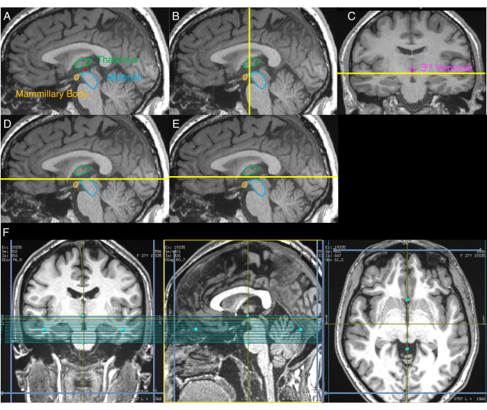
- 识别显示中脑和丘脑之间最大分离的矢状图像(图1A)。为此,目视检查重新格式化的T1w图像的矢状切片,直到确定显示最大分离的切片。
- 使用步骤2.3结束时的矢状面,目视识别描绘中脑最前侧的冠状平面(图1B)。
- 使用步骤2.4结束时的冠状图像,直观地识别描绘第三心室下侧的轴向平面(图1C)。
- 在步骤2.3结束时的矢状图像上,将NM-MRI体积的上边界与步骤2.5中确定的轴向平面对齐(图1D)。
- 将NM-MRI体积的上边界沿上方向移动3 mm(图1E)。
- 将NM-MRI体积与轴向和冠状图像中的中线对齐(图1F)。
- 获取NM-MRI图像。

图 1: 显示分步 NM-MRI 体积放置程序的图像。黄线表示用于卷放置的切片的位置,如协议中所述。(A)首先,确定中脑和丘脑之间分离最大的矢状图像(协议的步骤2.3)。(B)其次,使用 来自A的图像,识别描绘中脑最前侧的冠状平面(步骤2.4)。(C)第三,在B中识别的平面的冠状图像上 , 识别描绘第三脑室下侧的轴向平面(步骤2.5)。(D)第四,在 C 中识别的轴向平面 显示在A的 矢状图像上(步骤2.6)。(E)第五,轴向平面从 D 向上方向移动3 mm,该平面表示NM-MRI体积的上边界(步骤2.7)。(F)最终的NM-MRI体积放置,其中冠状图像对应于 C,矢状图像对应于 A,轴向图像对应于 E中的轴向平面。NM-MRI体积与冠状和轴向图像中的大脑中线以及矢状图像中的AC-PC线对齐(步骤2.8)。该数字的一部分已从 30年开始经爱思唯尔许可转载。缩写:NM-MRI = 神经黑色素敏感磁共振成像;AC-PC = 前连合-后连合。 请点击此处查看此图的大图。
3. 质量控制检查
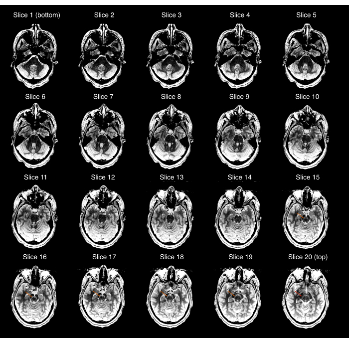
- 确保采集的NM-MRI图像覆盖整个SN,并且SN在中心图像中可见,但在NM-MRI体积的最优越或最差的图像中不可见。否则(图2),重复步骤2.3-2.9以确保正确的NM-MRI体积放置。如果参与者自获得高分辨率T1w扫描以来有显着移动,请重复步骤2.1-2.9。

图 2: 未通过第一次质量控制检查(协议的步骤 3.1)的 NM-MRI 采集示例。从最差(左上图)到最上级(右下图)显示的20个NM-MRI切片中的每一个;图像窗口/水平被设置为夸大黑质和脑碎之间的对比度。切片 15-19 中的橙色箭头显示了这些切片中黑质的位置。最高级切片(切片 20)中的红色箭头表示黑质在该切片中仍然可见,因此,采集未通过质量检查。缩写:NM-MRI = 神经黑色素敏感磁共振成像。 请点击此处查看此图的大图。
- 通过目视检查获取的NM-MRI扫描的每个切片来检查伪影,特别是通过SN和周围白质的伪影。
- 寻找信号强度的突然变化,其线性模式不尊重正常的解剖边界。例如,这可能显示为一个低强度区域,两侧有两个高强度区域。
- 如果伪影是血管的结果(图3A),请保留NM-MRI图像,因为这些伪影很可能始终存在。
- 如果伪影是参与者头部运动的结果(图3B),提醒参与者尽可能保持静止,并根据步骤3.2.5重新获取NM-MRI图像。
- 如果伪影不明确(图3C),请根据步骤3.2.5重新获取NM-MRI图像。重新采集后,如果伪影仍然存在,请继续处理这些图像,因为它们可能是生物学的,而不是采集问题的结果。
- 如果NM-MRI图像通过了步骤3.1中的质量控制检查,则复制先前的NM-MRI体积放置。如果NM-MRI图像未通过步骤3.1中的质量控制检查,请重复步骤2.3-2.9以确保正确的NM-MRI体积放置(如果参与者显着移动,则重复步骤2.1-2.9)。

图 3:未通过第二次质量控制检查(协议的步骤 3.2)的 NM-MRI 采集示例。每种情况仅显示一个代表性切片。(A) 由于由蓝色箭头识别的血管造成的血管伪影(红色箭头)而导致质量控制检查失败的 NM-MRI 采集。(B) 由于运动伪影(红色箭头)而未能通过质量控制检查的 NM-MRI 采集。(C) 由于模糊的伪影(红色箭头)而未能通过质量控制检查的 NM-MRI 采集。缩写:NM-MRI = 神经黑色素敏感磁共振成像。请点击此处查看此图的大图。
Access restricted. Please log in or start a trial to view this content.
结果
图4 显示了一名没有精神或神经系统疾病的28岁女性参与者的代表性结果。NM-MRI方案通过遵循 图1中概述的方案步骤2确保SN的完全覆盖,并通过遵循协议的步骤3确保NM-MRI图像令人满意。可以看到SN与NM浓度可忽略不计的相邻白质区域(即脑壳)之间的出色对比度。采集后立即检查这些图像,以确保SN的适当覆盖并检查伪影。由于SN的完全覆盖没有任何伪影?...
Access restricted. Please log in or start a trial to view this content.
讨论
多巴胺能系统在健康的认知和神经精神疾病中起着至关重要的作用。开发可用于重复研究 体内多 巴胺能系统的无创方法对于开发具有临床意义的生物标志物至关重要。此处描述的协议提供了获取SN的高质量NM-MRI图像的分步说明,包括NM-MRI体积的放置和质量控制检查,以确保可用数据。
尽管在其他地方已经讨论了用于分析NM-MRI数据的详细协议,但为了完整起见,我们简?...
Access restricted. Please log in or start a trial to view this content.
披露声明
Horga和Wengler博士分别报告拥有分析和使用神经黑色素成像在中枢神经系统疾病中的专利(WO2021034770A1,WO2020077098A1),授权给Terran Biosciences,但没有收到特许权使用费。
致谢
Horga博士得到了NIMH的支持(R01-MH114965,R01-MH117323)。Wengler博士得到了NIMH的支持(F32-MH125540)。
Access restricted. Please log in or start a trial to view this content.
材料
| Name | Company | Catalog Number | Comments |
| 3T Magnetic Resonance Imaging | General Electric | GE SIGNA Premier with 48-channel head coil |
参考文献
- Zecca, L., et al. New melanic pigments in the human brain that accumulate in aging and block environmental toxic metals. Proceedings of the National Academy of Sciences of the United States of America. 105 (45), 17567-17572 (2008).
- Zucca, F. A., et al. The neuromelanin of human substantia nigra: physiological and pathogenic aspects. Pigment Cell Research. 17 (6), 610-617 (2004).
- Sulzer, D., et al. Neuromelanin biosynthesis is driven by excess cytosolic catecholamines not accumulated by synaptic vesicles. Proceedings of the National Academy of Sciences of the United States of America. 97 (22), 11869-11874 (2000).
- Cowen, D. The melanoneurons of the human cerebellum (nucleus pigmentosus cerebellaris) and homologues in the monkey. Journal of Neuropathology & Experimental Neurology. 45 (3), 205-221 (1986).
- Zecca, L., et al. The absolute concentration of nigral neuromelanin, assayed by a new sensitive method, increases throughout the life and is dramatically decreased in Parkinson's disease. FEBS Letters. 510 (3), 216-220 (2002).
- Sulzer, D., et al. Neuromelanin detection by magnetic resonance imaging (MRI) and its promise as a biomarker for Parkinson's disease. NPJ Parkinson's Disease. 4 (1), 11(2018).
- Zucca, F. A., et al. Neuromelanin organelles are specialized autolysosomes that accumulate undegraded proteins and lipids in aging human brain and are likely involved in Parkinson's disease. NPJ Parkinson's Disease. 4 (1), 17(2018).
- Chen, X., et al. Simultaneous imaging of locus coeruleus and substantia nigra with a quantitative neuromelanin MRI approach. Magnetic Resonance Imaging. 32 (10), 1301-1306 (2014).
- Sasaki, M., et al. Neuromelanin magnetic resonance imaging of locus ceruleus and substantia nigra in Parkinson's disease. Neuroreport. 17 (11), 1215-1218 (2006).
- Trujillo, P., et al. Contrast mechanisms associated with neuromelanin-MRI. Magnetic Resonance in Medicine. 78 (5), 1790-1800 (2017).
- Kitao, S., et al. Correlation between pathology and neuromelanin MR imaging in Parkinson's disease and dementia with Lewy bodies. Neuroradiology. 55 (8), 947-953 (2013).
- Cassidy, C. M., et al. Neuromelanin-sensitive MRI as a noninvasive proxy measure of dopamine function in the human brain. Proceedings of the National Academy of Sciences of the United States of America. 116 (11), 5108-5117 (2019).
- Abi-Dargham, A., et al. Increased striatal dopamine transmission in schizophrenia: confirmation in a second cohort. American Journal of Psychiatry. 155 (6), 761-767 (1998).
- Laruelle, M., et al. Single photon emission computerized tomography imaging of amphetamine-induced dopamine release in drug-free schizophrenic subjects. Proceedings of the National Academy of Sciences of the United States of America. 93 (17), 9235-9240 (1996).
- Breier, A., et al. Schizophrenia is associated with elevated amphetamine-induced synaptic dopamine concentrations: evidence from a novel positron emission tomography method. Proceedings of the National Academy of Sciences of the United States of America. 94 (6), 2569-2574 (1997).
- Abi-Dargham, A., et al. Increased baseline occupancy of D-2 receptors by dopamine in schizophrenia. Proceedings of the National Academy of Sciences of the United States of America. 97 (14), 8104-8109 (2000).
- Hietala, J., et al. Presynaptic dopamine function in striatum of neuroleptic-naive schizophrenic patients. Lancet. 346 (8983), 1130-1131 (1995).
- Lindström, L. H., et al. Increased dopamine synthesis rate in medial prefrontal cortex and striatum in schizophrenia indicated by L-(β-11C) DOPA and PET. Biological Psychiatry. 46 (5), 681-688 (1999).
- Meyer-Lindenberg, A., et al. Reduced prefrontal activity predicts exaggerated striatal dopaminergic function in schizophrenia. Nature Neuroscience. 5 (3), 267-271 (2002).
- McGowan, S., Lawrence, A. D., Sales, T., Quested, D., Grasby, P. Presynaptic dopaminergic dysfunction in schizophrenia: a positron emission tomographic [18F] fluorodopa study. Archives of General Psychiatry. 61 (2), 134-142 (2004).
- Bose, S. K., et al. Classification of schizophrenic patients and healthy controls using [18F] fluorodopa PET imaging. Schizophrenia Research. 106 (2-3), 148-155 (2008).
- Kegeles, L. S., et al. Increased synaptic dopamine function in associative regions of the striatum in schizophrenia. Archives of General Psychiatry. 67 (3), 231-239 (2010).
- Toru, M., et al. Neurotransmitters, receptors and neuropeptides in post-mortem brains of chronic schizophrenic patients. Acta Psychiatrica Scandinavica. 78 (2), 121-137 (1988).
- Perez-Costas, E., Melendez-Ferro, M., Rice, M. W., Conley, R. R., Roberts, R. C. Dopamine pathology in schizophrenia: analysis of total and phosphorylated tyrosine hydroxylase in the substantia nigra. Frontiers in Psychiatry. 3, 31(2012).
- Howes, O. D., et al. Midbrain dopamine function in schizophrenia and depression: a post-mortem and positron emission tomographic imaging study. Brain. 136 (11), 3242-3251 (2013).
- Bernheimer, H., Birkmayer, W., Hornykiewicz, O., Jellinger, K., Seitelberger, F. Brain dopamine and the syndromes of Parkinson and Huntington Clinical, morphological and neurochemical correlations. Journal of the Neurological Sciences. 20 (4), 415-455 (1973).
- Hirsch, E., Graybiel, A. M., Agid, Y. A. Melanized dopaminergic neurons are differentially susceptible to degeneration in Parkinson's disease. Nature. 334 (6180), 345(1988).
- Fearnley, J. M., Lees, A. J. Ageing and Parkinson's disease: substantia nigra regional selectivity. Brain. 114 (5), 2283-2301 (1991).
- Damier, P., Hirsch, E., Agid, Y., Graybiel, A. The substantia nigra of the human brain: II. Patterns of loss of dopamine-containing neurons in Parkinson's disease. Brain. 122 (8), 1437-1448 (1999).
- Horga, G., Wengler, K., Cassidy, C. M. Neuromelanin-sensitive magnetic resonance imaging as a proxy marker for catecholamine function in psychiatry. JAMA Psychiatry. 78 (7), 788-789 (2021).
- Wengler, K., et al. Cross-scanner harmonization of neuromelanin-sensitive MRI for multisite studies. Journal of Magnetic Resonance Imaging. , (2021).
- Wengler, K., He, X., Abi-Dargham, A., Horga, G. Reproducibility assessment of neuromelanin-sensitive magnetic resonance imaging protocols for region-of-interest and voxelwise analyses. NeuroImage. 208, 116457(2020).
- Griswold, M. A., et al. Generalized autocalibrating partially parallel acquisitions (GRAPPA). Magnetic Resonance in Medicine. 47 (6), 1202-1210 (2002).
- vander Pluijm, M., et al. Reliability and reproducibility of neuromelanin-sensitive imaging of the substantia nigra: a comparison of three different sequences. Journal of Magnetic Resonance Imaging. 53 (5), 712-721 (2020).
- Cassidy, C. M., et al. Evidence for dopamine abnormalities in the substantia nigra in cocaine addiction revealed by neuromelanin-sensitive MRI. American Journal of Psychiatry. 177 (11), 1038-1047 (2020).
- Wengler, K., et al. Association between neuromelanin-sensitive MRI signal and psychomotor slowing in late-life depression. Neuropsychopharmacology. 46, 1233-1239 (2020).
- Biondetti, E., et al. Spatiotemporal changes in substantia nigra neuromelanin content in Parkinson's disease. Brain. 143 (9), 2757-2770 (2020).
- Shibata, E., et al. Use of neuromelanin-sensitive MRI to distinguish schizophrenic and depressive patients and healthy individuals based on signal alterations in the substantia nigra and locus ceruleus. Biological Psychiatry. 64 (5), 401-406 (2008).
- Fabbri, M., et al. Substantia nigra neuromelanin as an imaging biomarker of disease progression in Parkinson's disease. Journal of Parkinson's Disease. 7 (3), 491-501 (2017).
- Matsuura, K., et al. Neuromelanin magnetic resonance imaging in Parkinson's disease and multiple system atrophy. European Neurology. 70 (1-2), 70-77 (2013).
- Watanabe, Y., et al. Neuromelanin magnetic resonance imaging reveals increased dopaminergic neuron activity in the substantia nigra of patients with schizophrenia. PLoS One. 9 (8), 104619(2014).
Access restricted. Please log in or start a trial to view this content.
转载和许可
请求许可使用此 JoVE 文章的文本或图形
请求许可探索更多文章
This article has been published
Video Coming Soon
版权所属 © 2025 MyJoVE 公司版权所有,本公司不涉及任何医疗业务和医疗服务。