Method Article
Inducción de lesiones corneales y alcalinas mediante una técnica de punzón y trefina en un modelo de ratón
En este artículo
Resumen
Este protocolo describe un método para inducir una lesión precisa y reproducible de la córnea y el álcali limbal en un modelo de ratón. El protocolo es ventajoso, ya que permite una lesión distribuida uniformemente en la córnea y el limbo del ratón altamente curvados.
Resumen
La córnea es fundamental para la visión, y la curación de la córnea después de un traumatismo es fundamental para mantener su transparencia y función. A través del estudio de modelos de lesiones corneales, los investigadores pretenden mejorar su comprensión de cómo se cura la córnea y desarrollar estrategias para prevenir y controlar las opacidades corneales. La lesión química es uno de los modelos de lesión más populares que se ha estudiado ampliamente en ratones. La mayoría de los investigadores anteriores han utilizado un papel plano empapado en hidróxido de sodio para inducir lesiones en la córnea. Sin embargo, la inducción de lesiones en la córnea y las extremidades con papel de filtro plano no es fiable, ya que la córnea del ratón es muy curvada. Aquí, presentamos un nuevo instrumento, un punzón de biopsia modificado, que permite a los investigadores crear una lesión alcalina bien circunscrita, localizada y distribuida uniformemente en la córnea y el limbo murinos. Este método de punzón y trépano permite a los investigadores inducir una quemadura química precisa y reproducible en toda la córnea murina y el limbo, al tiempo que deja otras estructuras, como los párpados, sin verse afectadas por el producto químico. Además, este estudio introduce una técnica de enucleación que preserva la carúncula medial como punto de referencia para identificar el lado nasal del globo. La conjuntiva bulbar y palpebral, y la glándula lagrimal también se mantienen intactas con esta técnica. Los exámenes oftalmológicos se realizaron mediante biomicroscopio con lámpara de hendidura y tinción con fluoresceína en los días 0, 1, 2, 6, 8 y 14 después de la lesión. Los hallazgos clínicos, histológicos e inmunohistoquímicos confirmaron la deficiencia de células madre limbales y el fracaso de la regeneración de la superficie ocular en todos los ratones experimentales. El modelo de lesión corneal alcalina presentado es ideal para estudiar la deficiencia de células madre limbales, la inflamación de la córnea y la fibrosis. Este método también es adecuado para investigar la eficacia preclínica y clínica de los medicamentos oftalmológicos tópicos en la superficie corneal murina.
Introducción
La córnea es fundamental para la visión y exhibe características únicas, incluida la transparencia, que es un requisito previo para una visión clara. Además de desempeñar un importante papel protector, la córnea representa 2/3 del poder refractivo del ojo1. Debido a su importante papel en la visión, las lesiones corneales y la opacidad causan un deterioro visual significativo y son responsables de la segunda causa de ceguera prevenible en todo el mundo 2,3. En las lesiones corneales con disfunción limbal grave, la función barrera del limbo disminuye, lo que resulta en la migración de las células conjuntivales hacia la superficie corneal y la conjuntivalización corneal 4,5, lo que compromete drásticamente la visión. Por lo tanto, se requieren estrategias preventivas y terapéuticas eficaces para hacer frente a la carga mundial de la ceguera corneal y la discapacidad relacionada.
La comprensión actual del proceso de cicatrización de heridas corneales humanas se basa en estudios previos que han investigado las respuestas corneales a diversas lesiones. Se han empleado varias técnicas y modelos animales para inducir diversas lesiones corneales químicas o mecánicas 6,7,8,9 y para investigar diversos aspectos del proceso de cicatrización de heridas corneales.
El modelo de quemaduras alcalinas es un modelo de lesión bien establecido que se realiza aplicando hidróxido de sodio (NaOH) sobre la superficie de la córnea directamente o utilizando papel de filtro plano10. Una lesión alcalina provoca la liberación de mediadores proinflamatorios y la infiltración de células polimorfonucleares no solo en la córnea y la cámara anterior del ojo, sino también en la retina. Esto induce la apoptosis no intencionada de las células ganglionares de la retina y la activación de las células CD45+ 11. Por lo tanto, es fundamental localizar el sitio de la lesión con precisión para evitar lesiones excesivas no intencionales utilizando un modelo de lesión alcalina.
La longitud axial del globo ocular murino es de aproximadamente 3 mm12. Debido a esta corta distancia entre la córnea y la retina, existe una curvatura corneal pronunciada que proporciona un alto poder de refracción para enfocar la luz en la retina (Figura 1A). Como informamos anteriormente13, es difícil inducir lesiones químicas en esta superficie altamente curvada utilizando un papel de filtro plano, particularmente en el limbo (Figura 1B). La inducción de lesiones en el limbo requiere inclinar el papel de filtro, lo que tiene el potencial de causar lesiones no intencionales en el fórnix y la conjuntiva adyacente14. Otro enfoque consiste en aplicar directamente el agente químico en forma de gotas sobre la superficie de la córnea. Sin embargo, este método carece de control sobre el tiempo de exposición y existe un riesgo potencial de inducir lesiones en la conjuntiva, el fórnix y los párpados debido a la difusión del líquido en estas áreas.
Para superar estas limitaciones, este estudio presenta un novedoso método de punzón y trépano para inducir lesiones. Esta técnica tiene varias ventajas, entre las que se incluyen (i) la inducción de una lesión química eficaz en toda la superficie de la córnea y el limbo en el modelo de ratón, (ii) la inducción de una lesión localizada y bien circunscrita en la córnea, (iii) la capacidad de aplicar cualquier líquido de interés durante un tiempo predeterminado, y (iv) la capacidad de inducir diferentes tamaños de lesiones corneales mediante la selección de punzones de biopsia adecuados. Este método también es factible para modelos de lesiones de ratas y conejos, que también exhiben una superficie corneal curvada y son modelos animales comunes utilizados para estudiar la cicatrización de heridas en la superficie ocular.
Protocolo
Todos los procedimientos se llevaron a cabo de acuerdo con el APLAC de Stanford para el cuidado de animales de laboratorio número 33420, uso de animales con fines científicos, y la Declaración ARVO para el uso de animales en la investigación oftálmica y de la visión. Un total de 10 ratones machos y hembras C57BL/6 de entre 8 y 12 semanas de edad fueron generosamente proporcionados por el laboratorio Irving L. Weissman. Los animales se aclimataron a un ciclo de luz-oscuridad de 12 h y se les proporcionó agua y alimento ad libitum. Se indujo una lesión en un ojo del animal.
1. Preparación para el experimento
- Preparación de materiales
NOTA: Todos los reactivos deben mantenerse a temperatura ambiente.- Preparar la punzón-trépana: Prepare una punción de biopsia con un diámetro de 3,5 mm y marque el eje de la punzón a 5 mm de distancia de su borde distal. Fije el punzón firmemente y corte la parte distal marcada de su eje con una herramienta rotativa de dos velocidades. Use protección ocular durante este proceso. Corta el eje a 3,5 mm de profundidad y deja el último 1,5 mm unido. Doble la punta 90°, como se muestra en la Figura 1.
- Prepare una solución de NaOH 0,5 M disolviendo 1 g de NaOH en 50 ml de agua destilada.
- Prepare 10 ml de cóctel anestésico combinando 2 ml de 100 mg/ml de ketamina y 1 ml de xilacina de 20 mg/ml. Antes de la inyección, diluya esta mezcla con 7 ml de NaCl al 0,9% (solución salina normal).
- Prepare una solución de fluoresceína sódica al 0,1 % añadiendo 0,1 ml de líquido fluorescente AK-Fluor al 10 % en 9,9 ml de solución salina estéril tamponada con fosfato (1x PBS).
- Prepare PBS (1x) disolviendo 8 g de NaCl, 0,2 g de KCl, 1,44 g de Na2HPO4 y 0,23 g de NaH2PO4en 900 ml de agua destilada. Ajuste el pH de la solución a 7,4. Lleve la solución a un volumen final de 1 L agregando agua destilada.
- Prepare una solución de paraformaldehído (PFA) al 4% para la fijación. Bajo una cubierta química, disuelva 2 g de PFA en 45 ml de PBS. Calentar la mezcla a 65 °C y ajustar su pH a 7,4. Una vez que el PFA se haya disuelto, lleve la solución a un volumen final de 50 ml.
2. Preparación de los animales
- Pesar el ratón para determinar el volumen apropiado de cóctel anestésico inyectable para la inyección. Después de manipular y sujetar adecuadamente al ratón como se describe en Machholz et al.15, administrar el cóctel anestésico por vía intraperitoneal (IP) a una dosis de 0,01 mL/g16,17. Apuntar a los cuadrantes abdominales inferiores para la inyección de IP para prevenir daños en los órganos.
- Inyecte buprenorfina suspensión inyectable de liberación prolongada (3,25 mg/kg) por vía subcutánea para obtener analgesia adicional durante y después de la cirugía, ya que la lesión por álcali corneal es extremadamente dolorosa.
- Espere hasta que se logre un plano profundo de anestesia. Verifique si hay una falta de respuesta al pellizco del dedo del pie como una indicación de un plano profundo exitoso de anestesia. Aplique un ungüento ocular simple para el ojo contralateral para evitar que se seque antes de comenzar la cirugía.
- Después de la anestesia, coloque el ratón sobre la mesa quirúrgica preparada de acuerdo con los principios estándar de la cirugía de roedores18. Coloque una almohada de 5 mm de altura debajo de la cabeza del roedor, colocada en decúbito lateral. La almohada ayuda a sostener la cabeza del roedor durante la cirugía.
- Administrar gotas oftálmicas de clorhidrato de tetracaína (0,5%) para una mayor anestesia. Seque adecuadamente la superficie ocular con una lanza quirúrgica y recorte las pestañas.
3. Inducción de lesiones alcalinas
- Antes de comenzar la cirugía, organice la mesa de cirugía según los principios estándar de cirugía con roedores18,19 y mantenga el campo quirúrgico estéril durante la cirugía. Coloque el microscopio quirúrgico para visualizar correctamente el ratón anestesiado y ajuste el temporizador a 30 s. Coloque una toalla de papel retorcida en el hocico del animal para evitar la aspiración nasal involuntaria durante el proceso de enjuague.
- Determine la circunferencia del área limbal con el microscopio quirúrgico mientras se asegura de que los párpados del ratón estén bien abiertos con los dedos pulgar e índice. Sostenga suavemente la trépano limpia paralela al eje del ojo, sin aplicar ninguna presión hacia abajo. Evite girar el instrumento y mantenga el eje del punzón-trépano paralelo al eje del globo.
- Pídale al asistente quirúrgico que deje caer 3 gotas de solución de NaOH (equivalente a 40 μL) en el orificio del punzón-trépano para llenar la herramienta y cubrir la superficie corneal de manera adecuada. La tensión superficial del líquido evita cualquier fuga fuera del instrumento (Figura 1).
NOTA: Utilizamos una jeringa de 1 mm con una aguja de 27G con una punta aplanada en un ángulo de 50° para dejar caer con cuidado la solución de NaOH. - Después de 30 s, enjuague inmediatamente la córnea y el fórnix con 5 ml de PBS (1x). El video 1 muestra el procedimiento para inducir lesiones químicas.
- Utilice un papel indicador de pH universal para garantizar un pH de 7 a 7,5 en la superficie corneal del ojo lesionado. A continuación, aplique la pomada oftálmica triple antibiótica sobre la superficie ocular químicamente lesionada.
NOTA: El movimiento de la cola durante la cirugía es un signo de baja profundidad anestésica. Aplicar un cóctel anestésico adicional (ketamina + xilacina) inyectando un anestésico [bolo IP (50% del volumen inicial)]. - Después de la cirugía, confirme que el ratón se encuentra en una condición estable. Administre la segunda buprenorfina si el animal siente dolor. Utilice la misma técnica de manipulación que en 2.2. Comience el examen postoperatorio mientras el animal está bajo anestesia.
- Después del procedimiento, lavar, secar y desinfectar el punzón-trépano y la mesa quirúrgica con etanol al 70%.
4. Evaluación clínica
- Para el examen, anestesiar al ratón como se describió anteriormente en el paso 2.1.
- Examinar los ojos (lesionados y no lesionados) bajo un biomicroscopio con lámpara de hendidura. Usar una cámara para capturar fotos (en este estudio, se utilizó la cámara de un teléfono en modo cinemático).
- Puntúe la opacidad corneal según el sistema de clasificación de Yoeruek20: 0 = córnea normal y clara; 1 = opacidad leve; 2 = mayor opacidad, pero el iris y la pupila son fácilmente distinguibles; 3 = el iris y la pupila apenas se distinguen; 4 = la córnea es completamente opaca con una pupila invisible.
- Aplicar gotas oftálmicas fluoresceínicas al 0,1%. Seque el exceso de líquido fluorescente con un aplicador de algodón y evalúe la presencia de defectos epiteliales corneales utilizando el filtro azul de cobalto. Captura fotos.
- Vigile al animal hasta que recupere la conciencia suficiente para mantener la decúbito esternal. No vuelva a introducir al animal a otros animales hasta que se haya recuperado por completo.
5. Enucleación
- Sacrificar ratones 2 semanas después de la inducción de la lesión química por luxación cervical en 3-5 s21.
- Enuclear el ojo conservando la carúncula medial y toda la conjuntiva palpebral en el siguiente orden, como se muestra en el Video 2.
- Bajo el microscopio quirúrgico, diseccione cuidadosamente la unión de la carúncula y la piel. Con unas pinzas dentales, retraiga la carúncula y guíe la punta de las tijeras quirúrgicas por debajo de la conjuntiva palpebral hacia su unión con la placa tarsal.
- Cortar la conjuntiva a lo largo de la línea de adherencia hacia el canto lateral. Luego, gire las tijeras quirúrgicas hacia la unión de la conjuntiva y la placa tarsiana inferior en el plano subconjuntival.
- Después de completar la disección conjuntival, retraiga los párpados superior e inferior del lado nasal con los dedos pulgar e índice. Mientras se retrae, guíe la punta de una pinza de punta curva detrás del lagrimal protuberante hacia el nervio óptico. Sujete firmemente el nervio óptico y extraiga el globo terráqueo (Figura 2).
- Enjuague el globo con PBS (1x) y transfiéralo a la solución de fijación.
6. Tinción de hematoxilina y eosina (H&E) y ácido peryódico-Schiff (PAS)
- Fije los globos en una solución de formalina al 10% durante la noche a temperatura ambiente.
- Deshidratar los tejidos utilizando una serie secuencial de alcohol graduado con concentraciones de 70% de etanol, 80% de etanol, 90% de etanol y 100% de etanol, cada una durante 10-15 min. Posteriormente, sumergir los ojos en xileno durante 10 min. Por último, envuelva los ojos en parafina.
- Seccione los bloques de parafina en rodajas de 6 μm de grosor y móntelos en portaobjetos de microscopio de vidrio para la tinción de hematoxilina y eosina (H&E) y ácido peryódico-Schiff (PAS), como se describe en el Archivo Suplementario 1.
7. Imágenes y análisis de inmunofluorescencia
- Para estudios de inmunohistoquímica (IHQ), fijar los ojos en 1 ml de PBS (1x) que contenga PFA al 4% a 4 °C durante la noche. Lave la muestra 3 veces en 1 ml de PBS (1x) durante 5 minutos cada una.
- Para evitar la formación de cristales de hielo y proteger las estructuras moleculares de las proteínas, realice la saturación de sacarosa en serie con concentraciones de sacarosa al 10%, 20% y 30% de sacarosa en PBS. Incrustar los globos en una solución de tomografía de coherencia óptica (OCT; Figura 2) y guárdelos a -80 °C.
- Divida el bloque de OCT en rodajas de 12 μm de grosor y móntelo en portaobjetos de microscopio de vidrio para la tinción IHQ.
- Permeabilizar secciones de tejido en portaobjetos de microscopio en Triton X −100 % al 0,1 % en PBS (1x) durante 15 min. A continuación, bloquee los antígenos inespecíficos con BSA al 5% en PBS (1x) durante 1 h adicional a temperatura ambiente.
- Incubar secciones de tejido en portaobjetos de microscopio a 4 °C durante la noche en el cóctel de anticuerpos primarios dirigidos preparados en PBS (1x) que contengan un 1% de BSA. En este experimento, los anticuerpos primarios fueron anticuerpos anti-queratina 13 (K13) y anti-queratina 12 (K12) de conejo diluidos 1:100 con concentraciones de 2,24 μg/mL y 1,56 μg/mL, respectivamente.
- Después de la incubación, lave las secciones de tejido en los portaobjetos del microscopio 3 veces con PBS (1x). Posteriormente, detectar anticuerpos primarios unidos con 1:500 IgG anti-conejo de burro diluida. Incubar con el anticuerpo secundario a 4 °C durante 1 h en la oscuridad. A continuación, lavar en PBS (1x) 3 veces durante 5 minutos cada una.
- Aplique una gota de medio de montaje de fluorescencia antidecoloración que contenga DAPI en las secciones de tejido del microscopio y cúbralas con un cubreobjetos. Deje que las muestras se sequen en la oscuridad y examínelas bajo un microscopio fluorescente con la misma configuración de láser para ojos normales y lesionados.
Resultados
La eficacia del método en la inducción de la deficiencia de células madre limbales (LSCD) se evaluó mediante la evaluación de los signos clínicos e histológicos de la LSCD. La evaluación clínica se realizó mediante microscopía con lámpara de hendidura y tomografía de coherencia óptica del segmento anterior (AS-OCT) (Figura 3 y Figura 4).
La reepitelización se produjo de forma centrípeta y fue más rápida en la parte temporal de la córnea en comparación con su parte nasal. Los ojos lesionados desarrollaron una neblina corneal 2+-3+ inmediatamente después de la lesión química (Figura 3). Las células epiteliales migraron de la conjuntiva a la superficie corneal después de una lesión limbal. El defecto epitelial corneal grande se reepitelizó completamente en los días 12-14, lo que tomó más tiempo en comparación con una lesión epitelial corneal de tamaño similar y membrana basal y estroma intactos, que generalmente se curaron dentro de los 5 días posteriores a la lesión 8,9. Debido a la LSCD, el 50% de los ojos lesionados desarrollaron defectos epiteliales persistentes al final de la segunda semana (Figura 3). El edema corneal fue más prominente durante los primeros días (Figura 3, Figura 4), mientras que la fibrosis corneal fue significativa en la segunda semana y resultó en una opacidad corneal 4+ en el 100% de los ojos lesionados.
Los primeros signos de neovascularización (NV) se observaron clínica e histológicamente, 24 h después de la inducción de la lesión química, como se ilustra en la Figura 5, en consonancia con la línea de tiempo de la NV identificada por el estudio de Kvanta et al. que mostró signos de NV limbal 24 h después de la lesión22. Durante el proceso de curación, los nuevos vasos maduraron y al día 14 después de la lesión, NV cruzó el limbo y llegó a la córnea central. El limbo, que define el límite entre la conjuntiva y la córnea, fue destruido.
La evidencia histológica de deficiencia de células madre limbales y conjuntivalización fue observada por la aparición de células caliciformes PAS+ y vasos sanguíneos estromales 23,24,25,26. Las células caliciformes se observaron en el presente modelo de lesión y se indicaron con la flecha de la Figura 6.
Los epitelios conjuntivales y corneales expresan principalmente queratinas únicas, K13 y K12, respectivamente27. Después de la lesión limbal, nuevas células epiteliales que se originaron en la conjuntiva cubrieron la córnea desnuda, y K12 no se expresó en la superficie corneal de ningún animal lesionado durante 2 semanas después de la lesión. Este hallazgo, consistente con otros estudios28, indicó LSCD completa y ausencia de células epiteliales corneales en la superficie corneal. Sin embargo, en el estudio de Park et al.29, detectaron la expresión de K12 20 y 32 semanas después de la lesión, lo que sugiere una posible transdiferenciación de las células epiteliales.
En consecuencia, observamos que la lesión química destruyó el limbo y las células madre limbales, lo que resultó en la migración de las células epiteliales conjuntivales al centro de la córnea para cubrir la superficie corneal desnuda. Esto se valida aún más por el marcador de células epiteliales conjuntivales, K13, que se expresó en toda la conjuntiva y las superficies corneales, como se muestra en la Figura 7.
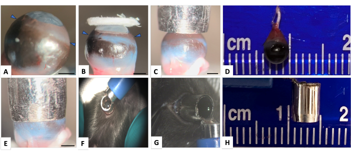
Figura 1: Ojo derecho normal de ratón y la trépano en puñetazo para inducir lesiones corneales y limbales. (A) Vista lateral que muestra el ojo de ratón con córnea muy curvada (las puntas de flecha indican el limbo). (B) La imagen demuestra que incluso un papel de filtro grande es insuficiente para cubrir adecuadamente el área limbal. El diámetro de limbo a limbo del ojo del ratón es de casi 4 mm y una biopsia por punción con un diámetro externo de 4,5 mm y un diámetro interno de 3,5 mm (paneles D y H), cubre adecuadamente la córnea y la superficie limbal como se muestra en los paneles (C) y (E). (F) La trépano punzón se sostiene apropiadamente sobre el globo alrededor del área limbal. (G) Para asegurarse de que no haya fugas a través del borde de la trépano punzón, después de colocar adecuadamente la trépano perforadora en un eje paralelo con el globo, el orificio se llena con azul de metileno. No se detecta ninguna fuga de azul de metileno. Barra de escala = 1 mm. Haga clic aquí para ver una versión más grande de esta figura.
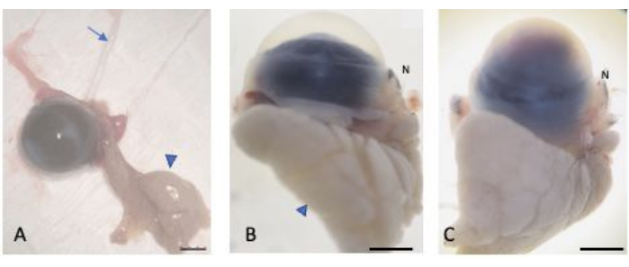
Figura 2: Ojos enucleados. (A) Los ojos fueron enucleados preservando la conjuntiva bulbar y palpebral, la glándula lagrimal (punta de flecha) y el nervio óptico (flecha). Los ojos normales (B) y lesionados (C) estaban saturados con sacarosa al 30% para protegerlos contra la formación de criocristales. La parte nasal del globo terráqueo es reconocible a través de la carúncula nasal (etiquetada con N). Barra de escala = 1 mm. Haga clic aquí para ver una versión más grande de esta figura.
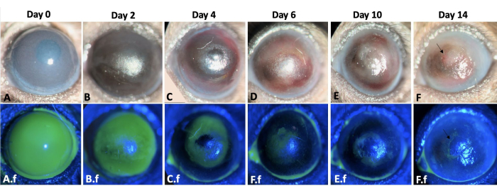
Figura 3: Cicatrización de heridas del ojo izquierdo. Aquí se muestra el proceso de cicatrización de la herida del ojo izquierdo del ratón a lo largo de 2 semanas después de la lesión corneal y alcalina limbal en un modelo de ratón (A-F). El examen del ojo con lámpara de hendidura. El edema corneal es más prominente en los días 0 y 2 (A,B), mientras que la fibrosis es más evidente durante la segunda semana después de la lesión (E-F). A.f-F.f muestran el proceso de reepitelización del mismo ojo. Se observa un defecto epitelial corneal y limbal total inmediatamente después de la inducción de la lesión en A.f. El defecto epitelial se curó por migración de células epiteliales conjuntivales en un patrón centrípeto a los 12-14 días (A.f-F.f). Sin embargo, el 50% de los ojos lesionados desarrollaron un defecto epitelial persistente al final de la segunda semana, como se muestra en las imágenes F y F.f. Barra de escala = 1 mm (panel C). Haga clic aquí para ver una versión más grande de esta figura.

Figura 4: OCT del segmento anterior del ojo del ratón. (A) La AS-OCT ilustra la curvatura normal de la córnea y la cámara anterior. La estructura del iris está bien definida y es reconocible. No se detecta adherencia iridocorneal en la periferia media del iris. (B) Inmediatamente después de la lesión, el grosor de la córnea aumenta debido a la formación de edema y se desarrolla adhesión iridocorneal en la periferia media del iris. (C) Dos semanas después de la lesión, la curvatura de la córnea ha cambiado y la adhesión iridocorneal total con destrucción de la cámara anterior es visible. Barra de escala = 1 mm. Haga clic aquí para ver una versión más grande de esta figura.
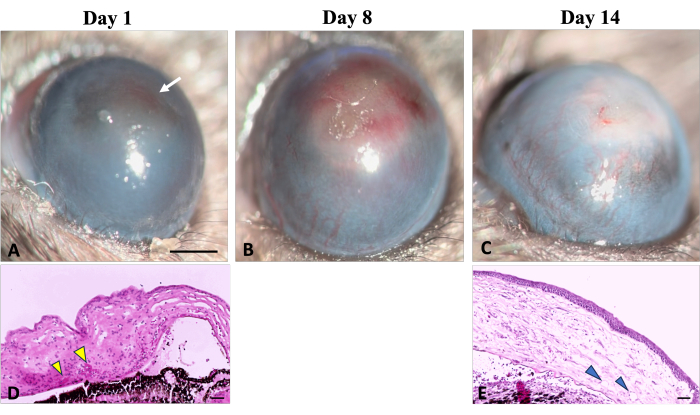
Figura 5: Neovascularización corneal. Se pueden observar signos clínicos e histológicos de neovascularización corneal durante el proceso de cicatrización de heridas después de una lesión por hidróxido de sodio (NaOH). (A) Los signos iniciales de neovascularización se hacen detectables el primer día después de la lesión, caracterizados por una coloración rojiza de la córnea (indicada por una flecha blanca). Esta decoloración es el resultado de la agregación de glóbulos rojos en el estroma, como se ilustra en la imagen histológica correspondiente (D) (indicada por puntas de flecha amarillas). (B) Durante la primera semana de regeneración, los nuevos vasos aumentan progresivamente y se extienden por toda la córnea. (C) Al final de las 2 semanas, el área limbal está destruida y los nuevos vasos continúan evolucionando. (E) La sección histológica de la córnea ilustra aún más la presencia de neovascularización estromal profunda (mostrada por puntas de flecha). Lámpara de hendidura Barra de escala de imagen = 1 mm, barra de escala de imagen histológica = 50 μm. Haga clic aquí para ver una versión más grande de esta figura.
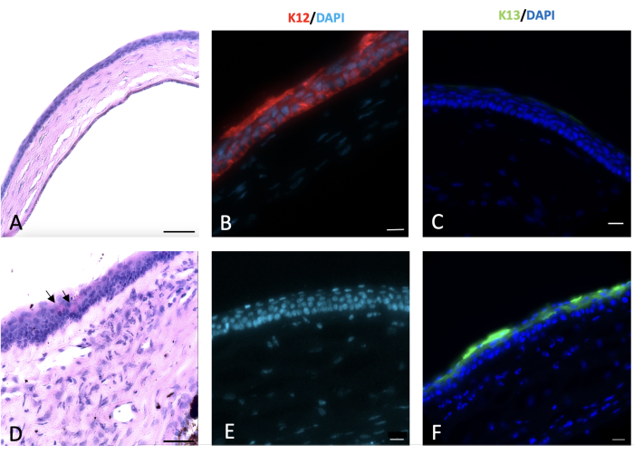
Figura 6: Tinción de córnea con ácido peryódico-Schiff e inmunohistoquímica (IHQ). La tinción inmunohistoquímica y de ácido peryódico de la córnea normal y lesionada se realizó 2 semanas después de la lesión. Epitelio corneal normal de ratón compuesto por 4-5 capas de células (A). La lesión alcalina de la córnea y el limbo condujo a la conjuntivalización de la córnea con la aparición de células caliciformes en la superficie corneal, como se muestra en las flechas negras en (D). Las células epiteliales corneales normales expresan K12 (B), que no es expresada por las células conjuntivales que cubren la córnea lesionada (E). La K13, un marcador característico de las células epiteliales conjuntivales, no se expresa en las células epiteliales corneales normales (C). Sin embargo, está presente en la superficie corneal lesionada por hidróxido de sodio (NaOH) que es un signo de conjuntivalización corneal (F). Barra de escala de imagen histológica = 50 μm, barra de escala de imagen teñida con IHQ = 20 μm. Haga clic aquí para ver una versión más grande de esta figura.
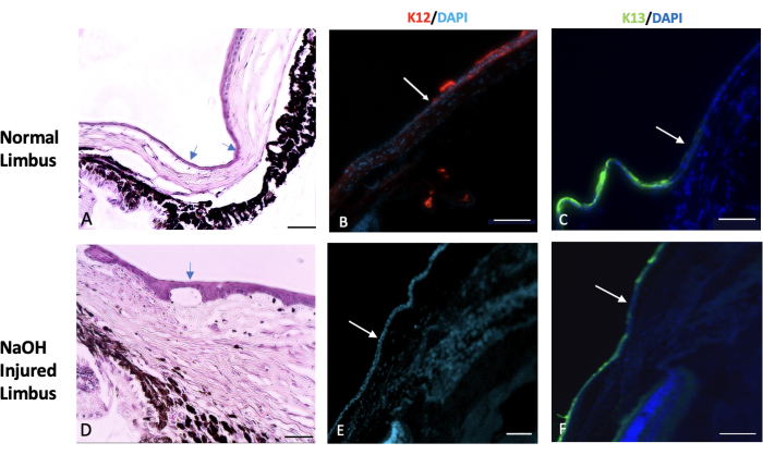
Figura 7: Hematoxilina y eosina y tinción inmunohistoquímica. Se realizó hematoxilina y eosina (H&E) y tinción inmunohistoquímica del tejido normal y lesionado del limbo. (A) El limbo normal marca el área de transición entre el final de la esclerótica y el comienzo de la córnea. Esta región suele estar cubierta por una o dos capas de células epiteliales conjuntivales (indicadas por las flechas). En un ojo sano, la expresión de un marcador epitelial corneal específico llamado K12 comienza en el limbo y se extiende a la superficie de la córnea (que se muestra en la imagen B). Por otro lado, la expresión de un marcador conjuntival conocido como K13 está restringida al limbo y no se extiende más allá de él (indicado por la flecha blanca en la imagen C). En los ojos lesionados por hidróxido de sodio (NaOH), los límites del limbo se alteran. Esto conduce a la migración de las células conjuntivales hacia la córnea lesionada. (D) Las imágenes del limbo lesionado por NaOH demuestran la presencia de neovascularización tanto debajo de la capa epitelial como dentro del tejido estromal. Después de la lesión, la superficie corneal lesionada carece de la presencia de K12 (E), mientras que K13 se expresa abundantemente en la superficie corneal (F). Barra de escala de imagen histológica = 50 μm, barra de escala de imagen teñida con IHQ = 100 μm. Haga clic aquí para ver una versión más grande de esta figura.
Archivo complementario 1: Protocolo de tinción. Haga clic aquí para descargar este archivo.
Video 1: Lesión corneal y limbal de NaOH en un modelo de ratón con una trépano punzante. El video muestra el procedimiento de inducción de lesiones corneales y limbales de NaOH en un modelo de ratón con una trépano de punzón. Es crucial sostener la trépano en un eje paralelo con el globo y aplicar una presión mínima en el limbo. Esta técnica adecuada es esencial para evitar fugas y lograr resultados óptimos. Haga clic aquí para descargar este video.
Vídeo 2: Ilustración de la técnica de enucleación preservando la conjuntiva bulbar. Para diferenciar el lado nasal del globo del lado temporal, la carúncula nasal se conserva junto con el globo. Se diseca toda la conjuntiva a partir de su unión con la placa tarsiana. Con una presión mínima, el contenido orbital sobresale hacia afuera. Al guiar las pinzas hacia la parte posterior del globo, se agarra el nervio óptico y se extrae el tejido. El tejido enucleado incluye el globo, la grasa orbitaria y la glándula lagrimal orbitaria. Haga clic aquí para descargar este video.
Discusión
Este estudio propone un dispositivo innovador, la trépano de punzón, que se puede utilizar para inducir con éxito una lesión corneal y limbal efectiva y reproducible en un modelo de ratón. Este modelo de deficiencia de células madre limbales es ideal para investigar la dinámica de la cicatrización de heridas corneales y la conjuntivalización después de una lesión.
La evidencia sugiere que tanto el nicho limbal como la parte central de la córnea murina contienen células madre30. Por lo tanto, se requiere una lesión corneal y limbal eficiente para producir un modelo de deficiencia de células madre, y el modelo de lesión presentado aquí permite la exposición del limbo corneal curvo a un agente químico durante un período específico. Para determinar la mejor concentración y duración de la lesión por NaOH, se infligieron lesiones con varias concentraciones y duraciones de NaOH. Las concentraciones más altas de NaOH o las exposiciones de mayor duración dieron lugar a un aumento del daño tisular y la fibrosis. Por lo tanto, los investigadores pueden ajustar estos parámetros en función de los objetivos específicos de su estudio y la gravedad deseada de la lesión.
Para reproducir con éxito este modelo de lesión corneal y limbal, se deben tener en cuenta varias consideraciones clave. En primer lugar, es imperativo medir el diámetro limbal a limbo del ojo objetivo para determinar el tamaño apropiado del punzón. Se recomienda seleccionar un punzón de biopsia con un diámetro externo que sea de 0,5 a 1 mm más grande que este diámetro.
La tensión superficial del líquido utilizado es un factor importante para evitar fugas en la interfaz entre la superficie ocular y el borde de la trépano del punzón, como se muestra en la Figura 1G. Por lo tanto, no es necesario aplicar presión en la punta de la biopsia por punción.
Para evitar causar daños mecánicos al tejido, es fundamental sostener la trépano del punzón en un eje paralelo con el ojo y abstenerse de aplicar presión en el limbo. Un ajuste incorrecto del eje de trépano del punzón puede aumentar el riesgo de fugas y dar lugar a un sitio descentrado de lesiones y resultados inexactos.
Algunas limitaciones potenciales de esta técnica incluyen la necesidad de seleccionar el tamaño de punzón adecuado, adquirir competencia en la sujeción de la trépano del punzón y el riesgo potencial de causar lesiones mecánicas. Sin embargo, estas limitaciones se pueden superar a través de la práctica y siguiendo las instrucciones descritas en este protocolo. La cepa y el rango de edad de los ratones son otros factores que afectan el proceso de reepitelización y deben ser considerados en el estudio.
Además, el protocolo propuesto es ventajoso ya que detalla un método de enucleación que preserva la conjuntiva bulbar y palpebral y permite la determinación de la parte nasal del globo terráqueo sin la aplicación de suturas quirúrgicas como marcador. Investigaciones previas han indicado que la región nasal del ojo posee la menor inervación neural en comparación con otras áreas de la córnea, lo que la hace más vulnerable a la neovascularización y a la reducción de la eficacia regenerativa31,32.
En resumen, los signos clínicos de LSCD, como la opacidad corneal (CO), los defectos epiteliales persistentes y la neovascularización corneal (NV), junto con los cambios histológicos observados, incluida la metaplasia de células caliciformes, la expresión de K13 en la superficie corneal y la ausencia de K12 en la superficie corneal, confirman la presencia de LSCD en este modelo. Estos hallazgos proporcionan evidencia de que esta nueva técnica es efectiva en la inducción de LSCD. Este modelo de lesión química se puede emplear en estudios preclínicos para investigar nuevos medicamentos y tratamientos farmacéuticos en el campo de la lesión y regeneración corneal.
Divulgaciones
Ninguno de los autores tiene ningún interés financiero en ninguna de las empresas o productos descritos en este estudio. Los autores declaran no tener ningún otro conflicto de intereses.
Agradecimientos
Reconocemos que NEI P30-EY026877 apoya esta investigación. Agradecemos enormemente a Charlene Wang y al Laboratorio del Dr. Irv Weissman en el Instituto de Biología de Células Madre y Medicina Regenerativa de la Universidad de Stanford por toda su amable ayuda en el suministro de animales de experimentación. Agradecemos la ayuda de Hirad Rezaeipoor en la preparación y edición de las imágenes.
Materiales
| Name | Company | Catalog Number | Comments |
| Anti-K12 antibody | ABCAM | ab185627 | |
| Anti-K13 antibody | ABCAM | ab92551 | |
| Bovine serum albumin (BSA) | ThermoFisher Scientific | B14 | |
| C57BL/6 mice | Dr Weissman Lab, Stanford University | ||
| Curved forceps | Storz | E1885 | |
| Disposable 90 degree bent needle | |||
| Disposable biopsy punch | Med blades | ||
| Donkey anti-rabbit IgG H&L | ABCAM | ab150073 | |
| Ethanol | ThermoFisher Scientific | T038181000CS | |
| Ethiqa XR (Buprenorphine extended-release injectable suspension) | Fidelis Animal Health | ||
| Heating pad for mouse | |||
| Ketamine hydrochloride | Ambler | ANADA 200-055 | |
| OCT | Tissue-Tek 4583 | ||
| Ophthalmic surgical scissors | |||
| pH Indicator Sticks | Whatman | ||
| Phosphate buffered saline (PBS) | ThermoFisher Scientific | AM9624 | |
| Prolong gold antifade reagent with DAPI | Invitrogen | P36935 | |
| Slit-lamp microscope | NIDEK | SL-450 | |
| Sodium fluorescein AK-fluor 10% | Dailymed | NDC17478-253-10 | |
| Sterile irrigation solution (BSS) | Alcon | 9017036-0119 | |
| Sterile syringe, 1 and 5 ml | |||
| Straight forceps | Katena K5 | 4550- Storz E1684 | |
| Surgical eye spears | American White 17240 Cross | ||
| Surgical microscope | Zeiss S5 microscope | ||
| Tetracaine ophthalmic drop | Alcon | NDC0065-0741-14 | |
| Timer | |||
| Triple antibiotic ophthalmic ointment | Bausch and Lomb | ||
| TritonX -100 | Fisher Scientific | 50-295-34 | |
| Two-speed rotary tool | 200-1/15 Two Speed Rotary Toolkit | ||
| Xylazine | AnaSed | NADA#139-236 |
Referencias
- Sridhar, M. S. Anatomy of cornea and ocular surface. Indian Journal of Ophthalmology. 66 (2), 190-194 (2018).
- Robaei, D., Watson, S. Corneal blindness: a global problem. Clinical & experimental Ophthalmology. 42 (3), 213-214 (2014).
- Lamm, V., Hara, H., Mammen, A., Dhaliwal, D., Cooper, D. K. C. Corneal blindness and xenotransplantation. Xenotransplantation. 21 (2), 99-114 (2014).
- Danjo, S., Friend, J., Thoft, R. A. Conjunctival epithelium in healing of corneal epithelial wounds. Investigative Ophthalmology & Visual Science. 28 (9), 1445-1449 (1987).
- Shapiro, M. S., Friend, J., Thoft, R. A. Corneal re-epithelialization from the conjunctiva. Investigative Ophthalmology & Visual Science. 21 (1 Pt 1), 135-142 (1981).
- Shah, D., Aakalu, V. K., Das, H. Murine Corneal Epithelial Wound Modeling. Wound Regeneration: Methods and Protocols. , 175-181 (2021).
- Rittié, L., Hutcheon, A. E., Zieske, J. D. Mouse models of corneal scarring. Fibrosis: Methods and Protocols. 1627, 117-122 (2017).
- Stepp, M. A., et al. Wounding the cornea to learn how it heals. Experimental Eye Research. 121, 178-193 (2014).
- Akowuah, P. K., De La Cruz, A., Smith, C. W., Rumbaut, R. E., Burns, A. R. An Epithelial Abrasion Model for Studying Corneal Wound Healing. Journal of Visualized Experiments. (178), 63112 (2021).
- Bai, J. Q., Qin, H. F., Zhao, S. H. Research on mouse model of grade II corneal alkali burn. International journal of Ophthalmology. 9 (4), 487-490 (2016).
- Paschalis, E. I., et al. The Role of Microglia and Peripheral Monocytes in Retinal Damage after Corneal Chemical Injury. The American Journal of Pathology. 188 (7), 1580-1596 (2018).
- Jiang, M., et al. Single-Shot Dimension Measurements of the Mouse Eye Using SD-OCT. Ophthalmic Surgery, Lasers and Imaging Retina. 43 (3), 252-256 (2012).
- Shadmani, A., Razmkhah, M., Jalalpoor, M. H., Lari, S. Y., Eghtedari, M. Autologous Activated Omental versus Allogeneic Adipose Tissue-Derived Mesenchymal Stem Cells in Corneal Alkaline Injury: An Experimental Study. Journal of Current Ophthalmology. 33 (2), 136-142 (2021).
- Swarup, A., et al. PNP Hydrogel Prevents Formation of Symblephara in Mice After Ocular Alkali Injury. Translational Vision Science & Technology. 11 (2), 31-31 (2022).
- Machholz, E., Mulder, G., Ruiz, C., Corning, B. F., Pritchett-Corning, K. R. Manual restraint and common compound administration routes in mice and rats. Journal of Visualized Experiments. (67), 2771 (2012).
- Jaber, S. M., et al. Dose Regimens, Variability, and Complications Associated with Using Repeat-Bolus Dosing to Extend a Surgical Plane of Anesthesia in Laboratory Mice. Journal of the American Association for Laboratory Animal Science. 53 (6), 684-691 (2014).
- Navarro, K. L., et al. Mouse Anesthesia: The Art and Science. ILAR Journal. 62 (1-2), 238-273 (2021).
- Hoogstraten-Miller, S. L., Brown, P. A. Techniques in Aseptic Rodent Surgery. Current Protocols in Immunology. 1, 1.12.1-1.12.14 (2008).
- ACLAM Medical Records Committee. Medical Records for Animals Used in Research, Teaching, and Testing: Public Statement from the American College of Laboratory Animal Medicine. ILAR Journal. 48 (1), 37-41 (2007).
- Yoeruek, E., et al. Safety, penetration and efficacy of topically applied bevacizumab: evaluation of eyedrops in corneal neovascularization after chemical burn. Acta Ophthalmologica. 86 (3), 322-328 (2008).
- Shomer, N. H., et al. Review of rodent euthanasia methods. Journal of the American Association for Laboratory Animal Science. 59 (3), 242-253 (2020).
- Kvanta, A., Sarman, S., Fagerholm, P., Seregard, S., Steen, B. Expression of Matrix Metalloproteinase-2 (MMP-2) and Vascular Endothelial Growth Factor (VEGF) in Inflammation-associated Corneal Neovascularization. Experimental Eye Research. 70 (4), 419-428 (2000).
- Tseng, S. C., Hirst, L. W., Farazdaghi, M., Green, W. R. Goblet cell density and vascularization during conjunctival transdifferentiation. Investigative Ophthalmology & Visual Science. 25 (10), 1168-1176 (1984).
- Huang, A. J., Tseng, S. C. Corneal epithelial wound healing in the absence of limbal epithelium. Investigative ophthalmology & visual science. 32 (1), 96-105 (1991).
- Rama, P., et al. Limbal stem-cell therapy and long-term corneal regeneration. New England Journal of Medicine. 363 (2), 147-155 (2010).
- Deng, S. X., et al. Global consensus on the definition, classification, diagnosis and staging of limbal stem cell deficiency. Cornea. 38 (3), 364-375 (2019).
- Wei, Z. G., Wu, R. L., Lavker, R. M., Sun, T. T. In vitro growth and differentiation of rabbit bulbar, fornix, and palpebral conjunctival epithelia: Implications on conjunctival epithelial transdifferentiation and stem cells. Investigative Ophthalmology and Visual Science. 34 (5), 1814-1828 (1993).
- Kao, W. W. Y. Keratin expression by corneal and limbal stem cells during development. Experimental Eye Research. 200, 108206 (2020).
- Park, M., et al. Plasticity of ocular surface epithelia: Using a murine model of limbal stem cell deficiency to delineate metaplasia and transdifferentiation. Stem Cell Reports. 17 (11), 2451-2466 (2022).
- Li, J., et al. Identification for Differential Localization of Putative Corneal Epithelial Stem Cells in Mouse and Human. Scientific Reports. 7 (1), 5169 (2017).
- McKenna, C. C., Lwigale, P. Y. Innervation of the Mouse Cornea during Development. Investigative Ophthalmology & Visual Science. 52 (1), 30-35 (2011).
- He, J., Bazan, H. E. P. Neuroanatomy and Neurochemistry of Mouse Cornea. Investigative Ophthalmology & Visual Science. 57 (2), 664-674 (2016).
Reimpresiones y Permisos
Solicitar permiso para reutilizar el texto o las figuras de este JoVE artículos
Solicitar permisoExplorar más artículos
This article has been published
Video Coming Soon
ACERCA DE JoVE
Copyright © 2025 MyJoVE Corporation. Todos los derechos reservados