
Esame obiettivo dell'orecchio
Panoramica
Fonte: Richard Glickman-Simon, MD, Assistant Professor, Dipartimento di Sanità Pubblica e Medicina di Comunità, Tufts University School of Medicine, MA
Questo video descrive l'esame dell'orecchio, iniziando con una revisione della sua superficie e dell'anatomia interna (Figura 1). Il padiglione auricolare cartilagineo è costituito da elica, antielica, lobo dell'orecchio e trago. Il processo mastoideo è posizionato appena dietro il lobo dell'orecchio. Il canale uditivo leggermente curvo termina alla membrana timpanica, che trasmette le onde sonore raccolte dall'orecchio esterno all'orecchio medio pieno d'aria. La tromba di Eustachio si collega all'orecchio medio con il rinofaringe. Le vibrazioni della membrana timpanica si trasmettono ai tre ossicini collegati dell'orecchio medio (malleo, incus e fette). Le vibrazioni vengono trasformate in segnali elettrici nell'orecchio interno e quindi trasportate al cervello dal nervo cocleare. L'udito, quindi, comprende una fase conduttiva che coinvolge l'orecchio esterno e medio, e una fase neurosensoriale che coinvolge l'orecchio interno e il nervo cocleare.
Il canale uditivo e la membrana timpanica vengono esaminati con l'otoscopio, uno strumento portatile con una sorgente luminosa, una lente d'ingrandimento e uno speculum a forma di cono monouso. È importante avere familiarità con i punti di riferimento della membrana timpanica (Figura 2). Solo due dei tre ossicini - il malleus e l'incus - possono essere visti normalmente; il malleus è vicino al centro, e l'uncus è solo posteriore. Un cono di luce può essere visto emanare verso il basso e anteriormente dall'umbo, o punto di contatto tra la membrana e la punta del malleo. Il breve processo delimita approssimativamente il confine tra le due regioni della membrana timpanica: la pars flaccida, che giace superiore e posteriore, e la pars tensamolto più grande , che giace anteriore e inferiore. Normalmente, la membrana timpanica è di colore rosa-grigio e riflette facilmente la luce dell'otoscopio.
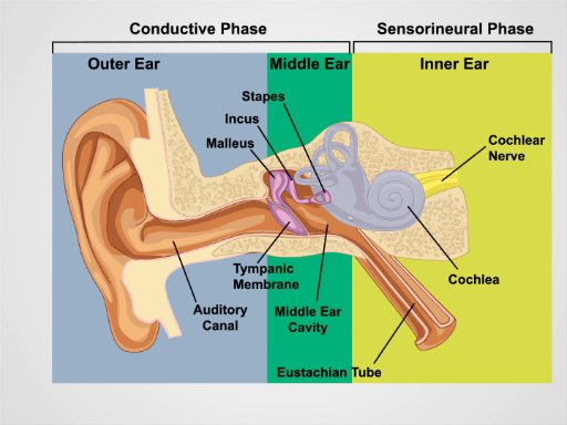
Figura 1. Anatomia dell'orecchio. Un disegno schematico dell'orecchio umano in sezione frontale con strutture dell'orecchio esterno, medio e interno etichettate.
Procedura
1. Esame dell'orecchio e udito
- Ispezionare i padiglioni auricolari e il tessuto circostante per i cambiamenti della pelle, noduli e deformità.
- Afferrare l'elica in modo superiore tra il pollice e l'indice uno alla volta e tirare delicatamente verso l'alto e all'indietro per verificare la mente di disagio in qualsiasi punto dell'orecchio esterno.
- Palpare il processo trago e mastoideo per tenerezza.
- Esegui il test vocale sussurrato per l'acutezza uditiva.
- Assicurati che la st
Applicazione e Riepilogo
Una corretta valutazione dell'orecchio richiede un controllo dell'udito e un esame otoscopico. La perdita dell'udito conduttiva deriva da disturbi dell'orecchio esterno e medio. L'impazione di cerume, l'otite esterna, il trauma, i corpi estranei e (meno comunemente) le esostosi possono portare alla perdita dell'udito ostruendo il canale uditivo. Le cause dell'orecchio medio della perdita dell'udito includono otite media, disfunzione della tromba di Eustachio, barotrauma e otosclerosi. La perdita dell'udito neurosensorial
Vai a...
Video da questa raccolta:

Now Playing
Esame obiettivo dell'orecchio
Physical Examinations II
55.2K Visualizzazioni

Esame obiettivo dell'occhio
Physical Examinations II
77.2K Visualizzazioni

Esame oftalmoscopico
Physical Examinations II
68.0K Visualizzazioni

Esame di naso, seni, cavità orale e faringe
Physical Examinations II
65.8K Visualizzazioni

Esame obiettivo della tiroide
Physical Examinations II
105.1K Visualizzazioni

Esame obiettivo dei linfonodi
Physical Examinations II
387.6K Visualizzazioni

Esame obiettivo dell'addome I: ispezione e auscultazione
Physical Examinations II
202.8K Visualizzazioni

Esame obiettivo dell'addome II: percussione
Physical Examinations II
248.2K Visualizzazioni

Esame obiettivo dell'addome III: palpazione
Physical Examinations II
138.5K Visualizzazioni

Esame obiettivo dell'addome IV: valutazione del dolore addominale acuto
Physical Examinations II
67.3K Visualizzazioni

Esame rettale maschile
Physical Examinations II
114.6K Visualizzazioni

Esame completo del seno
Physical Examinations II
87.7K Visualizzazioni

Esame obiettivo ginecologico I: valutazione dei genitali esterni
Physical Examinations II
307.3K Visualizzazioni

Esame obiettivo ginecologico II: esame con lo speculum
Physical Examinations II
150.4K Visualizzazioni

Esame obiettivo ginecologico III: palpazione rettovaginale e bimanuale
Physical Examinations II
147.8K Visualizzazioni
Personale delle biblioteche
Copyright © 2025 MyJoVE Corporation. Tutti i diritti riservati