JoVE 비디오를 활용하시려면 도서관을 통한 기관 구독이 필요합니다. 전체 비디오를 보시려면 로그인하거나 무료 트라이얼을 시작하세요.
Method Article
높은 처리량 응용 프로그램에 대한 성인 Zebrafish의 마음의 복구
요약
Use of zebrafish for cardiovascular research is expanding towards research on adult hearts. For these applications, quick and simple isolation of cardiac tissues is key to avoid post-mortem changes and to obtain an adequate number of samples. Here, we describe a fast and reproducible method for dissecting adult zebrafish hearts.
초록
개발, 재생을 공부하고, 질병이 세포 분리 및 RNA, DNA, 단백질의 정제를위한 성인 마음의 사용으로 확대되고의 제브라 피쉬 모델 시스템의 사용. 이러한 응용 프로그램의 모든 규제 유전자, 대사, 그리고 죽음 이후 시작하는 다른 변경을 방지하기 위해 제브라 피쉬 마음의 상당한 숫자의 신속한 복구를 요구하고있다. 성인 zebrafish의 마음은 돌연변이의 다양한 심장 구조를 공부하고 심장 재생을 공부해야합니다. 그러나, 기존의 제브라 피쉬의 심장 해부는 느리고 어려운 성인 zebrafish의 마음의 대규모 해부 지루한 제작, 특수 도구가 필요합니다. 전통적인 방법은 해부 동안 심장 손상의 위험을 항구. 여기, 우리는 빠른 재현, 심장 구조를 유지 성인 zebrafish의 마음의 절개하는 방법을 설명합니다. 또한,이 방법은 특수 도구가 필요하지 않습니다, 제브라 피쉬에 대한 통증이수 신선 또는 고정 시험편상에서 수행 될, 1 개월 이전처럼 어린 지브라 피쉬 수행 가능하다. 기재된 접근 방법은 심혈관 연구 성인 지브라 피쉬의 사용을 확장한다.
서문
Zebrafish are an excellent model for studying heart development and human disease1,2. Specific advantages include the translucent nature of zebrafish embryos, the availability of many genetic mutants and transgenic reporter lines, and the availability of genome editing technologies. In addition to their advantages for studying early heart development, zebrafish are an ideal system for studying vertebrate heart regeneration3.
More recently, adult zebrafish are playing an important part in bioinformatics approaches to studying cardiovascular development and disease, due to their relatively large clutch size and relatively quick and inexpensive breeding compared to other vertebrate models. Promising techniques include ribosome profiling, RNA-Seq, and cell dissociation and FACS sorting4-7. However, for these techniques the quality of the data can depend on obtaining a large number of samples in a rapid, efficient, and reproducible manner, before gene regulatory, metabolic, transcriptional, and other changes occur.
Dissection of adult zebrafish organs has been described in the past8,9. However, previous approaches to dissection of the heart were slow, ran the risk of damaging the heart during dissection, required special tools, and/or required fixation of the zebrafish prior to dissection; for these reasons, past approaches to zebrafish adult heart dissection were not optimized for high-throughput applications and/or applications requiring fresh tissue.
Here, we describe a method for adult zebrafish heart dissection that is simple, fast, efficient, and reproducible, while preserving cardiac morphology. This method does not include cutting into the pericardial space and therefore does not risk damaging the heart during dissection. Instead, this method relies on anatomical landmarks of the zebrafish, and therefore, it is highly reproducible. This dissection method is also versatile in that it can be used on fresh or fixed fish, and on zebrafish as young as one month old. Finally, this method results in minimal suffering to the zebrafish because after anesthesia and/or rapid cooling, the fish is additionally decapitated and pithed in the course of the dissection procedure.
프로토콜
참고 : 항상 IACUC 또는 윤리위원회의 승인이 제브라 피쉬를 사용하여 실험 절차를 시작하기 전에 제자리에 있는지 확인하십시오.
1. 시약 및 설치 준비
- 다음과 같은 솔루션을 준비합니다. 표준 제브라 피쉬 매뉴얼 (10)에서 이러한 모든 솔루션에 대한 조리법을 찾을 수 있습니다.
- 계란 물 500㎖
- 계란 물에 0.03 % Tricaine 200 ㎖
- 1X 인산염 완충 식염수 100 ㎖ (PBS)
- RBC 용해 버퍼 (선택 사항)
- 얼음에 1.5 ml의 미세 원심 튜브에 선택의 대상 버퍼 (정착액, 트리 졸, PBS 등)
주 : 피부와 접촉하면 정착액 및 트리 졸은 독성. 이 물질을 취급 할 때는 장갑을 착용 할 것.
- 해부 설치를 준비합니다 :
- 해부 현미경을 통해 구즈넥 광원을 배치합니다. 해부 현미경 무대에서 9cm 페트리 접시의 뚜껑을 놓고에 1X PBS의 얇은 층을 부어그것은 (요리 자체가 쉽게 해부 너무 깊은 때문에 뚜껑이 사용됩니다).
- 해부 근처의 수조에 페트리 접시 자체 (안 뚜껑)을 놓고 그 안에 1X PBS의 약 15 ml에 붓는다. 절개 한 후 심장을 씻어 사용합니다. 또한, RBC 용해 버퍼를 사용합니다.
- 장소 면도날, 두 집게, microscissors (옵션)의 쌍, 및 현미경 옆에 전송 피펫. 급속 냉각에 의한 안락사 선택하면 얼음의 컨테이너를 가져옵니다.
- Tricaine 의해 안락사 선택하면 250 ㎖의 유리 비이커에 달걀 물 0.03 % Tricaine 200㎖를 붓고. 제브라 피쉬의 사체의 처분을위한 작은 생물 학적 가방을 준비합니다.
2. Zebrafish의 준비
- 고정 물고기를 들어, 현미경 설정에, PBS에 세척 고정 물고기를 가져온다.
- > 10 분 동안 얼음과 급속 냉각하여 전체 성인 제브라 피쉬를 안락사.
- 복막을 통해 피부에 구멍을 만들기 위해 집게를 사용하여(멀리 마음에서) 고정액의 침투를 돕기 다음 4 ° C에서 RT 또는 O / N에서 2 시간 동안 수정 버퍼 (10)에 4 % 파라 포름 알데히드에 넣어합니다.
- 또한, 신선한 물고기, 장소 물고기는 작은 저장 탱크에 안락사하고 현미경 설정에 가져다. 개별 시설의 물고기 프로토콜과 해부 물고기 마음의 다운 스트림 용도에 따라 Tricaine 물고기를 마취 또는 잘린 전에 급속 냉각에 의해 안락사.
- 물고기가 움직이지 않을 때까지 얼음 물에 물고기 그물과 장소에 약 5 분을 한 물고기를 선택, 얼음을 사용합니다.
- 또한, 아가미의 움직임이 멈출 때까지 용액의 비커에 물고기 그물과 장소 하나 물고기를 선택, Tricaine를 사용합니다.
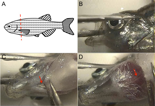
- 빠르게 꼬리 지느러미에 의해 안락사 물고기를 선택하고 1X PBS로 가득 차 있었다 페트리 접시 덮개에 옆으로 누워. razo를 사용하는 동안, 한 손으로 가슴 지느러미를 들어 올려 집게를 사용하여R 블레이드는 가슴 지느러미 (그림 1A)의 부착 후부, 다른 손으로 물고기를 목을 벨 수 있습니다. 현미경을 통해보고하거나 직접 현미경 무대에서 물고기를 시각화하여이 작업 단계를 수행합니다.
참고 : 갓 안락사 물고기 여전히 잘린 후 피합니다.

그림 1. Zebrafish의 성인 심장 해부는 제브라 피쉬의 해부학 적 구조를 사용한다. (A)는 물고기 목을 벨 집게와 가슴 지느러미를 들어 올려 그림과 같이 빨간색 점선을 따라 날카로운 깨끗한 면도날을 사용합니다. (B), 생선 머리를 안정 물고기 입 동안 포셉 한 가지를 배치하려면 다른 타인의 눈을 통해 거짓말을하고, 복부면이 위를하고 집게의 두 타인 페트리 접시의 바닥에 안정되도록하고 물고기 머리를 켜십시오. (C) 무료 그렇게해야 사용EPS는 아가미 (화살표). (D)이 리프팅의 첨부 파일을 잘라, 지느러미 대동맥 내강 혈액 (화살표)를 나타내는 핑크 스트라이프 흰색 구조로 볼 수있다. 이 그림의 더 큰 버전을 보려면 여기를 클릭하십시오.
3. 심장을 해부하다
- 현미경으로 생선 머리를 시각화. 다른 타인이 눈 (그림 1B)를 통해, 머리 외부에있는 동안 하나의 집게로, 뇌를 통해 물고기의 입에 집게 중 하나 타인을 배치하여 물고기의 머리를 안정. 집게의 두 타인 페트리 접시의 바닥에 대한 안정세를 확인합니다. 물고기 머리를 이런 식으로 잡아서 복부 표면 위쪽으로 향해야합니다.
- 다른 손으로, 몸 (그림 1C 화살표)에 아가미의 복부 첨부 파일을 잘라 두 번째 집게를 사용합니다. 집게와 약간이 리프팅, Dorsal 대동맥은 대동맥 루멘 (그림 1D 화살표)에 혈액의 빨간 줄무늬와 흰색 구조로 아래에 나타납니다. 대동맥 곤란 위쪽으로 잡아 당겨 집게 등의 대동맥을 잘라; 대안 등의 대동맥을 잘라 microscissors를 사용합니다.
- 이제, 지느러미의 기지에서 몸 연골을 포함하여, 그 기지에서 가슴 지느러미를 파악하기 위해 두 번째 집게를 사용하고이 들어 올립니다. 다른 가슴 지느러미와 반복합니다. 때때로, 마음은 가슴 지느러미와 함께 나오는, 그래서 마음을 폐기하기 전에 연결되어 있지 않은 확인이 조각을 검사합니다.
참고 : (때때로는 가슴 지느러미와 벗기 있지만)이 시점에서, 마음이, 눈에 보이는 그대로 남은 생선의 머리에 연결해야합니다. 심장은 그 형상, 그 핑크 색상 및 색소 심낭에 의해 포위 됨으로써 확인할 수있다. 물고기의 안락사가 신속하게 수행되기 때문에, 갓 안락사 물고기 마음은 여전히 SLO를 구타 할 수있다wly. - 양손 집게를 보유하고 무료 있도록, 먼저 집게로 물고기의 머리 가자. 멀리 심낭의 마음을 애타게 두 집게를 사용합니다. 나머지 심낭 멀리 당겨 동안 지느러미 대동맥의 나머지에 의해 마음을 잡고.
참고 : 고정 또는 신선한 여부, 심장 주위의 결합 조직이 더 부서지기 쉬운 반면 당기 부드러운 강인한. 따라서, 모든 주변의 심낭 조직은 심장이 그대로 남아 멀리 해부 할 수 있습니다. - 생물 학적 가방에 남은 물고기 시체를 버리십시오.
4. 다운 스트림 응용 프로그램에 대한 마음을 준비
- 집게를 사용하여, 지느러미 대동맥 / bulbus 동맥의 가장자리에 마음을 잡고 1X PBS의 신선한 접시에 마음을 놓습니다. 또한, 전송 피펫을 사용합니다. 앞뒤로 PBS의 마음을 움직 약 10 배 가능한 한 많은 피를 씻어.
- 응용 프로그램의 모든 possib을 제거하는 것이 중요하다있는르 혈액 세포는 동일한 전후 모션을 사용하는 대신에 PBS 15 ml의 용해 완충액으로 RBC 9cm 페트리 접시에 씻는다. 심장의 구조를 유지하는 (예를 들어, RNA 제조), 심장 공동을 열고 멀리 혈구 린스 용이하게 집게 나 microscissors를 사용 하류 애플리케이션에 중요하지 않은 경우.
- 별도의 심장 챔버가 요구되는 응용 프로그램의 경우, 서로 떨어져 bulbus 개존증, 심방 및 심실을 해부 집게 또는 microscissors를 사용합니다.
- 얼음에 대상 버퍼에 심장, 또는 별도의 챔버를 전송합니다.
참고 : 일반 대상은 세포 분리, 4 % 파라 포름 알데히드, 또는 트리 졸에 대한 버퍼를 포함한다.
결과
이 방법을 사용하여, 성인 심장 지브라 피쉬는 전통적인 방법을 사용하여 8 분에 걸쳐 5에 비해, 1 분 이하로 해부 될 수있다. 전통적인 방법 (8) 심낭에 맹목적으로 절단하기 때문에 일반적으로 심방 또는 bulbus 동맥 (그림 2B)의 손상이나 손실이 발생할 필요로하지만이 방법을 사용하여 해부 하트 (그림 2A) 안정적으로 그대로입니다. 해부 하트 그들?...
토론
성인 zebrafish의 심장을 해부하는 방법이 설명되어 있지만,이 방법은 시간이 많이 소요되었고 일반적으로 해부하는 동안 심장에 손상을 일으키는 원인이되었다. 성인 하트 다수 필요할 수있는 실험을 수행하기 위해 및 / 또는 심장 조직의 열화를 피하는 하류 응용에 중요 할 때, 전통적인 절개 기술을 사용하여 요구되는 시간은 금지된다. 마찬가지로, 재현성 손상되지 않은 그대로의 마음을 얻는 ?...
공개
The authors have no disclosures.
감사의 말
The authors would like to thank Dr. Shaun Coughlin for hosting the filming of this procedure in his laboratory, and for general support. R.A. was supported by the NIH (F32HL110489) and the Sarnoff Cardiovascular Research Foundation. S.R. was supported by a Research Fellowship of the Deutsche Forschungsgemeinschaft (DFG) and the American Heart Association (AHA). D.Y.R.S was supported by the NIH (RO1HL54737), the Packard Foundation, and the Max Planck Society.
자료
| Name | Company | Catalog Number | Comments |
| Small tank for transporting fish | Aquaneering | ZHCT100 | |
| Fish net | Petsmart | 36-16731 | |
| 250 ml glass beaker | Kimble | 14005-250 | |
| 9 cm polystyrene Petri dish | Nunc | 172958 | |
| Razor blade | Personna American Safety Razor Company | 94-120-71 | |
| 2 Dumont #5SF forceps | Fine Science Tools | 11252-00 | |
| Dissecting microscope | Olympus | SZX16 | |
| Tricaine | Sigma | A-5040 | |
| Plastic transfer pipette | Thermo Scientific | 202-20S | |
| Gooseneck light source | Dolan-Jenner Industries, Inc | Fiber-Lite 180 Illuminator, 181 Dual Gooseneck System | |
| Fluorescent light source | Lumen Dynamics | X-Cite 120Q | optional |
| Micro-scissors | Biomedical Research Instruments, Inc | 11-1000 | optional |
| RBC lysis buffer | eBioscience | 00-4333-57 | optional |
참고문헌
- Arnaout, R., et al. Zebrafish model for human long QT syndrome. Proceedings of the National Academy of Sciences of the United States of America. 104 (27), 11316-11321 (2007).
- Chi, N. C., et al. Genetic and physiologic dissection of the vertebrate cardiac conduction system. PLoS Biology. 6 (5), 109 (2008).
- Poss, K. D. Getting to the heart of regeneration in zebrafish. Seminars in Cell & Developmental Biology. 18 (1), 36-45 (2007).
- Fang, Y., et al. Translational profiling of cardiomyocytes identifies an early Jak1/Stat3 injury response required for zebrafish heart regeneration. Proceedings of the National Academy of Sciences of the United States of America. 110 (33), 13416-13421 (2013).
- Manoli, M., Driever, W. Fluorescence-activated cell sorting (FACS) of fluorescently tagged cells from zebrafish larvae for RNA isolation. Cold Spring Harbor Protocols. 2012 (8), (2012).
- Cannon, J. E., et al. Global analysis of the haematopoietic and endothelial transcriptome during zebrafish development. Mechanisms of Development. 130 (2-3), 122-131 (2013).
- Wang, Z., Gerstein, M., Snyder, M. RNA-Seq: a revolutionary tool for transcriptomics. Nature Reviews Genetics. 10 (1), 57-63 (2009).
- Gupta, T., Mullins, M. C. Dissection of organs from the adult zebrafish. Journal of Visualized Experiments. 37, (2010).
- Singleman, C., Holtzman, N. G. Heart dissection in larval, juvenile and adult zebrafish, Danio rerio. Journal of Visualized Experiments. 55, (2011).
- Westerfield, M. . The zebrafish book: a guide for the laboratory use of zebrafish (Brachydanio rerio. , (1993).
재인쇄 및 허가
JoVE'article의 텍스트 или 그림을 다시 사용하시려면 허가 살펴보기
허가 살펴보기더 많은 기사 탐색
This article has been published
Video Coming Soon
Copyright © 2025 MyJoVE Corporation. 판권 소유