
Aby wyświetlić tę treść, wymagana jest subskrypcja JoVE. Zaloguj się lub rozpocznij bezpłatny okres próbny.
Method Article
Sperm Collection and Computer-Assisted Sperm Analysis in the Teleost Model Japanese Medaka (Oryzias latipes)
W tym Artykule
Podsumowanie
This article describes two quick and efficient methods for collecting sperm from the small model fish medaka (Oryzias latipes), as well as a protocol for reliably assessing sperm quality using computer-assisted sperm analysis (CASA).
Streszczenie
Japanese medaka (Oryzias latipes) is a teleost fish and an emerging vertebrate model for ecotoxicology, developmental, genetics, and physiology research. Medaka is also used extensively to investigate vertebrate reproduction, which is an essential biological function as it allows a species to perpetuate. Sperm quality is an important indicator of male fertility and, thus, reproduction success. Techniques for extracting sperm and sperm analysis are well documented for many species, including teleost fish. Collecting sperm is relatively simple in larger fish but can be more complicated in small model fish as they produce less sperm and are more delicate. This article, therefore, describes two methods of sperm collection in the small model fish, Japanese medaka: testes dissection and abdominal massage. This paper demonstrates that both approaches are feasible for medaka and shows that abdominal massage can be performed a repeated number of times as the fish quickly recover from the procedure. This article also describes a protocol for computer-assisted sperm analysis in medaka to objectively assess several important indicators of medaka sperm quality (motility, progressivity, duration of motility, relative concentration). These procedures, specified for this useful small teleost model, will greatly enhance understanding of the environmental, physiological, and genetic factors influencing fertility in vertebrate males.
Wprowadzenie
Japanese medaka is a small, egg-laying freshwater teleost fish native to East Asia. Medaka has become an excellent vertebrate model system for ecotoxicology, developmental genetics, genomics, and evolutionary biology and physiology studies1,2. Similar to the popular zebrafish, they are relatively easy to breed and highly resistant to many common fish diseases1,2. There are several advantages of using medaka as a model, including a short generation time, transparent embryos1,2, and a sequenced genome3. Unlike zebrafish, medaka has a sex-determining gene4 as well as a high temperature (from 4-40 °C) and salinity (euryhaline species) tolerance5. Also, many genetic and anatomical tools, as well as protocols6,7,8,9,10,11,12, have been developed in medaka to facilitate the study of its biology.
Reproduction is an essential physiological function as it allows a species to perpetuate. Vertebrate reproduction requires a myriad of precisely orchestrated events, including the production of oocytes in females and the production of sperm in males. Sperm are unique cells, produced through the complex process of spermatogenesis, in which there are a number of checkpoints in place to guarantee delivery of a high-quality product13. Gamete quality has become a focus in aquaculture and fish population studies due to its impact on fertilization success and larval survival. Sperm quality is, therefore, an important indicator of male fertility in vertebrates.
Three useful factors for assessing fish sperm quality are motility, progressivity, and longevity. Percent motility and progressive motility are common indicators of sperm quality as progressive motion is necessary for and correlates strongly with fertilization success14,15. Duration of movement is also an important indicator in fish as sperm remain fully motile for less than 2 min in most teleost species and the trajectory of sperm is generally less linear than in mammals15. However, many studies assessing sperm motility in the past relied on subjective or semi-quantitative methods of analyzing sperm15,16. For instance, sperm motility in medaka has been estimated in the past visually under a microscope17. It has also been estimated by recording sperm movement and using imaging software to merge frames and measure swimming path and velocity18,19,20. Such approaches often lack robustness, providing different results according to the person performing the analysis15,21.
Computer-assisted sperm analysis (CASA) was initially developed for mammals. CASA is a fast quantitative method to assess sperm quality by recording and measuring velocity and trajectory in an automated manner15. In fishes, it has been used in different species to monitor the effects of several water pollutants on sperm quality, for identifying interesting progenitors to improve broodstock, to improve the efficiency of cryopreservation and storage, and to optimize conditions for fertilization15. Therefore, it is a powerful tool for reliably assessing sperm quality in different vertebrate species. However, due to the important diversity in reproductive strategies between fishes, the sperm of teleost fish differs from that of mammals and from one fish species to another. Teleost fish, which primarily fertilize eggs externally by releasing gametes into water, have highly concentrated sperm that are relatively simple in structure with no acrosome, unlike mammals, which fertilize internally and therefore do not have to compensate for dilution in water, but do have to withstand more viscous fluids14. Additionally, sperm from most fish move rapidly but are fully motile for less than 2 min after activation, although there are several exceptions15,22. Because motility can decrease rapidly in most fish, extreme care should be taken with the timing of analysis after activation when determining a sperm analysis protocol for fish.
Reproduction is one of the fields in biology in which teleosts and medaka have been extensively used as model organisms. Indeed, medaka males show interesting reproductive and social behaviors, such as mate guarding23,24. In addition, several transgenic lines exist to study the neuroendocrine control of reproduction in this species25,26,27. Sperm sampling, a procedure that is relatively simple in larger fish, can be more complicated in small model fish as they produce less sperm and are more delicate. For this reason, most studies involving sperm sampling in medaka extract milt (fish semen) by crushing dissected testes17,28,29,30. A few studies also use a modified abdominal massage to express the milt directly into activating medium18,19,20; however, with this method it is difficult to visualize the amount and color of milt extracted. In zebrafish, abdominal massage is commonly used to express milt, which is immediately collected in a capillary tube31,32,33. This method enables estimation of the volume of milt, as well as observation of ejaculate color, which is a quick and simple indicator of sperm quality32,33. Therefore, a clear and well described protocol for sperm collection and analysis is lacking for medaka.
This article therefore describes two methods of sperm collection in the small model fish Japanese medaka: testes dissection and abdominal massage with capillary tubes. It demonstrates that both approaches are feasible for medaka and shows that abdominal massage can be performed a repeated number of times as the fish quickly recovers from the procedure. It also describes a protocol for computer-assisted sperm analysis in medaka to reliably measure several important indicators of medaka sperm quality (motility, progressivity, longevity, and relative sperm concentration). These procedures, specified for this useful small teleost model, will greatly enhance understanding of the environmental, physiological, and genetic factors influencing fertility in vertebrate males.
Protokół
All experimentation and animal handling were conducted in accordance with the recommendations on the experimental animal welfare at Norwegian University of Life Sciences (NMBU). The experiments were performed using adult (6-9-month-old) male Japanese medaka (Hd-rR strain) raised at NMBU (Ås, Norway). The methods were also briefly tested in 9-month-old male Japanese medaka (CAB strain) raised at the National Research Institute for Agriculture, Food, and the Environment (INRAE, Rennes, France).
1. Instrument and solution preparation
- Prepare anesthetic stock solution (0.6% Tricaine).
- Dilute 0.6 g of Tricaine (MS-222) in 100 mL of 10x Phosphate Buffer Saline (PBS).
- Aliquot 2 mL of the anesthetic stock solution into 50 2 mL plastic tubes and store at -20 °C until use for anesthesia or euthanasia.
- Prepare recovery water (0.9% sodium chloride [NaCl] solution).
- Add 27 g of NaCl into 3 L of aquarium water.
- Store the solution at room temperature (RT) until use.
- Adjust the activation medium if necessary (Hank's balanced salt solution [HBSS]).
NOTE: HBSS can be purchased commercially or made in the laboratory (Table of Materials).- Measure the pH of the HBSS using a pH meter. Adjust the pH if necessary, using hydrochloric acid or sodium hydroxide, so the final pH is 7.1-7.3.
- Measure the osmolality of the HBSS using an osmometer for future reporting.
NOTE: The range on the commercial product is 266-294 mOsmol/kg; in the current study, the osmolality was 287 mOsmol/kg. It may be diluted with distilled water to reduce the osmolality if desired, but this is not necessary as there is not a large difference in the activation of medaka sperm in 150-300 mOsmol/kg HBSS. - Store the solution at RT until use.
- Prepare the holding sponge.
- Cut a soft sponge to fit snugly in a Petri dish.
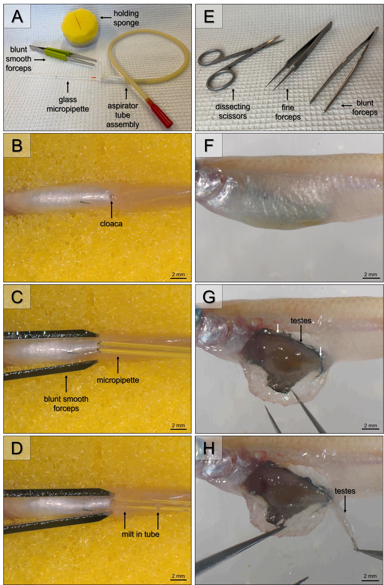
- Cut a straight line in the middle of the sponge that is long enough to receive the fish (3-4 cm) and about 1 cm deep (Figure 1A). This slit in the sponge will hold the fish ventral side up to expose the cloaca.
2. Sperm collection
NOTE: Sperm collection can be achieved by two different methods: abdominal massage or testes dissection.
- Sperm collection by abdominal massage
- Prepare 0.03% anesthetic solution by diluting one tube of Tricaine stock (0.6%) in 38 mL of aquarium water in a 100 mL glass container.
- Prepare the instruments, including blunt end smooth forceps and a 10 µL disposable calibrated glass micropipette and aspirator tube assembly (Figure 1A). Dampen the holding sponge prepared in step 1.4 with the anesthetic solution.
- Prepare tubes with 36 µL of the activating solution for immediate analysis. Preheat the activating solution in a water bath or incubator set to 27 °C for at least 5 min.
NOTE: Although samples can be analyzed from individual fish, individual variation can be reduced by pooling samples from multiple males in the same activating solution. When pooling samples from multiple fish, use 36 µL of activating solution per fish. This dilution may need to be adjusted depending on the strain or rearing conditions of the medaka used as these factors can impact the sperm concentration and volume. The CASA program will indicate whether the concentration is too high to identify sperm. - Anesthetize the fish by putting it in anesthetic solution for 30-90 s.
NOTE: The anesthesia duration must be adapted as it varies according to the size of the fish. To ensure that the fish is fully anesthetized, gently pinch the caudal peduncle with forceps. If the fish does not react, the massage can be started. - Take the fish out of the anesthetic solution and use a paper towel or gently wipe to dry the abdomen of the fish. Place the fish in the trough of the damp holding sponge ventral side up so its gills are exposed to the anesthetic solution in the sponge (Figure 1B).
- If the area around the cloaca is wet, gently dry the underside of the fish with a disposable tissue wipe.
- Place the fish in the holding sponge under a dissecting microscope and place the micropipette with an aspirator tube attached against the cloaca of the fish (Figure 1C).
- Massage the abdomen of the fish by gently squeezing with blunt end smooth forceps in a rostral to caudal motion while simultaneously sucking to collect the expelled milt into the pipette (Figure 1D).
- Release the fish from the sponge into the recovery water. Allow them to recover in the solution for at least 15 min before returning them to the aquarium system.
- Transfer the milt into a prepared tube with preheated activating solution and pipette up and down several times by sucking and blowing on the aspirator tube assembly.
- Homogenize the diluted sperm gently by flicking the tubes before analysis.
NOTE: For best results, analyze samples immediately (e.g., 5 s) after activation. In medaka, the analysis may be delayed, if necessary, as sperm remain motile for several hours, but the time should remain consistent between samples as motility decreases with time.
- Sperm collection by testes dissection
- Prepare 0.08% euthanasia solution by diluting two tubes of Tricaine stock (0.6%) in 26 mL of aquarium water in a 100 mL glass container.
- Prepare dissection tools, including blunt and fine forceps and small dissecting scissors (Figure 1E).
- Prepare a tube for each sample with 120 µL of activating solution for immediate analysis. Preheat the activating solution in a water bath or incubator set to 27 °C for at least 5 min.
NOTE: Although samples can be analyzed from individual fish, individual variation can be reduced by pooling samples from multiple males in the same activating solution. For pooling samples from multiple fish, use 120 µL of activating solution per fish. This dilution may need to be adjusted depending on the strain or rearing conditions of the medaka used as these factors can impact the sperm concentration and volume. The CASA program will indicate whether the concentration is too high to identify sperm. - Euthanize the fish by putting it into the 0.08% anesthetic solution for 30-90 s.
NOTE: The duration depends on the size of the fish. To ensure that the fish is euthanized, wait for operculum movements to cease. The fish should not react to the touch of forceps. - Remove the fish from the euthanasia solution and gently dry the fish with a paper towel or gently wipe.
NOTE: At this step, the fish can be weighed to later calculate the gonadosomatic index (GSI, gonadal weight / body weight). - Place the fish under a dissecting microscope with its left lateral side facing up (Figure 1F).
- Using small dissecting scissors, cut a flap dorsally from the cloaca, and then across the ribs to the gills to expose the internal organs (Figure 1G).
- Locate the testes, cut the attachment at both ends with fine forceps, and remove the testes (Figure 1H).
NOTE: To calculate the GSI, the testes can be weighed at this step. Work quickly to avoid drying of the tissue. - Transfer the testes to a prepared tube with the preheated activating solution.
- Use forceps to crush the testes several times against the side of the tube to release the sperm. Sperm release can usually be visualized and will make the solution slightly cloudy.
- Homogenize the diluted sperm gently by flicking the tubes before analysis.
NOTE: For best results, analyze samples immediately (e.g., 5 s) after activation. In medaka, the analysis may be delayed, if necessary, as sperm remain motile for several hours, but the time should remain consistent between samples as motility decreases with time.

Figure 1: Milt collection by abdominal massage (A-D) and testes dissection (E-H). (A) Instruments for abdominal massage: holding sponge, blunt smooth forceps, and 10 µL disposable calibrated glass micropipette with aspirator tube assembly; (B) Position of fish in the holding sponge, with gills exposed to anesthesia in the sponge and cloaca facing up; (C) Position of blunt smooth forceps on abdomen and micropipette against cloaca; (D) Milt in micropipette after gentle massage and sucking. (E) Instruments for testes dissection: blunt forceps, fine forceps, and small dissecting scissors; (F) Position of fish for testes dissection; (G) Lateral view of internal organs; (H) Remove the testes by cutting the attachment at both ends with fine forceps. Scale bar: 2 mm. Please click here to view a larger version of this figure.
3. Sperm analysis with CASA system
- The CASA system (SCA Evolution) should be set up according to the manual with a microscope using a green filter and 10x objective with phase contrast.
- Prepare disposable 20 µm counting chamber slides by preheating on a warming plate or in an incubator set to 27 °C for at least 5 min.
- Open the sperm analysis software and select the motility module.
- Set the configuration for the medaka as shown in Figure 2B.
- Place a prewarmed disposable 20 µm counting chamber slide under the microscope on a heated stage set to 27 °C.
- Pipette the sample into the chamber on the slide until it fills the chamber without overfilling. Carefully wipe away excess samples from the entrance of the chamber with a cotton tip or gently wipe to prevent floating cells.
- Select Analyze to look at the sample under the microscope.
NOTE: If the microscope icon is red, the microscope lighting needs to be adjusted for the program to track sperm accurately. Adjust the brightness of the microscope, so the tail movement of the sperm is clearly seen. The icon should be blue. - Ensure the microscope is focused and select Analyze again to record the sperm in the field. Move the slide so a new area of the sample is in the frame and repeat to capture for 3-5 different fields of view. Avoid fields with air bubbles, cell masses, or artifacts.
- Select Results to view the results.
NOTE: If the fields on the results page are outlined in red, follow the system's prompts to delete the fields that vary too much in concentration or motility. - Double-click on a field to view the results for the individual field or to manually check for any mislabeled or untracked spermatozoa. Right-click on individual spermatozoa to relabel the motility, if necessary (Figure 2A).

Figure 2: SCA Evolution software screenshot. (A) Sperm tracking results for one field. View field data on the right side and double-click spermatozoa to view individual data; (B) Results summary for all fields with configuration menu open. Please click here to view a larger version of this figure.
Wyniki
Type of data obtained
Sperm motility analysis from the SCA Evolution software provides data on motility (percentage of motile and immotile sperm), as well as progressivity (percentage of progressive and non-progressive sperm), and velocity (percentage of rapid, medium, and slow-moving sperm). It also combines progressivity and velocity (rapid progressive, medium progressive, non-progressive). These labels are based on measurements (Figure 3A) and calculations (
Dyskusje
Osmolality is an important factor in the activation of fish sperm36,37. Generally, sperm are immotile in the testes and become motile in media that is hyperosmotic relative to seminal fluid for marine fishes, and hypo-osmotic relative to seminal fluid for freshwater fishes37. Similar to blood, seminal plasma in freshwater fishes is typically lower than that of marine fishes (about 300 mOsmol/kg compared to 400 mOsmol/kg)2...
Ujawnienia
The authors have nothing to disclose.
Podziękowania
This work has been funded by the Norwegian University of Life Sciences and the U.S. Fulbright program. The authors would like to thank Anthony Peltier and Lourdes Carreon G Tan at NMBU for fish facility maintenance and Guillaume Gourmelin from the ISC LPGP at INRAE (France) for providing fish and lab space to further test these methods.
Materiały
| Name | Company | Catalog Number | Comments |
| 1.5 mL tubes | Axygen | MCT-150-C | Any standard brand can be used |
| 10 µL disposable calibrated glass micropipette and aspirator tube assembly | Drummond | 2-000-010 | |
| 10x objective with phase contrast | Nikon | MRP90100 | |
| 2 mL tubes | Axygen | MCT-200-c-s | Any standard brand can be used |
| Blunt forceps | Fine Science Tools | 11000-12 | |
| Blunt smooth forceps | Millipore | XX6200006P | |
| Disposable 20 micron counting chamber slide | Microptic | 20.2.25 | Leja 2 chamber slides |
| Dissecting microscope | Olympus | SZX7 | Any standard brand can be used |
| Fine forceps | Fine Science Tools | 11253-20 | |
| HBSS | Sigmaaldrich | H8264-1L | |
| Holding sponge | self-made | ||
| Inverted microscope | Nikon | Eclipse Ts2R | |
| SCA Evolution | Microptic | ||
| Small dissecting scissors | Fine Science Tools | 14090-09 | |
| Sodium Chloride (NaCl) | Sigmaaldrich | S9888 | |
| Tabletop vortex | Labnet | C1301B | |
| Tricaine | Sigmaaldrich | A5040 |
Odniesienia
- Shima, A., Mitani, H. Medaka as a research organism: past, present and future. Mechanisms of Development. 121 (7-8), 599-604 (2004).
- Wittbrodt, J., Shima, A., Schartl, M. Medaka - a model organism from the far east. Nature Reviews Genetics. 3 (1), 53-64 (2001).
- Kasahara, M., et al. The medaka draft genome and insights into vertebrate genome evolution. Nature. 447 (7145), 714-719 (2007).
- Matsuda, M., et al. DMY is a Y-specific DM-domain gene required for male development in the medaka fish. Nature. 417 (6888), 559-563 (2002).
- Sakamoto, T., Kozaka, T., Takahashi, A., Kawauchi, H., Ando, M. Medaka (Oryzias latipes) as a model for hypoosmoregulation of euryhaline fishes. Aquaculture. 193 (3-4), 347-354 (2001).
- Royan, M. R., et al. 3D atlas of the pituitary gland of the model fish medaka (Oryzias latipes). Frontiers in Endocrinology. 12, 719843 (2021).
- Fontaine, R., Hodne, K., Weltzien, F. A. Healthy brain-pituitary slices for electrophysiological investigations of pituitary cells in teleost fish. Journal of Visualized Experiments. (138), e57790 (2018).
- Fontaine, R., Weltzien, F. -. A. Labeling of blood vessels in the teleost brain and pituitary using cardiac perfusion with a dii-fixative. Journal of Visualized Experiments. (148), e59768 (2019).
- Ager-Wick, E., et al. Preparation of a high-quality primary cell culture from fish pituitaries. Journal of Visualized Experiments. (138), e58159 (2018).
- Porazinski, S. R., Wang, H., Furutani-Seiki, M. Microinjection of medaka embryos for use as a model genetic organism. Journal of Visualized Experiments. (46), e1937 (2010).
- Wiley-Blackwell. . Medaka: Biology, Management, and Experimental Protocols. , (2019).
- Royan, M. R., et al. Gonadectomy and blood sampling procedures in the small size teleost model japanese medaka (Oryzias latipes). Journal of Visualized Experiments. (166), e62006 (2020).
- Bhat, I. A., et al. Testicular development and spermatogenesis in fish: insights into molecular aspects and regulation of gene expression by different exogenous factors. Reviews in Aquaculture. 13 (4), 2142-2168 (2021).
- vander Horst, G., Garcia Alvarez, O., Garde, J. J., Soler, A. J., Jones, D. Status of sperm functionality assessment in wildlife species: From fish to primates. Animals. 11 (6), 1491 (2021).
- Kime, D. E., et al. Computer-assisted sperm analysis (CASA) as a tool for monitoring sperm quality in fish. Comparative Biochemistry and Physiology Part C: Toxicology & Pharmacology. 130 (4), 425-433 (2001).
- Rurangwa, E., Kime, D. E., Ollevier, F., Nash, J. P. The measurement of sperm motility and factors affecting sperm quality in cultured fish. Aquaculture. 234 (1-4), 1-28 (2004).
- Yang, H., Tiersch, T. R. Sperm motility initiation and duration in a euryhaline fish, medaka (Oryzias latipes). Theriogenology. 72 (3), 386-392 (2009).
- Hashimoto, S., et al. Effects of ethinylestradiol on medaka (Oryzias latipes) as measured by sperm motility and fertilization success. Archives of Environmental Contamination and Toxicology. 56 (2), 253-259 (2009).
- Hara, Y., Strüssmann, C. A., Hashimoto, S. Assessment of short-term exposure to nonylphenol in Japanese medaka using sperm velocity and frequency of motile sperm. Archives of Environmental Contamination and Toxicology. 53 (3), 406-410 (2007).
- Kawana, R., Strüssmann, C. A., Hashimoto, S. Effect of p-Nonylphenol on sperm motility in Japanese medaka (Oryzias latipes). Fish Physiology and Biochemistry. 28, 213-214 (2003).
- Gallego, V., Herranz-Jusdado, J. G., Rozenfeld, C., Pérez, L., Asturiano, J. F. Subjective and objective assessment of fish sperm motility: when the technique and technicians matter. Fish Physiology and Biochemistry. 44 (6), 1457-1467 (2018).
- Browne, R. K., et al. Sperm motility of externally fertilizing fish and amphibians. Theriogenology. 83 (1), 1-13 (2015).
- Arias Padilla, L. F., et al. Cystic proliferation of germline stem cells is necessary to reproductive success and normal mating behavior in medaka. eLife. 10, 62757 (2021).
- Okuyama, T., Yokoi, S., Takeuchi, H. Molecular basis of social competence in medaka fish. Development, Growth, and Differentiation. 59 (4), 211-218 (2017).
- Okubo, K., et al. Forebrain Gonadotropin-releasing hormone neuronal development: Insights from transgenic medaka and the relevance to X-linked Kallmann syndrome. Endocrinology. 147 (3), 1076-1084 (2006).
- Hodne, K., Fontaine, R., Ager-Wick, E., Weltzien, F. A. Gnrh1-induced responses are indirect in female Medaka Fsh cells, generated through cellular networks. Endocrinology. 160 (12), 3018-3032 (2019).
- Karigo, T., et al. Whole brain-pituitary in vitro preparation of the transgenic Medaka (Oryzias latipes) as a tool for analyzing the differential regulatory mechanisms of LH and FSH release. Endocrinology. 155 (2), 536-547 (2014).
- Kowalska, A., Kowalski, R., Zakęś, Z. The effect of selective cyclooxygenase (COX) inhibitors on japanese medaka (Oryzias latipes) reproduction parameters. World Academy of Science, Engineering and Technology. 77, 19-23 (2011).
- Kowalska, A., Siwicki, A. K., Kowalski, R. K. Dietary resveratrol improves immunity but reduces reproduction of broodstock medaka Oryzias latipes (Temminck & Schlegel). Fish Physiology and Biochemistry. 43 (1), 27-37 (2007).
- Tan, E., Yang, H., Tiersch, T. R. Determination of sperm concentration for small-bodied biomedical model fishes by use of microspectrophotometry. Zebrafish. 7 (2), 233-240 (2010).
- Harvey, B., Kelley, R. N., Ashwood-Smith, M. J. Cryopreservation of zebra fish spermatozoa using methanol. Canadian Journal of Zoology. 60 (8), 1867-1870 (1982).
- Wasden, M. B., Roberts, R. L., DeLaurier, A. Optimizing sperm collection procedures in Zebrafish. Journal of the South Carolina Academy of Science. 15 (2), 7 (2017).
- Draper, B. W., Moens, C. B. A High-throughput method for Zebrafish sperm cryopreservation and in vitro fertilization. Journal of Visualized Experiments. (29), e1395 (2009).
- Castellini, C., Dal Bosco, A., Ruggeri, S., Collodel, G. What is the best frame rate for evaluation of sperm motility in different species by computer-assisted sperm analysis. Fertility and Sterility. 96 (1), 24-27 (2011).
- Acosta, I. B., et al. Effects of exposure to cadmium in sperm cells of zebrafish, Danio rerio. Toxicology Reports. 3, 696-700 (2016).
- Wilson-Leedy, J. G., Kanuga, M. K., Ingermann, R. L. Influence of osmolality and ions on the activation and characteristics of zebrafish sperm motility. Theriogenology. 71 (7), 1054-1062 (2009).
- Alavi, S. M. H., Cosson, J. Sperm motility in fishes. (II) Effects of ions and osmolality: A review. Cell Biology International. 30 (1), 1-14 (2006).
- Kowalska, A., Kamaszews ki, M., Czarnowska-Kujawska, M., Podlasz, P., Kowalski, R. K. Dietary ARA improves COX activity in broodstock and offspring survival fitness of a model organism (Medaka Oryzias latipes). Animals. 10 (11), 2174 (2020).
- Inoue, K., Takei, Y. Asian medaka fishes offer new models for studying mechanisms of seawater adaptation. Comparative Biochemistry and Physiology Part B: Biochemistry and Molecular Biology. 136 (4), 635-645 (2003).
- Zadmajid, V., Myers, J. N., Sørensen, S. R., Ernest Butts, I. A. Ovarian fluid and its impacts on spermatozoa performance in fish: A review. Theriogenology. 132, 144-152 (2019).
- Poli, F., Immler, S., Gasparini, C. Effects of ovarian fluid on sperm traits and its implications for cryptic female choice in zebrafish. Behavioral Ecology. 30 (5), 1298-1305 (2019).
- Cosson, J., Groison, A. L., Suquet, M., Fauvel, C., Dreanno, C., Billard, R. Studying sperm motility in marine fish: An overview on the state of the art. Journal of Applied Ichthyology. 24 (4), 460-486 (2008).
- Beirão, J., Soares, F., Herráez, M. P., Dinis, M. T., Cabrita, E. Sperm quality evaluation in Solea senegalensis during the reproductive season at cellular level. Theriogenology. 72 (9), 1251-1261 (2009).
- Beirão, J., et al. Sperm handling in aquatic animals for artificial reproduction. Theriogenology. 133, 161-178 (2019).
- Yang, H., Tiersch, T. R. Current status of sperm cryopreservation in biomedical research fish models: Zebrafish, medaka, and Xiphophorus. Comparative Biochemistry and Physiology Part C: Toxicology & Pharmacology. 149 (2), 224-232 (2009).
- Yang, H., Tiersch, T. R. Sperm cryopreservation in biomedical research fish models. Cryopreservation in Aquatic Species. 2, 439-454 (2011).
- Viveiros, A., Fessehaye, Y., ter Veld, M., Schulz, R., Komen, H. Hand-stripping of semen and semen quality after maturational hormone treatments, in African catfish Clarias gariepinus. Aquaculture. 213 (1-4), 373-386 (2002).
- Ransom, D. G., Zon, L. I. Appendix 3 collection, storage, and use of Zebrafish sperm. Methods in Cell Biology. 60, 365-372 (1998).
- Cosson, J. Frenetic activation of fish spermatozoa flagella entails short-term motility, portending their precocious decadence. Journal of Fish Biology. 76 (1), 240-279 (2010).
- Kowalski, R. K., Cejko, B. I. Sperm quality in fish: Determinants and affecting factors. Theriogenology. 135, 94-108 (2019).
Przedruki i uprawnienia
Zapytaj o uprawnienia na użycie tekstu lub obrazów z tego artykułu JoVE
Zapytaj o uprawnieniaThis article has been published
Video Coming Soon
Copyright © 2025 MyJoVE Corporation. Wszelkie prawa zastrzeżone