A subscription to JoVE is required to view this content. Sign in or start your free trial.
Method Article
Ex Vivo Culture of Patient Tissue & Examination of Gene Delivery
In This Article
Summary
This article describes the culture of patient tissue slices for gene delivery studies and subsequent analysis of gene expression using IVIS bioluminescence imaging.
Abstract
This video describes the use of patient tissue as an ex vivo model for the study of gene delivery. Fresh patient tissue obtained at the time of surgery is sliced and maintained in culture. The ex vivo model system allows for the physical delivery of genes into intact patient tissue and gene expression is analysed by bioluminescence imaging using the IVIS detection system. The bioluminescent detection system demonstrates rapid and accurate quantification of gene expression within individual slices without the need for tissue sacrifice. This slice tissue culture system may be used in a variety of tissue types including normal and malignant tissue and allows us to study the effects of the heterogeneous nature of intact tissue and the high degree of variability between individual patients. This model system could be used in certain situations as an alternative to animal models and as a complementary preclinical mode prior to entering clinical trial.
Protocol
Preparation of Media and Reagents
- Antibiotic Solution
Phosphate buffered saline (PBS) with 200 U/ml penicillin, 200 μg/ml streptomycin, 5 μg/ml fungizone - Collection and Treatment medium
Dulbecco's modified eagle medium (DMEM) with 200 U/ml penicillin, 200 μg/ml streptomycin, 5 μg/ml fungizone - Culture medium
Collection medium containing 10% heat-inactivated fetal bovine serum
I. Tissue collection and storage
Approval for patient tissue collection was obtained from the Clinical Research Ethics Committee of the Cork Teaching Hospitals and informed consent was obtained from the patients the day before surgery. Liver tissue was obtained from patients undergoing partial hepatectomy for malignant disease.
- Fresh patient tissue is obtained at the time of surgical resection.
- Tissue is stored in collection media at 4°C (Tissue may be stored refrigerated for up to 12 h without significant loss of viability, however immediate processing is recommended).
II. Tissue slice preparation and culture
Slicing was performed using a vibratome (Leica, Germany). The tissue slicing system was used according to the manufacturer's instructions. Tissue slice preparation was performed under a sterile hood using instruments cleaned with 70% 2-propanol. Slice thickness is set at 2000 microns and cut using a reciprocating blade at 22-26 rpm.
- Tissue is washed in antibiotic solution three times.
- Tissue is attached to mounting stage using non-toxic adhesive (Dermabond).
- Slice thickness is set and slicing is achieved at 22-26 rpm.
- Slices are maintained in culture medium in 6-well culture dishes (one slice per well) at 37°C with 5% CO2 in a humidified environment.
III. Gene delivery to tissue slices
- Slices are pre-incubated at 37°C for 2 h prior to treatment.
- Culture medium is replaced with treatment medium.
- Slices are directly injected with viral vector (25 μl of viral particles Ad5.CMVluc [1x10⁹]). Alternatively, particles may be added to the medium, for passive infusion.
- After two hours of incubation, serum is added to medium.
IV. Analysis of gene expression by bioluminescent analysis
- Slices are injected with 100 μl of luciferin substrate (3 mg/ml).
- 6-well culture dish is placed on stage and incubated for 10 min.
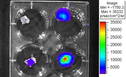
- Slices are imaged for 5 min at high sensitivity (Figure 1).
V. Representative Results

Figure 1. Bioluminescent imaging of patient liver slices using IVIS detection system.
Discussion
We describe an ex vivo patient tissue culture method and bioluminescent detection system for the assessment of gene delivery within non-fixed human tissue. The method offers a simple and reproducible way of culturing tissue slices. It has significant potential as it allows the study of gene delivery into intact human tissue, the analysis of gene delivery into a variety of human tissue including malignant tissue and can provide important information concerning the effects of the high degree of variability between...
Disclosures
No conflicts of interest declared.
Acknowledgements
This work was funded through the Cork South Infirmary Victoria University Hospital Breast fund, the Irish Cancer Society (CRI07TAN) and the Cork Cancer Research Centre. Replication incompetent recombinant Adenovirus 5 particles under the transcriptional control of the CMV promoter was a kind gift from Prof. Andrew Baker, University of Glasgow.
Materials
| Name | Company | Catalog Number | Comments |
| DMEM | Sigma-Aldrich | D6429 | |
| PBS | Sigma-Aldrich | D8537 | |
| Firefly Luciferin | Biosynth International, Inc | ||
| Dermabond | Johnson & Johnson | ||
| Vibrotome | Leica Microsystems | VT 1000 A |
References
- van de Bovenkamp, M., Groothuis, G. M., Draaisma, A. L., Merema, M. T., Bezuijen, J. I., van Gils, M. J. Precision-cut liver slices as a new model to study toxicity-induced hepatic stellate cell activation in a physiologic milieu. Toxicol Sci. 85, 632-638 (2005).
- Speirs, V., Green, A. R., Walton, D. S., Kerin, M. J., Fox, J. N., Carleton, P. J. Short-term primary culture of epithelial cells derived from human breast tumours. Br J Cancer. 78, 1421-1429 (1998).
- Kirby, T. O., Rivera, A., Rein, D., Wang, M., Ulasov, I., Breidenbach, M. A novel ex vivo model system for evaluation of conditionally replicative adenoviruses therapeutic efficacy and toxicity. Clin Cancer Res. 10, 8697-8703 (2004).
- Josserand, V., Texier-Nogues, I., Huber, P., Favrot, M. C., Coll, J. L. Non-invasive in vivo optical imaging of the lacZ and luc gene expression in mice. Gene Ther. 14, 1587-1593 (2007).
Reprints and Permissions
Request permission to reuse the text or figures of this JoVE article
Request PermissionThis article has been published
Video Coming Soon
Copyright © 2025 MyJoVE Corporation. All rights reserved