Method Article
Whole Animal Perfusion Fixation for Rodents
In This Article
Summary
Here we describe a low-cost, rapid, controlled and uniform fixation procedure using 4% paraformaldehyde perfused via the vascular system: through the heart of the rat to obtain the best possible preservation of the brain.
Abstract
The goal of fixation is to rapidly and uniformly preserve tissue in a life-like state. While placing tissue directly in fixative works well for small pieces of tissue, larger specimens like the intact brain pose a problem for immersion fixation because the fixative does not reach all regions of the tissue at the same rate 5,7. Often, changes in response to hypoxia begin before the tissue can be preserved 12. The advantage of directly perfusing fixative through the circulatory system is that the chemical can quickly reach every corner of the organism using the natural vascular network. In order to utilize the circulatory system most effectively, care must be taken to match physiological pressures 3. It is important to note that physiological pressures are dependent on the species used. Techniques for perfusion fixation vary depending on the tissue to be fixed and how the tissue will be processed following fixation. In this video, we describe a low-cost, rapid, controlled and uniform fixation procedure using 4% paraformaldehyde perfused via the vascular system: through the heart of the rat to obtain the best possible preservation of the brain for immunohistochemistry. The main advantage of this technique (vs. gravity-fed systems) is that the circulatory system is utilized most effectively.
Protocol
1. Prepare Fixative
(See table Fixative and Buffers.)
2. Prepare Perfusion Buffers
(See table Fixative and Buffers.)
3. Prepare Apparatus and Anesthesia
- Using the water bath, warm perfusion buffer to 37 °C. Place outlet valve in a beaker filled with buffer. Fill a 50 ml syringe with buffer and attach to fixative tubing. Flush tubing repeatedly by expelling and withdrawing buffer.
- Clear the line with buffer until all air bubbles in the tubing are eliminated. It is crucial for the success of a perfusion not to have air bubbles in any of the lines.
- Remove the syringe and connect the 4% paraformaldehyde fixative (room temperature) container. To avoid air bubbles at the tubing end, squeeze the tube while positioning the tube into the container so that a drop of buffer protrudes from the end to contact the fluid surface within the container.
- Close the outlet port (needle end). Turn buffer valve (blue) to the same position as the fixative valve (white). This will allow flow from the buffer line only.
- Open the outlet port and repeat step A1 through A3 for filling the buffer line. After all the air bubbles are eliminated close the outlet port, remove the syringe and connect the buffer container, taking care not to introduce air bubbles into the tubing.
- Test system for ability to hold pressure by pumping the rubber manometer blub. There is normally some resistance due to the compression of air in the system.
- Setup surgery tools in order for easy of access. Fill perfusion needle with buffer to eliminate the possibility of air bubbles.
- Prior to surgery, a ketamine/xylazine mixture (up to 80 mg/kg body weight ketamine and 10 mg/kg body weight xylazine) is administered via intraperitoneal injection (27 gauge needle and 1 cc syringe). Additional administration of anesthetic will be performed as necessary during the course of each operation to maintain a surgical plane of anesthesia.
4. Perfusion Surgery
- Once the animal has reached a surgical plane of anesthesia, place it on the shallow tray filled with crushed ice. (Use toe pinch-response method to determine depth of anesthesia. Animal must be unresponsive before continuing).
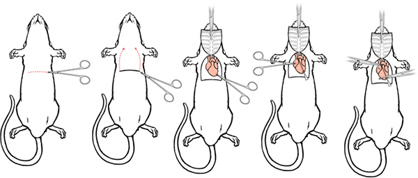
- Make a 5-6 cm lateral incision through the integument and abdominal wall just beneath the rib cage. Carefully separate the liver from the diaphragm.
- Make a small incision in the diaphragm using the curved, blunt scissors. The position and pressure of your finger can aid in the ability to cut the diaphragm.
- Continue the diaphragm incision along the entire length of the rib cage to expose the pleural cavity.
- Place curved, blunt scissors along one side of the ribs, carefully displacing the lungs, and make a cut through the rib cage up to the collarbone. Make a similar cut on the contralateral side.
- Lifting the sternum away, carefully trim any tissue connecting it to the heart. Clamp the tip of the sternum with the hemostat and place the hemostat over the head. When done properly, the thymus lifts away from the heart along with the sternum, providing a clear view of the major vessels.
- Make a small incision to the posterior end of the left ventricle using iris scissors.
Click here to view larger figure.
- Pass a 15-gauge blunt- or olive-tipped perfusion needle through the cut ventricle into the ascending aorta. The tip should be visible through the wall of the aorta, and should not reach the aortic arch where the brachial and carotid arteries diverge.
- Use a hemostat to clamp the heart, this secures the needle and prevents leakage. If desired, the modified hemostat can be used to clamp the aorta around the needle tip (these hemostats remain in place until dissection begins, but are omitted from future illustrations for clarity).
- Finally, make an incision to the animal's right atrium using iris scissors to create as large an outlet as possible without damaging the descending aorta. At this point the animal is ready to be perfused.
5. Perfusion
- Open and attach outlet port to needle base taking care not to introduce any air bubbles.
- Pump up the manometer bulb to a pressure of 80 mm Hg quickly & evenly. Maintain this pressure throughout the buffer infusion period. Start the timer.
- Adjust the needle angle. The angle of the needle is critical to the achievement of a maximum flow rate (note the flow change with angle adjustment).
- Switch the buffer valve (blue) once buffer is almost finished (200 ml). The fluid should be running clear. The clearing of the liver is an indicator of a good perfusion. The liver should be clear at this point. Indicate time for your records.
- Fixation tremors should be observed within seconds; this should be considered the true time of fixation. Indicate time for your records.
- The pressure can gradually be increase up to a maximum of 130 mm Hg 2 to maintain a steady flow rate.
- Close the outlet valve once the fixative is nearly finished. Indicate ending time for your records.
- The rat should be stiff at this stage.
- The used paraformaldehyde must be collected and stored for disposal according to the regulations of your institution.
6. Dissection 4,11
- Remove the head using a pair of scissors.
- Make a midline incision along the integument from the neck to the nose and expose the skull.
- Trim off the remaining neck muscle so that the base of the skull is exposed; remove any residual muscle using scissor or rongeurs.
- Place the sharp end of a pair of iris scissors into the foramen magnum on one side, carefully sliding the scissors along the inner surface of the skull.
- Next, make a cut extending to the distal edge of the posterior skull surface. Make an identical cut on the contralateral side. Use the rongeurs to clear away the skull around the cerebellum.
- Carefully slide the scissors along the inner surface of the skull as the tip travels from the dorsal distal posterior corner to the distal frontal edge of the skull, lifting up on the blade as you are cutting to prevent damage to the brain. Repeat for opposite side.
- Using rongeurs peel the dorsal surface of the skull away from the brain. Trim away the sides of the skull using rongeurs as well.
- Using a spatula, sever the olfactory bulbs and nervous connections along the ventral surface of the brain.
- Gently tease the brain away from the head, trimming any dura that still connects the brain to the skull using iris scissors.
- Remove the brain and place it in a vial of fixative containing fluid at least 10x the volume of the brain itself. Swirl the vial occasionally.
7. Post-fixation & Storage
- Keep the brain in fixative for 24 hours at 4 °C, swirling occasionally.
- After 24 hours, wash the brain with phosphate buffered saline by exchanging the media 3 times and swirling each time.
- Brains can then be stored in phosphate buffered saline or HBHS with sodium azide and kept at 4 °C.
8. Representative Results
An initial indicator of the success of the perfusion is the clearing of extremities such as the nose, ears, and paws and internal organs such as the thymus gland and liver (IHC World). Gross inspection of the brain reveals the blood vessel void of blood (white to pale yellow appearance). This will be also true within tissue sections destine for staining and immunohistochemistry. The final indicator of outcome of the perfusion is the condition of the ultrastructure in the tissue.1,6,7,8
| Prepare Fixative: |
| Prepare 8% Paraformaldehyde stock |
|
| Prepare 0.2 M Sodium Phosphate Buffer, pH 7.4 |
|
| Prepare 4% Paraformaldehyde Fixative |
|
| Prepare Perfusion and Storage Buffers: |
| Prepare Phosphate Buffered Saline, pH 7.4 |
|
| HEPES Buffered Hanks Solution (HBHS) with Sodium Azide pH 7.4 |
|
Table 1. Preparation of Fixative and Buffers.
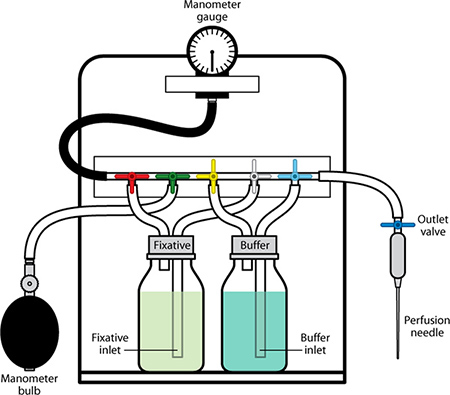
Figure 1. Perfusion Apparatus with labeled components.
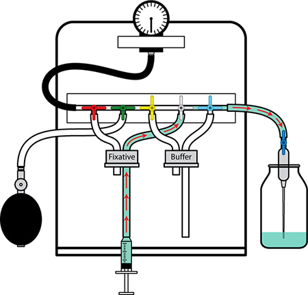
Figure 2. Preparation of the Perfusion Apparatus I. Begin with the fixative line. Flush the tubing of air bubbles and fill with buffer by rapidly expelling and withdrawing buffer into the syringe and through the line. Shut the valve on the needle end off. Place the fixative line into the fixative bottle without introducing air bubbles.
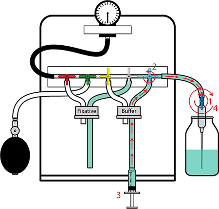
Figure 3. Preparation of the Perfusion Apparatus II. 1. Open valve on the needle end. 2. Turn buffer valve (blue) to position for flow. 3. Flush the tubing of air bubbles and fill with buffer by rapidly expelling and withdrawing buffer with the syringe. 4. Close valve on the needle end. Place tubing into the buffer bottle. Test pressure to make sure the system is sealed correctly by pumping up the manometer bulb and watching the gauge. The apparatus is now ready for the perfusion procedure.
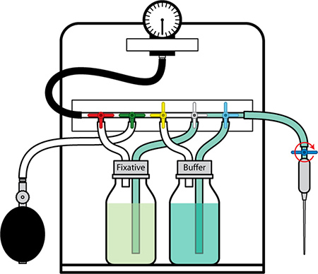
Figure 4. Perfusion Apparatus in position for buffer delivery.
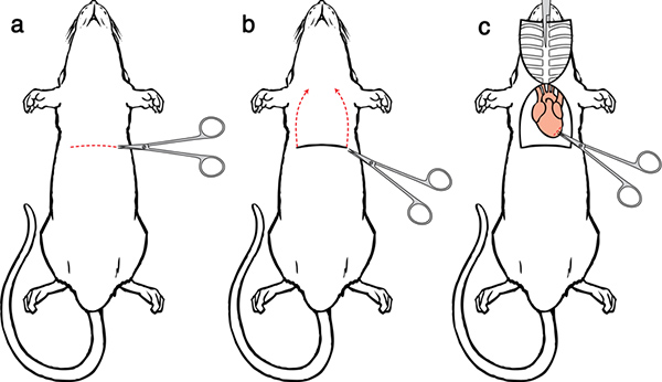
Figure 5. Perfusion Surgery I. a) Make a lateral incision through the integument and abdominal wall. b) Make an incision in the diaphragm and cut across the diaphragm exposing the heart. Make parallel cuts on either side of the ribs up to the collarbone. c) clamp the tip of the sternum with the hemostat and place the hemostat over the head.
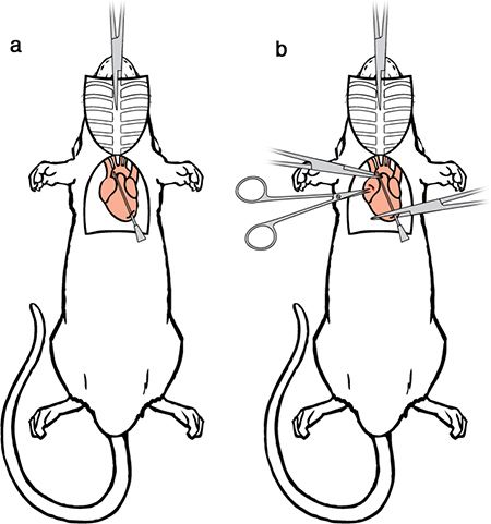
Figure 6. Perfusion Surgery II. a) Pass the perfusion needle through the cut ventricle into the ascending aorta. b) Secure the perfusion needle use one set of hemostats to clamp the heart. A second set of a modified hemostat can also be used to clamp the aorta around the needle tip to prevents leakage. Using iris scissors make a small incision to the posterior end of the left ventricle.
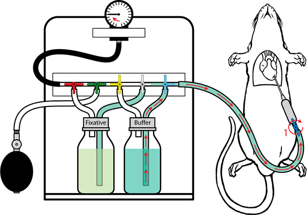
Figure 7. Perfusion with Buffer Pump the manometer bulb to create pressure in the line. Open up the valve (1) and attach to the needle hub. It is critical to make sure no air bubbles are introduced. Pump up the manometer bulb to a pressure of 80 mm Hg and maintain this pressure throughout the buffer infusion period.
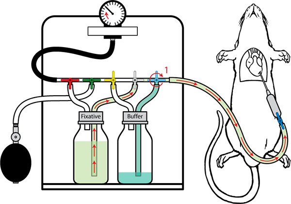
Figure 8. Perfusion with Fixative Once buffer is almost finished (200 ml) switch the buffer valve (1) to allow for the fixative to flow. The pressure can gradually be increased up to a maximum of 130 mm Hg.
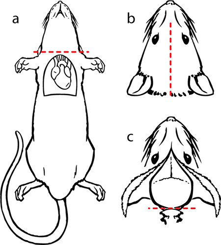
Figure 9. Dissection I. a) remove the head using a pair of scissor. b) make an incision in the skin along the midline from the neck to the nose and expose the skull. c) trim off the remaining neck muscle so that the base of the skull is exposed. Hold the head so that the large opening in the cranium surface (foramen magnum) is accessible. Carefully insert the sharp end of a pair of iris scissors into the foramen magnum and make a cut. Repeat the same maneuver for the contralateral side. Use the rongeurs to clear away the skull around the cerebellum.

Figure 10. Dissection II. a) carefully slide the scissors along the inner surface of the skull. Lift up on the tip as you are cutting to prevent damage to the brain. Repeat the same for opposite side. Use rongeurs peel the skull away from the brain. b) illustration with the skull removed and the brain exposed. c) use a spatula to sever the olfactory bulbs and nerve connections at the most anterior location of the brain. Carefully guide the spatula tip along the underside of the brain to sever connections for ease of remove. Remove the brain and place it in a vial of fixative.
Discussion
Additional notes for a successful surgery:
- Once the diaphragm is breached, precede rapidly as hypoxia and hypercapnia will initiate irreversible physiological changes that may confound subsequent analysis.
- When the needle is inserted and clamped in place, the residual pressure will push blood to the base of the needle (flashback). In fact, larger animals may vigorously expel blood. It is critical that blood at least reach the base of the needle to avoid introducing air bubbles that will present an obstacle to complete perfusion. If no blood is observed at the needle base, remove the needle and refill with buffer by attaching it to the outlet port of the perfusion rig. Once buffer has been run through the tip, the needle can be reinserted into the heart.
- Proper placement of the needle within the aorta is critical, it should not reach as far as the aortic arch.
- If multiple perfusions are taking place it is not necessary to setup from the beginning every time. The user needs to have the proper volume for both the buffer and fixative in each bottle (200mls animal/solution). The bottles can also be refilled if needed. The most important aspect to consider is flushing out the perfusion line that runs from the setup to the animal (output valve). After the completion of a perfusion, this line will have 4% paraformaldehyde in it. Remove the tubing from the buffer bottle and use a 50 ml syringe filled with water to flush out the fixative. Make sure the outlet valve is placed in a waste collection beaker for proper disposal of the paraformaldehyde. Repeat this three times. Refill with buffer using the same method shown in 2.3.
- This method for fixation is highly adaptable with the ability to change both buffer and fixative other fixatives (such as those used for EM) according to the requirements of the experiment.
- The system is also frequently used for the extraction of live tissue. For this process one simply uses the delivery of buffer only (the buffer bottle while still attached to entire system is placed in an ice bucket and the fixative bottle is setup as empty but sealed). This assures a cold buffer perfusion at the optimal pressure. From here the system can be adapted to the infusion of fixable dyes by either adding the dye directly to the buffer (if the quantity isn't an issue) or use a syringe with a minimal amount of buffer/dye solution in between the buffer – fixative step.
- In the case of vascular casting, the pressure monitoring buffer step is crucial however due to the viscosity and solidifying nature of the casting solution either a 50ml syringe must be used for direct delivery or a disposable secondary attachment to the apparatus (tubing, valve and holding container) made to interface with the current system be used. Our laboratories' vascular casting preceded the design and manufacturing of the perfusion apparatus. Therefore we cannot state that the pressure measurements will be an accurate measurement at the delivery site. The numbers may have to be adjusted to achieve successful delivery of viscous fluids.
Disclosures
The authors have nothing to disclose.
Acknowledgements
This work was funded by the Center for Neural Communication Technology (CNCT), a P41 Resource Center funded by the National Institute of Biomedical Imaging and Bioengineering (NIBIB, P41 EB002030) and supported by the National Institutes of Health (NIH).
Materials
| Name | Company | Catalog Number | Comments |
| Anesthetic: | |||
| Ketamine/xylazine mixture (Anesthetic may vary per laboratory / institution) | Ketaset | NDC 0856-2013-01 | 10 mL vial |
| Surgery: | |||
| Needle tip, 27 GA x 1.25" | Materiel Services | 25251 | |
| Large blunt/blunt curved scissors (~14.5 cm) | Fine Science Tools | 14519-14 | |
| Straight iris scissors | Fine Science Tools | 14058-11 | |
| Standard tweezers | Fine Science Tools | 11027-12 | |
| Pair of fine (Graefe) tweezers | Fine Science Tools | 11050-10 | |
| 1 large hemostat forceps - curved or straight (~19 cm) | surgicaltools.com | 17.21.51 | |
| 2 standard hemostat forceps - straight serrated (14 cm) | Fine Science Tools | 13013-14 | |
| 1 modified hemostat (with 15-gauge hole filed through the tip) | Fine Science Tools | 13013-14 | |
| 15-gauge blunt or olive-tipped needle (perfusion needle) | Fisnar | 5601137 | |
| Perfusion: | |||
| HyPerfusion system or equivalent | |||
| Phosphate buffered saline, pH 7.4 | |||
| 4% Paraformaldehyde in 0.1 M Phosphate Buffer, pH 7.4 | |||
| Shallow glass or plastic tray, approximately 10" x 10" | |||
| Crushed ice | |||
| Water bath (37 °C) | |||
| Timer | |||
| 50 ml syringe | Medline Industries | NPMJD50LZ | |
| Dissection: | |||
| Pair of standard sharp/blunt straight scissors (~12 cm) | Fine Science Tools | 14054-13 | |
| Medium curved or straight rongeurs (14-16 cm) or skull bone removal pliers | Fine Science Tools | 16020-14 | |
| Straight iris scissors (~9 cm) | Fine Science Tools | 14058-11 | |
| Micro-spatula (double 2" flat ends, one rounded, one tapered to 1/8") | Fine Science Tools | 10091-12 | |
| Post-Fixation & Storage: | |||
| 50 ml glass vial | |||
| 40 ml HEPES-Buffered Hanks Solution (HBHS) with sodium azide (90mg/l) | |||
References
- Cinar, O., Semiz, O., Can, A. A microscopic survey on the efficiency of well-known routine chemical fixatives on cryosections. Acta. Histochem. 108, 487-496 (2006).
- Fritz, M., Rinaldi, G. Blood pressure measurement with the tail-cuff method in Wistar and spontaneously hypertensive rats: Influence of adrenergic- and nitric oxide-mediated vasomotion. J. Pharmacol. Toxicol. Methods. 58, 215-221 (2008).
- Ikeda, K., Nara, Y., Yamorii, Y. Indirect systolic and mean blood pressure determination by anew tail cuff method in spontaneously hypertensive rats. Laboratory Animals. 25, 26-29 (1991).
- Jacobowitz, D. M. Removal of discrete fresh regions of rat brain. Brain Res. 80, 111-115 (1974).
- Jonkers, B. W., Sterk, J. C., Wouterlood, F. G. Transcardial perfusion fixation of the CNS by means of a compressed-air-driven device. Journal of Neurosci. Meth. 12, 141-149 (1984).
- Jung-Hwa, T. a. o. -. C. h. e. n. g., Gallant, J., Brightman, P. E., Dosemeci, M. W., A, ., Reese, T. S. Structural changes at the synapse after delayed perfusion fixation in different regions of the mouse brain. J. Comp. Neurol. 501, 731-740 (2007).
- Kasukurthi, R., Brenner, M. J., Morre, A. M., Moradzadeh, A., Wilson, Z. R., Santosa, K. B., Mackinnon, S. E., Hunter, D. A. Transcardial perfusion versus immersion fixation for assessment of peripheral nerve regeneration. J. Neurosci. Meth. 184, 303-309 (2009).
- Lamberts, R., Goldsmith, P. C. Fixation, fine structure, and immunostaining for neuropeptides: perfusion versus immersion of the neuroendocrine hypothalamus. J. Histochem. Cytochem. 34, 389-398 (1986).
- Ramos-Vara, J. A. Technical Aspects of Immunohistochemistry. Vet. Pathol. 42, 405-426 (2005).
- Shi, Z. R., Itzkowitz, S. H., Kim, Y. S. A comparison of three immunoperoxidase techniques for antigen detection in colorectal carcinoma tissues. J. Histochem. Cytochem. 36, 317-322 (1988).
- Walker, W. F. . Vertebrate dissection. , (1980).
- Zwienenberg, M., Gong, Q., Lee, L. L., Berman, R. F., Lyeth, B. G. J. Neurotrauma. Monitoring in the Rat: Comparison of Monitoring in the Ventricle, Brain Parenchyma, and Cisterna Magna. 16, 1095-1102 (1999).
Reprints and Permissions
Request permission to reuse the text or figures of this JoVE article
Request PermissionThis article has been published
Video Coming Soon
Copyright © 2025 MyJoVE Corporation. All rights reserved