A subscription to JoVE is required to view this content. Sign in or start your free trial.
Method Article
Functionalization of Atomic Force Microscope Cantilevers with Single-T Cells or Single-Particle for Immunological Single-Cell Force Spectroscopy
In This Article
Summary
We present a protocol to functionalize atomic force microscope (AFM) cantilevers with a single T cell and bead particle for immunological studies. Procedures to probe single-pair T cell-dendritic cell binding by AFM and to monitor the real-time cellular response of macrophages to a single solid particle by AFM with fluorescence imaging are shown.
Abstract
Atomic force microscopy based single cell force spectroscopy (AFM-SCFS) is a powerful tool for studying biophysical properties of living cells. This technique allows for probing interaction strengths and dynamics on a live cell membrane, including those between cells, receptor and ligands, and alongside many other variations. It also works as a mechanism to deliver a physical or biochemical stimulus on single cells in a spatiotemporally controlled manner, thus allowing specific cell activation and subsequent cellular events to be monitored in real-time when combined with live-cell fluorescence imaging. The key step in those AFM-SCFS measurements is AFM-cantilever functionalization, or in other words, attaching a subject of interest to the cantilever. Here, we present methods to modify AFM cantilevers with a single T cell and a single polystyrene bead respectively for immunological studies. The former involves a biocompatible glue that couples single T cells to the tip of a flat cantilever in a solution, while the latter relies on an epoxy glue for single bead adhesion in the air environment. Two immunological applications associated with each cantilever modification are provided as well. The methods described here can be easily adapted to different cell types and solid particles.
Introduction
Atomic force microscopy (AFM), a versatile tool, has found many applications in cell biology research1,2,3,4,5. Apart from its high-resolution imaging capability, the native force-probing feature allows biophysical properties of living cells to be investigated directly in situ at the single-cell level6,7. These include the rigidities of subcellular structures or even whole cells8,9,10,11,12, specific ligand/receptor binding strengths at the single-molecule level on the cell surface13, and adhesion forces between single-pairs of solid particles and cells or between two cells1,2,14,15. The latter two are often categorized as single-cell force spectroscopy (SCFS)16. Owing to the readily available cantilevers with various spring constant, the force range accessible to AFM is rather broad from a few piconewtons (pN) to micronewtons (µN), which adequately covers the entire range of cellular events involving forces from a few tens of pN, such as receptor-based single-molecule binding, to nN, such as phagocytic cellular events15. This large dynamic force range makes AFM advantageous over other force-probing techniques such as optical/magnetic tweezers and a biomembrane force probe, as they are more suitable for weak-force measurements, with force typically less than 200 pN17,18. In addition, AFM can function as a high-precision manipulator to deliver various stimuli onto single cells in a spatiotemporally defined manner4,19. This is desirable for the real-time single-cell activation studies. Combined with live-cell fluorescence imaging, the subsequent cellular response to the specific stimulus can be monitored concurrently, thus making AFM-based SCFS exceedingly robust as optical imaging providing a practical tool to probe cellular signaling. For instance, AFM was used to determine the strains required to elicit calcium transients in osteoblasts20. In this work, calcium transients were tracked fluorescently through calcium ratiometric imaging after the application of localized forces on cultured osteoblasts with an AFM tip. Recently, AFM was employed to stretching collagen fibrils on which hepatic stellate cells (HSC) were grown and this mechano-transduced HSC activation was real-time monitored by a fluorescent Src biosensor, whose phosphorylation as represented by the fluorescence intensity of the biosensor is correlated with HSC activation3.
In AFM-based SCFS experiments, proper functionalization of AFM cantilevers is a key step toward successful measurements. Since our research interest focuses on immune cells activation, we routinely functionalize cantilevers with particulate matters such as single solid particles that can trigger phagocytosis and/or strong immune responses4,14,15 and single T cells that can form an immune synapse with antigen presenting cells, such as activated dendritic cells (DC)2. Single solid particles are normally coupled to a cantilever via an epoxy glue in the air environment, whereas single T cells, due to their non-adhesive nature, are functionalized to a cantilever via a biocompatible glue in solution. Here, we describe the methods to perform these two types of cantilever modification and give two associated applications as well. The first application is to probe T cell/DC interactions with AFM-SCFS to understand the suppressive mechanism of regulatory T cells from the cell mechanics point of view. The second one involves combining AFM with live-cell fluorescence imaging to monitor the cellular response of macrophage to a solid particle in real-time to reveal the molecular mechanism of receptor-independent phosphatidylinositol 4,5-bisphosphate (PIP2)-Moesin mediated phagocytosis. The aim of this protocol is to provide a reference framework for interested researchers to design and implement their own experimental settings with AFM-based single-cell analysis for immunological research.
Protocol
The mouse experiment protocol follows the animal care guidelines of Tsinghua University
1. Cantilever functionalization with single T cells
- Mouse spleen cells preparation
- Sacrifice the mouse (8-16 weeks of age (either sex); e.g., C57BL/6 strain) using carbon dioxide, followed by cervical dislocation.
- Clean the mouse with 75% ethanol and make a midline skin incision followed by splenectomy.
- Homogenize the spleen in 4 mL of PBS containing 2% fetal bovine serum (FBS) using glass slides and remove aggregates and debris by passing the cell suspension through a 70 μm mesh nylon strainer.
- Centrifuge the cell suspension at 500 x g for 5 min, discard the supernatant and resuspend cells in 2 mL of red blood cell lysis buffer (balanced at room temperature) for 5 min. Terminate the lysis reaction by adding 8 mL of PBS solution.
- Centrifuge the cell suspension at 500 x g for 5 min and resuspend cells at a density of 1 x 10⁸ cells/mL in PBS containing 2% FBS and 1 mM EDTA (Labelled as Solution A), typically 0.25-2 mL depending on the cell density. Transfer the resuspended cells to a 5 mL (12 x 75 mm) polystyrene round bottom tube.
- Mouse CD4+ T cells preparation
- Add 50 μL/mL rat serum (see Table of Materials) and 50 μL/mL CD4+ T cell isolation cocktail (see Table of Materials) to the cell sample obtained from step 1.1.5. Mix and incubate for 10 min at room temperature.
- Vortex the stock streptavidin-coated magnetic particle solution (see Table of Materials) for 30 s or until the particles appear evenly dispersed.
- Add 75 μL/mL streptavidin-coated magnetic particles to the cell sample. Mix and incubate for 2.5 min at room temperature.
- Add Solution A to top up the cell sample to 2.5 mL and mix by gently pipetting up and down for 2-3 times.
- Place the sample tube (without lid) into the magnet (see Table of Materials) and incubate for 5 min at room temperature. Carefully pour the enriched cell suspension into a new 5 mL polystyrene round-bottom tube.
- Centrifuge the cell suspension at 500 x g for 5 min. Discard the supernatant and resuspend the enriched T cells in 500 μL of Solution A.
NOTE: The enriched CD4+ T cells contain both conventional and regulatory T cells.
- Regulatory T cells separation from conventional T cells
- Add 25 µL of FcR blocker (see Table of Materials) to the enriched T cell sample obtained from step 1.2.6. Mix and incubate for 5 min at room temperature.
- Add 25 μL of regulatory T cell positive selection cocktail (see Table of Materials) to the T cell sample. Mix and incubate for 10 min at room temperature.
- Add 10 μL of PE selection cocktail (see Table of Materials) to the T cell sample. Mix and incubate for 5 min at room temperature.
- Vortex the stock dextran-coated magnetic particle solution (see Table of Materials) for 30 s or until the particles appear evenly dispersed.
- Add 10 μL of dextran-coated magnetic particles to the T cell sample. Mix and incubate for 5 min at room temperature.
- Add the Solution A to top up the T cell sample to 2.5 mL and mix by gently pipetting up and down for 2-3 times.
- Place the T cell sample tube (without lid) into the magnet and incubate for 5 min at room temperature. Carefully pour the supernatant to a new tube.
NOTE: The supernatant contains the enriched conventional CD4+T cells. - Centrifuge the enriched conventional CD4+T cells at 500 x g for 5 min. Discard the supernatant and resuspend the cells in 4 mL of RPMI1640 containing 10% FBS, 0.05 mM β-Mercaptoethanol, 0.01 M HEPES and 1% penicillin/streptomycin (labeled as Medium B).
- Remove the tube in which regulatory T cells are enriched from the magnet. Add 2.5 mL of Solution A to the tube and mix by gently pipetting up and down for 2-3 times. Put the tube back into the magnet, incubate for 5 min, and then carefully pour off and discard the supernatant. Repeat this step three more times.
- Resuspend the enriched regulatory T cells in 2 mL of Medium B.
- Incubate both purified conventional T cells and regulatory T cells with 100 U/mL hIL-2 overnight or for at least 4 h at 37 °C in a humidified incubator with 5% CO2 before being used for cantilever functionalization.
- Dendritic cells preparation
- Prepare piranha solution, a mixture of 30% H2O2 (30%) and 70% H2SO4 (conc) (v/v). Slowly pour 3 mL of H2O2 into 7 mL of H2SO4 under constant stirring and cooling.
CAUTION: Piranha solution is highly corrosive, and it can burn and destroy body tissues. Therefore, it is safer to use the piranha solution under a hood and wear appropriate safety equipment, as the mixture will splash around the beaker. Neutralize the solution with NaOH to pH 7 after use. - Immerse the glass coverslip of 24 mm diameter into the piranha solution for 30 min and rinse thoroughly afterward with the sterile ultrapure water.
- Dip a pair of pointed tweezers in 75% ethanol for 30 min for cold disinfection.
- Introduce the cleaned glass coverslips into a 6-well culture plate by the tweezers.
- Tilt a 6 cm plastic culture dish in which DC2.4 cells were pre-cultured with 4 mL of Medium B and aspirate all the medium. Add 2 mL of PBS into the culture dish to rinse DC2.4 cells and discard PBS. Repeat this rinsing step two more times.
- Add 1 mL of 0.25% trypsin EDTA to the culture dish for 2 min. Add 1 mL of Medium B to this dish to end the enzyme digestion reaction. Transfer the digested cell suspension to a 15 mL tube.
- Centrifuge the cell suspension at 500 x g for 5 min and resuspend DC2.4 cells at a density of 2 x 10⁵ cells/mL in Medium B.
- Seed DC2.4 cells on the glass coverslips prepared at step 1.4.4 and incubate the cells overnight in a humidified chamber at 37 °C with 5% CO2.
NOTE: In order to measure interacting forces between two single cells, a relatively low concentration of DC2.4 cells (i.e., <10% confluency) is necessary to have a proper spacing among cells.
- Prepare piranha solution, a mixture of 30% H2O2 (30%) and 70% H2SO4 (conc) (v/v). Slowly pour 3 mL of H2O2 into 7 mL of H2SO4 under constant stirring and cooling.
- AFM cantilever preparation
NOTE: Cantilevers that are suitable for single cell force spectroscopy experiments are those with low spring constants, typically in the range of 0.01-0.06 N/m. Here, soft tip-less cantilevers are preferred for single cells and single solid particles functionalization.- Clean the cantilevers by Piranha treatment or plasma or UV-ozone cleaning.
- Mount the cleaned cantilever to the AFM scanning head.
- Prepare a clean sample chamber filled with pure water and calibrate the cantilever in water solution by first running a force curve on the glass substrate to obtain the sensitivity (the slope of the linear fit over the repulsive part of the approaching curve) and then recording a thermal noise spectrum to extract the spring constant according to the instruction manual of the AFM.
- Remove the AFM scanning head from the solution, wash the mounted cantilever with a few drops of pure ethanol, and keep the cantilever dry on the scanning head.
- Attaching single T cells to the cantilever
- Preheat the living cell environment enclosure with 5% CO2 at 37 °C.
- Mount the glass coverslip with DC2.4 cells grown on it from step 1.4.8 to a sample chamber assembly, add 600 µL of Medium B to the chamber immediately, and then put the assembly onto the AFM sample stage.
- Add hIL-2 incubated CD4+T cells (either conventional or regulatory T cells) into the sample chamber.
NOTE: The total sample volume should not exceed 1 mL. - Wait until the added CD4+T cells are fully settled down on the bottom of the coverslip.
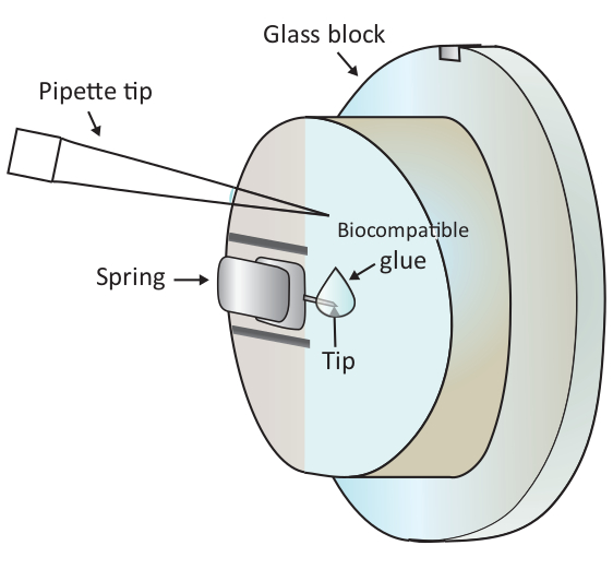
NOTE: Air bubbles will cause great disturbance to the experiment, therefore, it is advisable to avoid any air bubbles in step1.6.2 and 1.6.3. - Add a drop of 2 μL of biocompatible glue onto the end of the mounted cantilever with a pipette as shown in Figure 1 and then place the scanning head on the sample stage quickly, thus letting the cantilever coated with the biocompatible glue immerse in the solution.
CAUTION: Do not touch the glass block or the cantilever with the pipette tip. Since the biocompatible glue used here is prone to oxidation in air, this step should be done as quickly as possible. - Locate a healthy T cell beneath the tip of the cantilever coarsely under the microscope by moving the sample stage and then finely adjust the positioning by moving the scanning head.
NOTE: A healthy CD4+T cell typically has a relatively large size, smooth edges, and optically transmissive in the bright-field imaging. - Lower the cantilever manually with step-sizes starting from 50 μm then to 10, 5, 2 and 0.5 μm gradually by controlling the stepper motors. Hold the position of the stepper motors and adjust the positioning of the scanning head for better alignment between the cantilever tip and the cell, once the cantilever makes a firm contact with the target T cell as indicated by a small displacement of the laser beam position in the photodetector corresponding to a typical force range of 0.5-1.5 nN.
NOTE: This step can also be done by running a single force measurement in which set-point (the force applied to the cell) and contact time can be well defined in the software. However, due to the non-adhesive nature of T cells, the manual approach provides more flexibility in controlling aiming, positioning, and contact time than does the automatic approaching and it works reliably for T cell adhesion. Future experimentalists should try both the manual and automatic approaching to find out which works better for their systems of interest. - Retract the cantilever after 30 s of contact.
NOTE: If the cell moves with the cantilever, the attachment is successful. If not, repeat step 1.6.6 but on a different T cell. The biocompatible glue is easily oxidized. Step 1.6.5-1.6.7 should be completed within 5 min. In addition, if the same cantilever fails three times for the T cell attachment, a new cantilever should be used, and the attachment procedure should begin from Step 1.5.2 again.

Figure 1: Schematic representation of adding a small drop of biocompatible glue onto the mounted cantilever. The cantilever is mounted via a clamping spring on the glass-block holder that is installed on the AFM scanning head (not drawn here). When the scanning head stands on a leveled surface, the cantilever is vertically oriented as shown in the drawing. About 2 µL biocompatible glue can be added to the tip of the cantilever with a micro-pipette. Please click here to view a larger version of this figure.
- Force spectroscopy of single-pair T cell/ dendritic cell interaction
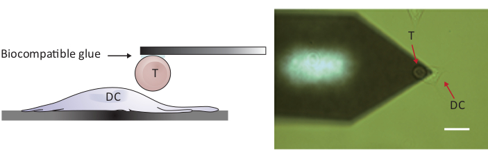
NOTE: To probe cell/cell interactions, an AFM with a Z-range larger than the conventional 10-15 μm is required in order to fully separate the two cells. The AFM used here has a Z-range of 100 μm, which is adequate to separate T cell from the dendritic cell after cell/cell contact.- Position the attached T cell above a separate DC2.4 cell by moving the sample stage and/or the scanning head (see Figure 2).
- Set proper parameters and run force spectroscopy.
NOTE: The following key settings are typically used: Setpoint 0.5 nN, Pulling Length 50 μm, Z movement Constant Speed, Extend Speed 5 μm/s, Contact Time 10 s, Delay Mode Constant Force. For each T-DC pairs, 20 repeats of force curves are collected and a minimum of 14 force curves are used for further analysis. - Mount a new cleaned cantilever, calibrate it in pure water as in Step 1.5.3, and go back to the same T-DC cells sample to repeat Step 1.6 and 1.7 for a different T-DC pair. Probe at least 5 pairs for each condition.

Figure 2: Experimental configuration of force-probing between a single T cell and DC. (A) Schematic drawing of the experimental configuration in which a T cell attached to the cantilever is brought to a DC grown on the substrate for force-probing. (B) Bright-field image of a T cell-functionalized cantilever and a DC. Scale bar, 20 µm. Please click here to view a larger version of this figure.
2. Cantilever functionalization with single polystyrene beads
- Single beads preparation
- Dilute the stock suspension of 6 μm polystyrene beads in 100% ethanol.
NOTE: The concentration of diluted beads solution should be low enough so that when added to a glass coverslip surface, individual beads are well separated without significant clustering after solvent evaporation. - Clean the 24 mm diameter glass coverslip with ethanol and remove any dust by N2 air flow.
- Mount the cleaned glass coverslip to a sample chamber assembly and put the assembly onto the microscope.
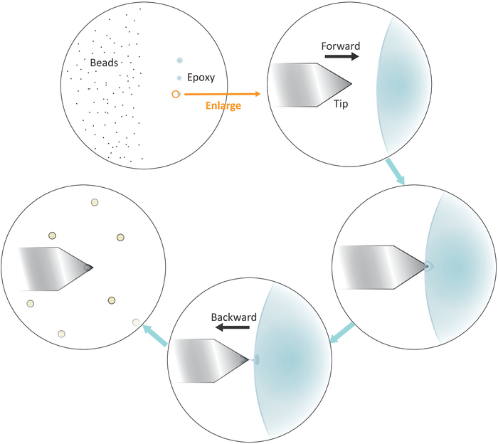
- Put a drop of diluted beads solution to the left side but close to the center of the coverslip (see Figure 3) and check the spacing among the beads after solvent evaporation in bright-field under the microscope with a 20x objective. Proceed to the next step if individual beads are well separated.
- Dip a micropipette tip or a toothpick into a well-mixed epoxy glue and then transfer a small amount of such glue to three separate spots with successive gentle touches on the right side but close to the center of the coverslip.
NOTE: The three glue spots should be vertically aligned (see Figure 3). The last spot with the least amount of the glue will be used later.
- Dilute the stock suspension of 6 μm polystyrene beads in 100% ethanol.

Figure 3: Schematic representation of work flow for single-beads functionalization on the cantilever. Well separated micron-sized beads are prepared on the left side of the substrate and a tiny amount of epoxy glue is transferred onto the right side of the substrate through 3 successive gentle touches, resulting in 3 glue spots. Only the last spot with the least amount of the glue (indicated by a circle) is used to coat the very end of the cantilever. Approach the cantilever into the glue from the left and then move the cantilever backward once it is immersed into the glue to confine the glue at the very end of the cantilever. Bring the target bead underneath the cantilever and align them properly before making a firm contact (typically 2-5 nN) for the bead adhesion. When the bead is successfully functionalized on the cantilever, a new cantilever can be mounted to start a new functionalization cycle. Please click here to view a larger version of this figure.
- AFM cantilever preparation
- Mount a cleaned tip-less cantilever to the AFM scanning head.
- Calibrate this cantilever in the air with a clean surface to obtain the spring constant.
- Attaching single beads to the cantilever
- Position the cantilever tip over the left boundary of the last epoxy glue spot as shown in Figure 3.
- Bring the cantilever close to the glue slowly by lowering the stepper motors with small step sizes.
- Pull the cantilever away swiftly from the glue laterally by moving the AFM scanning head backward (to the left) manually once the tip is immersed in the glue.
NOTE: Make sure that only a tiny amount of the glue adheres to the very end of the tip. If there is excessive glue on the tip, it is possible to reduce the amount of the glue by touching followed by sliding the tip on an empty surface. - Move the cantilever tip on top of a well isolated single bead.
- Approach the cantilever to the single bead slowly and make a firm contact with the bead (as indicated by the displacement of the laser beam position in the photodetector corresponding to a typical force range of 2-5 nN) for about 10 s during which a fine adjustment of the tip positioning laterally will help better locate the bead at the very end of the tip. Retract the tip at the end of the contact.
NOTE: The disappearance of the very bead from the original focal plane indicates a successful adherent event. - Demount the bead-modified cantilever carefully and store it in a cantilever box overnight for full solidification of the glue.
- Fluorescence imaging of cellular response of macrophage to a single bead delivered by AFM.
NOTE: Fluorescence imaging was performed on a home-made objective-type total internal reflection fluorescence microscope based on a commercial microscope stand. This imaging system is equipped with 4 laser sources (405 nm, 488 nm, 561 nm, 647 nm), a splitter viewer for two-color detection, and an electron multiplying charge coupled device (EMCCD) for wide-field imaging.- Grow RAW264.7 cells on a glass coverslip at 37 °C in a 5% CO2 humidified chamber.
- Transfect Moesin-EGFP and PLCδ-PH-mCherry to RAW264.7 cells using a transfection kit (see Table of Materials) as per the manufacturer’s protocol to fluorescently visualize Moesin and phosphatidylinositol 4,5-bisphosphate (PIP2) molecules respectively.
NOTE: Moesin has an ITAM motif that can activate Syk, a key player in phagocytosis. PIP2 is known to recruit Moesin to the cell membrane. - Put the glass coverslip with cells onto a sample chamber assembly and mount the assembly onto the AFM sample stage.
- Mount the bead-modified cantilever to the AFM scanning head.
- Run a force curve in an empty area and calibrate the force with the sensitivity from this curve and the spring constant measured in step 2.2.2.
- Find a well-isolated cell with proper fluorescence intensities in both green (Moesin-EGFP) and red (PLCδ-PH-mCherry) channels with 488/561 nm excitations.
- Deliver the naked 6 μm polystyrene bead with AFM to the cell surface with 1 nN constant force and 500 s contact time.
- Record fluorescence image series of the cell in contact with the bead for analysis (typically 10 frames/s).
NOTE: To reduce the photobleaching of the fluorophores, a relatively low excitation power should be used for searching the cells of interest. In addition, an intermittent excitation scheme can be employed to prolong the fluorescence time traces if the dynamics of cell responses is on a slow time scale.
Results
Figure 4A shows typical force-distance curves from the binding interaction between single-T cell and single-DC in one approach-retract cycle. The light red curve is the extension curve and the dark red one is the retraction curve. Since the extension curve is typically used for indentation or rigidity-analysis, here only the retraction curve is concerned for cell adhesion. The minimum value (the green circle) in the curve gives a measure of the maximum adhesi...
Discussion
AFM-based single-cell force spectroscopy has evolved to be a powerful tool to address the biophysical properties of living cells. For those applications, the cantilever needs to be functionalized properly in order to probe specific interactions or properties on the cells of interest. Here, the methods for coupling single T cell and single micron-sized bead to the tip-less cantilever are described respectively. To attach a single T cell to the cantilever, a biocompatible glue was chosen as cell adhesive. It is a specially...
Disclosures
The authors have nothing to disclose.
Acknowledgements
This work is supported by the National Natural Science Foundation of China General Program (31370878), State Key Program (31630023) and Innovative Research Group Program (81621002).
Materials
| Name | Company | Catalog Number | Comments |
| Material | |||
| 10 μl pipette tip | Thermo Fisher | 104-Q | |
| 15 ml tube | Corning | 430791 | |
| 6 cm diameter culture dish | NALGENE nunc | 150462 | |
| 6-well culture plate | JET | TCP011006 | |
| AFM Cantilever | NanoWorld | Arrow-TL1-50 | tipless cantilever |
| β-Mercaptoethanol | Sigma | 7604 | |
| Biocompatible glue | BD Cell-Tak | 354240 | |
| CD4+ T cell isolation Cocktail | STEMCELL | 19852C.1 | |
| DC2.4 cell line | A gift from K. Rock (University of Massachusetts Medical School, Worcester, MA) | ||
| Dextran-coated magnetic particles | STEMCELL | SV30010 | |
| EDTA | GENEray | Generay-E1101-500 ml | |
| Epoxy | ERGO | 7100 | |
| Ethanol | twbio | 00019 | |
| FBS | Ex Cell Bio | FSP500 | |
| FcR blocker | STEMCELL | 18731 | |
| Glass coverslip | local vender (Hai Men Lian Sheng) | HX-E37 | 24mm diameter, 0.17mm thinckness |
| Glass slides | JinTong department of laboratory and equipment management, Haimen | N/A | customized |
| H2O2 (30%) | Sino pharm | 10011218 | |
| H2SO4 | Sino pharm | 80120892 | |
| HEPES | Sigma | 51558 | |
| Magnet | STEMCELL | 18000 | |
| Mesh nylon strainer | BD Falcon | REF 352350 | |
| Moesin-EGFP | N/A | cloned in laboratory | |
| Mouse CD25 Treg cell positive isolation kit | STEMCELL | 18782 | Component: FcR Blocker,Regulatory T cell Positive Selection Cocktail, PE Selection Cocktail, Dextran RapidSpheres, |
| Mouse CD4+ Tcell isolation kit | STEMCELL | 19852 | Component:CD4+T cell isolation Cocktail, Streptavidin RapidSpheres, Rat Serum |
| NaOH | Lanyi chemical products co., LTD? Beijing | 1310-73-2 | |
| PBS | Solarbio | P1022-500 | |
| PE selection cocktail | STEMCELL | 18151 | |
| Penicillin-Streptomycin | Hyclone | SV30010 | |
| PLCδ-PH-mCherry | Addgene | 36075 | |
| Polystyrene microspheres 6.0μm | Polysciences | 07312-5 | |
| polystyrene round bottom tube | BD Falcon | 352054 | |
| Rat serum | STEMCELL | 13551 | |
| RAW264.7 | ATCC | ||
| Recombinant Human Interleukin-2 | Peprotech | Peprotech, 200-02-1000 | |
| Red blood cell lysis buffer | Beyotime | C3702 | |
| Regulatory T cell positive selection cocktail | STEMCELL | 18782C | |
| RPMI 1640 | Life | C11875500BT | |
| Sample chamber | Home made | ||
| Streptavidin-coated magnetic particles | STEMCELL | 50001 | |
| Transfection kit | Clontech | 631318 | |
| Trypsin 0.25% EDTA | Life | 25200114 | |
| Tweezers | JD | N/A | |
| Name | Company | Catalog Number | Comments |
| Equipment | |||
| 20x objective NA 0.8 | Zeiss | 420650-9901 | Plan-Apochromat |
| Atomic force microscope | JPK | cellHesion200 | |
| Centrifuge | Beckman coulter | Allegra X-12R | |
| Fluorescence imaging | home-made objective-type total internal reflection fluorescence microscop based on a Zeiss microscope stand | ||
| Humidified CO2 incubator | Thermo Fisher | HERACELL 150i | |
| Inverted light microscope | Zeiss | Observer A1 manual |
References
- Benoit, M., Gabriel, D., Gerisch, G., Gaub, H. E. Discrete interactions in cell adhesion measured by single-molecule force spectroscopy. Nature Cell Biology. 2 (6), 313-317 (2000).
- Chen, J., et al. Strong adhesion by regulatory T cells induces dendritic cell cytoskeletal polarization and contact-dependent lethargy. Journal of Experimental Medicine. 214 (2), 327-338 (2017).
- Liu, L., et al. Mechanotransduction-modulated fibrotic microniches reveal the contribution of angiogenesis in liver fibrosis. Nature Materials. 16 (12), 1252-1261 (2017).
- Mu, L. B., et al. A phosphatidylinositol 4,5-bisphosphate redistribution-based sensing mechanism initiates a phagocytosis programing. Nature Communications. 9, (2018).
- Qi, C., et al. Pathology-targeted cell delivery via injectable micro-scaffold capsule mediated by endogenous TGase. Biomaterials. 126, 1-9 (2017).
- Muller, D. J., Helenius, J., Alsteens, D., Dufrene, Y. F. Force probing surfaces of living cells to molecular resolution. Nature Chemical Biology. 5 (6), 383-390 (2009).
- Muller, D. J., Dufrene, Y. F. Atomic force microscopy: a nanoscopic window on the cell surface. Trends in Cell Biology. 21 (8), 461-469 (2011).
- Radotic, K., et al. Atomic force microscopy stiffness tomography on living Arabidopsis thaliana cells reveals the mechanical properties of surface and deep cell-wall layers during growth. Biophysics Journal. 103 (3), 386-394 (2012).
- Kuznetsova, T. G., Starodubtseva, M. N., Yegorenkov, N. I., Chizhik, S. A., Zhdanov, R. I. Atomic force microscopy probing of cell elasticity. Micron. 38 (8), 824-833 (2007).
- Scheuring, S., Dufrene, Y. F. Atomic force microscopy: probing the spatial organization, interactions and elasticity of microbial cell envelopes at molecular resolution. Molecular Microbiology. 75 (6), 1327-1336 (2010).
- Berdyyeva, T. K., Woodworth, C. D., Sokolov, I. Human epithelial cells increase their rigidity with ageing in vitro: direct measurements. Physics in Medicine and Biology. 50 (1), 81-92 (2005).
- Sokolov, I., Dokukin, M. E., Guz, N. V. Method for quantitative measurements of the elastic modulus of biological cells in AFM indentation experiments. Methods. 60 (2), 202-213 (2013).
- Bozna, B. L., et al. Binding strength and dynamics of invariant natural killer cell T cell receptor/CD1d-glycosphingolipid interaction on living cells by single molecule force spectroscopy. Journal of Biological Chemistry. 286 (18), 15973-15979 (2011).
- Flach, T. L., et al. Alum interaction with dendritic cell membrane lipids is essential for its adjuvanticity. Nature Medicine. 17 (4), 479-487 (2011).
- Ng, G., et al. Receptor-independent, direct membrane binding leads to cell-surface lipid sorting and Syk kinase activation in dendritic cells. Immunity. 29 (5), 807-818 (2008).
- Helenius, J., Heisenberg, C. P., Gaub, H. E., Muller, D. J. Single-cell force spectroscopy. Journal of Cell Science. 121 (11), 1785-1791 (2008).
- Litvinov, R. I., Shuman, H., Bennett, J. S., Weisel, J. W. Binding strength and activation state of single fibrinogen-integrin pairs on living cells. Proceedings of the National Academy of Sciences of the United States of America. 99 (11), 7426-7431 (2002).
- Evans, E., Ritchie, K., Merkel, R. Sensitive Force Technique to Probe Molecular Adhesion and Structural Linkages at Biological Interfaces. Biophysics Journal. 68 (6), 2580-2587 (1995).
- Lamprecht, C., Hinterdorfer, P., Ebner, A. Applications of biosensing atomic force microscopy in monitoring drug and nanoparticle delivery. Expert Opinion on Drug Delivery. 11 (8), 1237-1253 (2014).
- Charras, G. T., Horton, M. A. Single cell mechanotransduction and its modulation analyzed by atomic force microscope indentation. Biophysics Journal. 82 (6), 2970-2981 (2002).
- Sun, M. Z., et al. Multiple membrane tethers probed by atomic force microscopy. Biophysics Journal. 89 (6), 4320-4329 (2005).
- Yan, J. C., Liu, B., Shi, Y., Qi, H. Class II MHC-independent suppressive adhesion of dendritic cells by regulatory T cells in vivo. Journal of Experimental Medicine. 214 (2), 319-326 (2017).
- Hao, J. J., et al. Phospholipase C-mediated hydrolysis of PIP2 releases ERM proteins from lymphocyte membrane. Journal of Cell Biology. 184 (3), 451-462 (2009).
- Rodriguez, R. M., et al. Lymphocyte-T Adhesion to Fibronectin (Fn) - a Possible Mechanism for T-Cell Accumulation in the Rheumatoid Joint. Clinical and Experimental Immunology. 89 (3), 439-445 (1992).
- Kimura, A., Ersson, B. Activation of Lymphocytes-T by Lectins and Carbohydrate-Oxidizing Reagents Viewed as an Immunological Recognition of Cell-Surface Modifications Seen in the Context of Self Major Histocompatibility Complex Antigens. European Journal of Immunology. 11 (6), 475-483 (1981).
- Miller, K. The Stimulation of Human Lymphocyte-B and Lymphocyte-T by Various Lectins. Immunobiology. 165 (2), 132-146 (1983).
- Vitte, J., Pierres, A., Benoliel, A. M., Bongrand, P. Direct quantification of the modulation of interaction between cell- or surface-bound LFA-1 and ICAM-1. Journal of Leukocyte Biology. 76 (3), 594-602 (2004).
- Beaussart, A., et al. Quantifying the forces guiding microbial cell adhesion using single-cell force spectroscopy. Nature Protocols. 9 (5), 1049-1055 (2014).
- Shu, F., et al. Cholesterol Crystal-Mediated Inflammation Is Driven by Plasma Membrane Destabilization. Frontiers in Immunology. 9, (2018).
- Hosseini, B. H., et al. Immune synapse formation determines interaction forces between T cells and antigen-presenting cells measured by atomic force microscopy. Proceedings of the National Academy of Sciences of the United States of America. 106 (42), 17852-17857 (2009).
Reprints and Permissions
Request permission to reuse the text or figures of this JoVE article
Request PermissionThis article has been published
Video Coming Soon
Copyright © 2025 MyJoVE Corporation. All rights reserved