
A subscription to JoVE is required to view this content. Sign in or start your free trial.
Method Article
Procurement and Decellularization of Rat Hindlimbs Using an Ex Vivo Perfusion-Based Bioreactor for Vascularized Composite Allotransplantation
In This Article
Summary
We describe the surgical technique and decellularization process for composite rat hindlimbs. Decellularization is conducted using low-concentration sodium dodecyl sulfate through an ex vivo machine perfusion system.
Abstract
Patients with severe traumatic injuries and tissue loss require complex surgical reconstruction. Vascularized composite allotransplantation (VCA) is an evolving reconstructive avenue for transferring multiple tissues as a composite subunit. Despite the promising nature of VCA, the long-term immunosuppressive requirements are a significant limitation due to the increased risk of malignancies, end-organ toxicity, and opportunistic infections. Tissue engineering of acellular composite scaffolds is a potential alternative in reducing the need for immunosuppression. Herein, the procurement of a rat hindlimb and its subsequent decellularization using sodium dodecyl sulfate (SDS) is described. The procurement strategy presented is based upon the common femoral artery. A machine perfusion-based bioreactor system was constructed and used for ex vivo decellularization of the hindlimb. Successful perfusion decellularization was performed, resulting in a white translucent-like appearance of the hindlimb. An intact, perfusable, vascular network throughout the hindlimb was observed. Histological analyses showed the removal of nuclear contents and the preservation of tissue architecture across all tissue compartments.
Introduction
VCA is an emerging option for patients requiring complex surgical reconstruction. Traumatic injuries or tumor resections result in volumetric tissue loss that can be difficult to reconstruct. VCA offers the transplantation of multiple tissues such as the skin, bone, muscle, nerves, and vessels as a composite graft from a donor to a recipient1. Despite its promising nature, VCA is limited due to long-term immunosuppressive regimens. Lifelong use of such drugs results in increased risk for opportunistic infections, malignancies, and end-organ toxicity1,2,3. To help reduce and/or eliminate the need for immunosuppression, tissue-engineered scaffolds using decellularization approaches for VCA show great promise.
Tissue decellularization entails retaining the extracellular matrix structure while removing the cellular and nuclear contents. This decellularized scaffold can be repopulated with patient-specific cells4. However, preserving the ECM network of composite tissues is an added challenge. This is due to the presence of multiple tissue types with varying tissue densities, architectures, and anatomic locations within a scaffold. The present protocol offers a surgical technique and a decellularization method for a rat hindlimb. This is a proof-of-concept model for applying this tissue engineering technique to composite tissues. This can also prompt subsequent efforts to regenerate composite tissues through recellularization.
Access restricted. Please log in or start a trial to view this content.
Protocol
Cadaveric male Lewis rats (300-430 g) obtained from the Toronto General Hospital Research Institute were used for all experiments. For all surgical procedures, sterile instruments and supplies were used to maintain aseptic technique (see the Table of Materials). All procedures were performed in compliance with guidelines from the Animal Care Committee at Toronto General Hospital Research Institute, University Health Network (Toronto, ON, Canada). A total of four hindlimbs were decellularized.
1. Presurgical preparation
- Prepare 50 mL of 5% heparinized saline. From a 50 mL saline bag, take out 2.5 mL of saline solution using a 5 mL syringe and discard. Using a 5 mL syringe, add 2.5 mL of heparin to the saline bag. Invert the saline bag to mix its contents.
- Place a cadaveric rat in a supine position under a blue pad. Shave the hindlimb and groin area circumferentially using an electric shaver and remove hair.
- Bring the rat to the surgical station and apply Povidone iodine scrub solution using a gauze to the hindlimb and groin area. Subsequently, apply 70% isopropyl alcohol to wipe off the scrub solution using gauze.
- Discard the blue pad from step 1.2 and change into new, sterile gloves.
2. Procurement of rat hindlimb
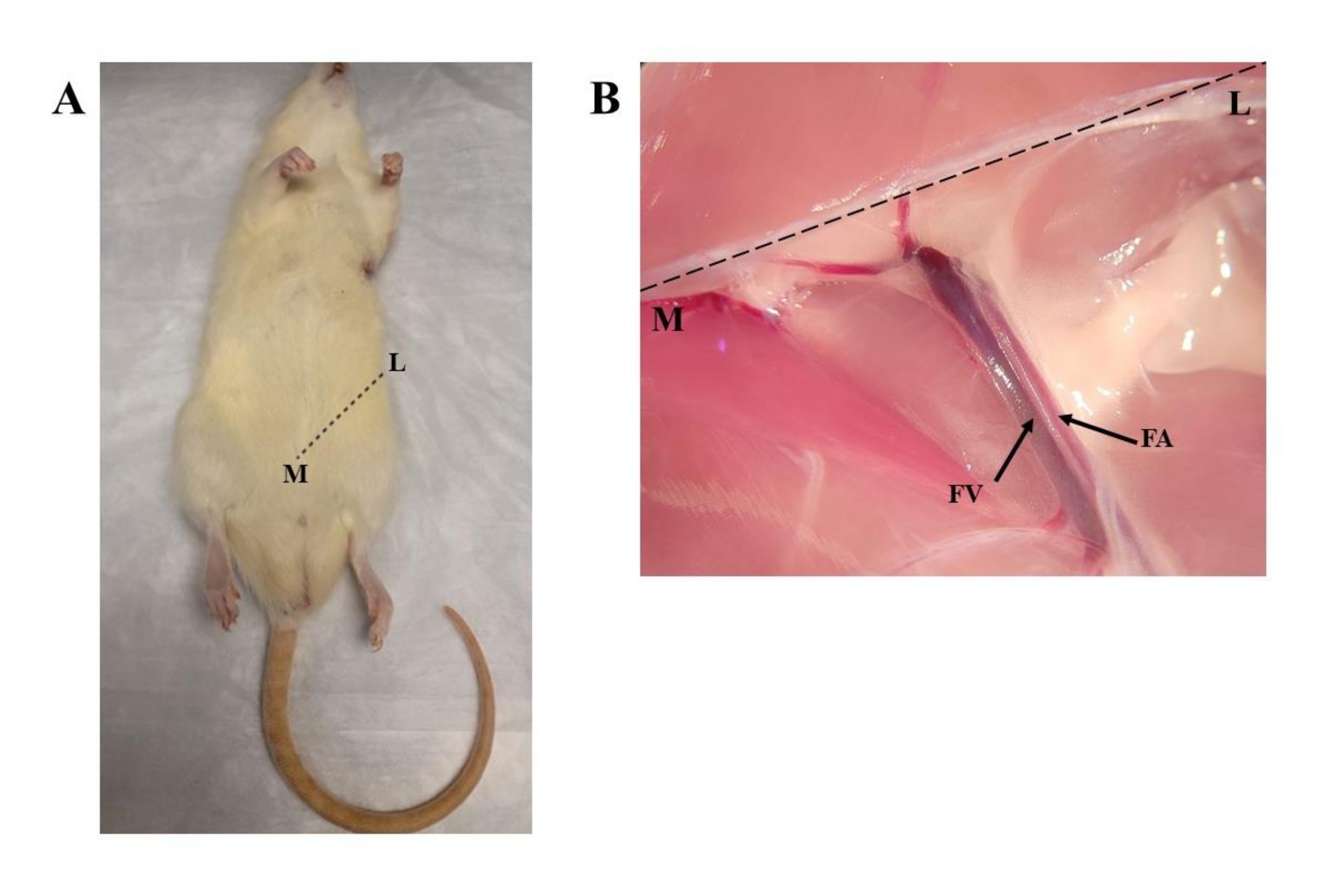
- Make a skin incision using a #10 surgical blade and a #3 blade runner along the inguinal ligament level, moving from a lateral to a medial direction (Figure 1A). Use Adson forceps to hold the surrounding skin to ensure a smooth incision.
- When the underlying fat is exposed, use blunt dissection to carefully dissect through the fat. Locate the superior epigastric vessels.
- Use microscissors to dissect proximally and expose the underlying femoral nerve, artery, and vein at the inguinal ligament level.
- Under a dissection microscope, identify the femoral vessels and dissect the artery and vein proximally using fine forceps to obtain sufficient length from the bifurcation points of the arterial network (Figure 1B).
- Ligate the femoral artery and vein separately using 6-0 sutures.
- Conduct circumferential dissection around the remainder of the hindlimb without disrupting the ligated femoral vessels.
- Transect the femoral bone mid-length using a bone cutter.
- To fully isolate the hindlimb, transect the ligated femoral vessels below the ligatures using microscissors.
- Cannulate the femoral artery using a 24 G angiocatheter under the dissection microscope. Use fine forceps to insert the cannula carefully. Flush with heparinized saline until clear outflow is observed from the femoral vein.
- Secure the cannula by tying one suture around the cannulated vessel and another suture distally around the cannula itself. Ensure the cannula is placed proximally to prevent blocking the bifurcation points.
- Submerge the procured hindlimbs in phosphate-buffered saline (PBS) until decellularization.

Figure 1: Procurement of rat hindlimb. (A) Marking of skin incision at the inguinal ligament level from lateral to medial. (B) View of the femoral vein and the femoral artery, which have been dissected proximally toward the inguinal ligament, indicated by the dotted line. Abbreviations: L = lateral; M = medial; FV = femoral vein; FA = femoral artery. Please click here to view a larger version of this figure.
3. Preparation of solutions
- In a 6 L glass flask, prepare a 5 L detergent reservoir of 0.25% sodium dodecyl sulfate (SDS) by dissolving 12.5 g of SDS powder in 5 L of ultrapure distilled water. Cover the opening of the flask with parafilm to seal it.
- In a 1 L glass jar, prepare 1 L of 0.25% SDS solution separately by dissolving 2.5 g of SDS in 1 L of ultrapure distilled water. Add a stir bar to mix the solution on a magnetic stirrer until all the SDS is dissolved.
- Prepare 1 L of 1x PBS wash solution with 1% Antibiotic-Antimycotic (AA) solution. In a 1 L flask, add 990 mL of 1x PBS solution and 10 mL of AA.
- Separately, in a 500 mL glass jar, prepare 1x PBS + 1% AA solution again using 495 mL of 1x PBS solution and 5 mL of AA.
- Prepare 200 mL of 1% peracetic acid (PAA)/4% ethanol (EtOH) solution. Prepare this solution under a fume hood.
4. Bioreactor and perfusion circuit construction
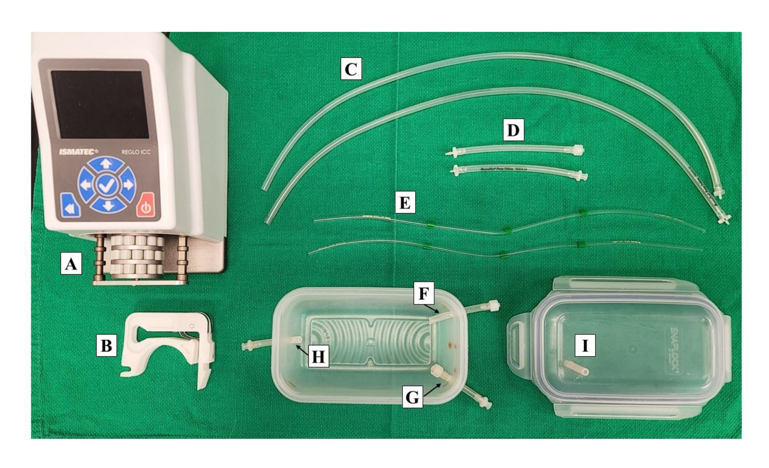
NOTE: Refer to Figure 2 for the configuration of the bioreactor and perfusion circuit throughout the listed steps.
- In a 500 mL chamber (plastic container), drill three holes (1/4 in) at the labeled locations in Figure 2: Port A is the inlet line, Port B is the replenishing line, and Port C is the outlet line. Remove and discard excess plastic. Spray and wipe the chamber with 70% ethanol.
- Cut three 5 cm long silicone tubes and insert one halfway into each port of the chamber. Connect a male Luer connector on the opening of Port A facing the inside of the chamber and a female Luer connector on the other end of the tube outside of the chamber.
- Connect a female Luer connector at Port B and Port C on the ends of the tubes facing out of the chamber.
- In an air-tight lid for the plastic container, drill one hole on the surface of the lid.
- Cut a 3 cm silicone tube and insert it into the hole of the lid. Ensure approximately 2 cm of the tube is located out of the lid, as shown in Figure 2.
- Sterilize both the chamber and the lid with the tubing.
- Cut two 30 cm silicone tubes using scissors. Connect a 1/16 in to a 1/8 in connector on one end of each tube.
- Separately, cut two 12 cm silicone tubes using scissors. Connect a 1/16 in to a 1/8 in connector on one end of both tubes. Connect a male Luer connector on the other end of one tube and a female Luer connector to the other tube.
- Sterilize all the tubing material from step 4.7 and step 4.8. Include two 3-stop pump tubings (1.85 mm) in the sterilization.
- Prepare three 3-way stopcocks, one syringe filter, two 1 mL serological pipettes, and one 10 mL syringe for the decellularization setup. Remove the filter from the 1 mL serological pipettes.

Figure 2: Preparation of bioreactor and perfusion circuit construction. Apparatus shown of the perfusion circuit including (A) peristaltic pump and (B) corresponding cassettes for both inlet and outlet lines. (C, D) Silicone tubings of 12 cm and 30 cm are also shown with respective connectors. (E) Tubing for peristaltic pump (1.85 mm). Bioreactor chamber with labeled ports for (F) inflow, (G) replenishing port, and (H) outflow. (I) Bioreactor lid shown with ventilation port. Please click here to view a larger version of this figure.
5. Decellularization of rat hindlimbs
- Place the sterilized chamber and screw on three single-use 3-way stopcocks at the inlet, outlet, and replenishing lines. Ensure the stopcock for the replenishing port is capped at its remaining two ports to prevent leakage.
- Attach the tubes made in step 4.8 to the stopcocks at the inlet and outlet lines.
- Connect peristaltic tubing to the tubes in the step above. Secure the cassette on the peristaltic tubing and place it on the peristaltic pump. Do not secure the cassettes with tubing in place yet.
- Connect one tube from step 4.7 to the end of the peristaltic tubing of the outlet line from the step above. Connect a 1 mL serological pipette on the other end. Suspend the end attached with a serological pipette in the waste reservoir flask.
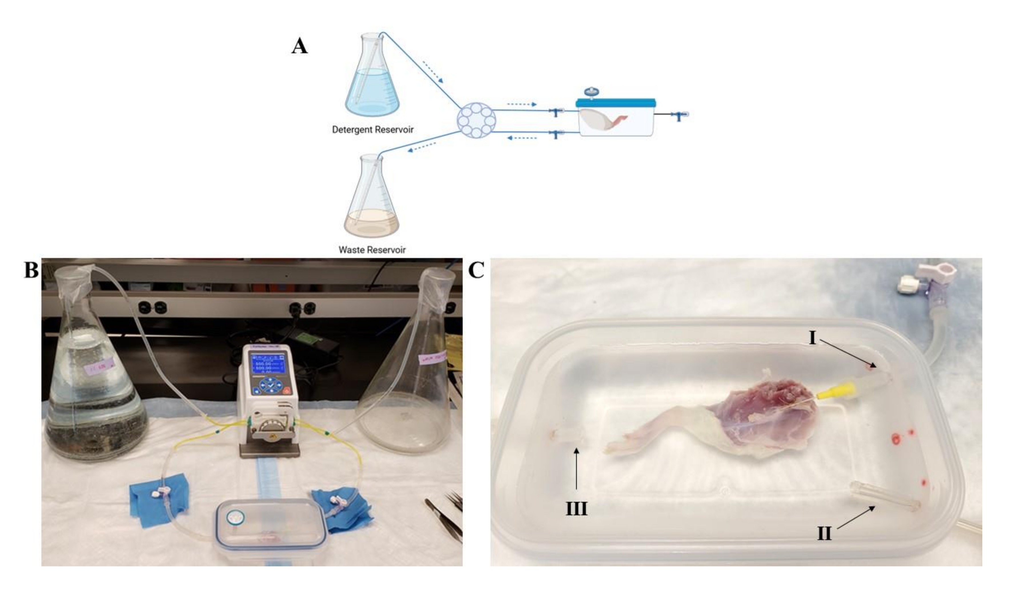
- Repeat step 4.7 for the inlet line. Suspend the end of the tube attached to the serological pipette into the detergent reservoir. Seal the opening of the detergent reservoir flask with parafilm immediately. See Figure 3A,B for an overview of the decellularization circuit.
- Add 0.25% SDS from the 1 L glass jar (step 3.2) into the bioreactor chamber at the halfway level.
- Take the procured hindlimb using Adson forceps and suspend it into the bioreactor chamber carefully.
- Use two pairs of Adson forceps to guide the cannulated portion of the hindlimb to the inlet line. While holding the cannula with one pair of forceps, use the other pair of forceps to twist and secure the inlet line to the cannula. Ensure the cannula is not pulled or twisted to prevent decannulation.
- Once secured, add more 0.25% SDS from the 1 L glass jar to fully submerge the limb, as needed. Ensure the outlet port is also submerged in the bioreactor reservoir to ensure that consistent outflow is maintained (Figure 3C).
- Attach a single-use syringe filter to the ventilation port on the lid of the bioreactor chamber, referring to step 4.4 and step 4.5.
- Secure the lid on the bioreactor and ensure the chamber is sealed from all sides (Figure 3B).
- To remove air from tubing and prime the perfusion circuit, use a new, single-use 10 mL syringe to draw detergent from the detergent reservoir using the 3-way stopcock at the inlet line.
- Once drawn, use the same fluid to insert into the 3-way stopcock at the outlet line. Ensure that there is detergent present throughout the tubing at both the inlet and outlet lines.
- Press down and secure the cassettes with the tubing into the peristaltic pump. Turn the peristaltic pump on using the power button.
- On the peristaltic pump screen, proceed to the second tab using the arrow key to set the perfusion rate for the first channel. Input flow rate as the mode of delivery and set the perfusion rate at 1 mL/min. Ensure the direction of flow is correct according to apparatus set-up. Repeat for the second channel.
- Calibrate the peristaltic pump to ensure the amount of fluid delivered through the inlet line and/or taken from the outlet line is flowing at a consistent rate between the two. Ensure that the tubing ID is set to 1.85 mm.
- Begin decellularization via machine perfusion at 1 mL/min for both the inlet and outlet lines by pressing the power button on the keypad. Monitor and ensure that the flow is consistent and ongoing at both the inlet and outlet lines.
- Continue decellularization and monitor daily. Use the 1 L of 0.25% SDS (step 3.2) to replenish the bioreactor reservoir through the replenishing port, as needed. Look for a white, translucent appearance of the tissue, which will appear by day 5, indicating decellularization of the rat hindlimbs.

Figure 3: Overview of perfusion decellularization bioreactor circuit of rat hindlimb. (A) Schematic representation of bioreactor perfusion circuit. Blue arrows indicate the direction of detergent and waste flow. (B) Overview of the decellularization circuit with bioreactor containing rat hindlimb. The SDS reservoir (left flask) leads into the peristaltic pump and into the inlet tubing of the bioreactor. The outflow is connected to the waste reservoir (right flask) through the peristaltic pump. (C) (I) Bioreactor containing rat hindlimb with inlet tubing connected to the cannulated femoral artery. (II) Replenishing port located in the corner for perfusing detergent. (III) Outflow tubing suspended in suspension reservoir. Abbreviation: SDS = sodium dodecyl sulfate. Please click here to view a larger version of this figure.
6. Post-decellularization washing and sterilization
- Following the confirmation of decellularization, replace the detergent reservoir with the 1x PBS + 1% AA reservoir. Seal the opening of the flask with parafilm. Begin 1x PBS + 1% AA perfusion at 1 mL/min and continue for 2 days.
- Replace the 1x PBS + 1% AA reservoir with 200 mL of 0.1% PAA/4% EtOH reservoir and begin perfusion at 1 mL/min for 2 h.
- Disconnect the hindlimb from the inlet line using two pairs of sterile Adson forceps, with one pair of forceps to twist the inlet line and the other holding the cannula. Ensure that the cannula is not pulled to prevent decannulation.
- Store the limb in a 500 mL glass jar containing 1x PBS + 1% AA at 4 °C until further use.
Access restricted. Please log in or start a trial to view this content.
Results
The procurement protocol was successful in isolating and cannulating the common femoral arteries for subsequent perfusion steps. The representative dissection images in Figure 1A,B show the incision location and exposure of the femoral vessels with sufficient distance from the bifurcation points. Figure 2 shows the apparatus required for preparing the bioreactor and perfusion circuit. The endpoint of decellularization was determined by obse...
Access restricted. Please log in or start a trial to view this content.
Discussion
Rat hindlimbs are useful as experimental models in VCA5. Tissue engineering of acellular scaffolds represents the first step in addressing the shortcomings of long-term immunosuppression regimens associated with VCA. The use of composite grafts poses an added challenge given the presence of multiple tissues, each having unique functional, immunogenic, and structural properties. The present protocol shows a successful method for obtaining acellular composite rat hindlimbs. These scaffolds can be fu...
Access restricted. Please log in or start a trial to view this content.
Disclosures
The authors have no conflicts of interest to declare.
Acknowledgements
Figure 3A was created in BioRender.com.
Access restricted. Please log in or start a trial to view this content.
Materials
| Name | Company | Catalog Number | Comments |
| 0.9% Sodium Chloride Injection USP 50 mL | Baxter Corporation | JB1308M | |
| 1 mL Disposable Serological Pipets | VWR | 75816-102 | |
| 10 cc Disposable Syringes | Obtained from Research Institution | ||
| 3-way Stopcock | Obtained from Research Institution | ||
| 5cc Disposable Syringes | Obtained from Research Institution | ||
| 70% Isopropyl Alcohol | Obtained from Research Institution | ||
| Acrodisc Syringe Filter 0.2 µm | VWR | CA28143-310 | |
| Adson Forceps, Straight | Fine Science Tools | 11006-12 | |
| Angiocatheter 24 G 19 mm (¾”) | VWR | 38112 | |
| Antibiotic-Antimycotic Solution (100x) 100 mL | Multicell | 450-115-EL | |
| Bone Cutter | Fine Science Tools | 12029-12 | |
| Connectors for 1/16" to 1/8" Tubes | McMasterCarr | 5117K52 | |
| Female Luer to barbed adapter (PVDF) - 1/8" ID | McMasterCarr | 51525K328 | |
| Fine Forceps | Fine Science Tools | 11254-20 | |
| Fine Forceps with Micro-Blunted Tips | Fine Science Tools | 11253-20 | |
| Heparin Sodium Injection 10,000 IU/10 mL | LEO Pharma Inc. | 006174-09 | |
| Male Luer to barbed adapter (PVDF) - 1/8" ID | McMasterCarr | 51525K322 | |
| Micro Needle Holder | WLorenz | 04-4125 | |
| Microscissors | WLorenz | SP-4506 | |
| Peracetic Acid | Sigma Aldrich | 269336-100ML | |
| Peristaltic Pump, 3-Channel | Cole Parmer | RK-78001-68 | |
| Phosphate Buffered Saline 1x 500 mL | Wisent | 311-425-CL | |
| Povidone Surgical Scrub Solution | Obtained from Research Institution | ||
| Pump Tubing, 3-Stop, Tygon E-LFL | Cole Parmer | RK-96450-40 | |
| Pump Tubing, Platinum-Cured Silicone | Cole Parmer | RK-96410-16 | |
| Scalpel Blade - #10 | Fine Science Tools | 10010-00 | |
| Scalpel Handle - #3 | Fine Science Tools | 10003-12 | |
| Sodium Dodecyl Sulfate Reagent Grade: Purity: >99%, 1 kg | Bioshop | SDS003.1 | |
| Surgical Suture #6-0 | Covidien | VS889 |
References
- Kueckelhaus, M., et al. Vascularized composite allotransplantation: Current standards and novel approaches to prevent acute rejection and chronic allograft deterioration. Transplant International. 29 (6), 655-662 (2016).
- Duisit, J., et al. Bioengineering a human face graft: The matrix of identity. Annals of Surgery. 266 (5), 754-764 (2017).
- Zhang, Q., et al. Decellularized skin/adipose tissue flap matrix for engineering vascularized composite soft tissue flaps. Acta Biomaterialia. 35, 166-184 (2016).
- Londono, R., Gorantla, V. S., Badylak, S. F. Emerging implications for extracellular matrix-based technologies in vascularized composite allotransplantation. Stem Cells International. 2016 (10), 1-16 (2016).
- Fleissig, Y. Y., Beare, J. E., LeBlanc, A. J., Kaufman, C. L. Evolution of the rat hind limb transplant as an experimental model of vascularized composite allotransplantation: Approaches and advantages. SAGE Open Medicine. 8, 205031212096871(2020).
- Tao, M., et al. Sterilization and disinfection methods for decellularized matrix materials: Review, consideration and proposal. Bioactive Materials. 6 (9), 2927-2945 (2021).
- Chen, Y., Geerts, S., Jaramillo, M., Uygun, B. E. Preparation of decellularized liver scaffolds and recellularized liver grafts. Methods in Molecular Biology. 1577, 255-270 (2018).
- Ahmed, S., Chauhan, V. M., Ghaemmaghami, A. M., Aylott, J. W. New generation of bioreactors that advance extracellular matrix modelling and tissue engineering. Biotechnology Letters. 41 (1), 1-25 (2019).
- Cohen, S., et al. Generation of vascular chimerism within donor organs. Scientific Reports. 11 (1), 13437(2021).
- Lupon, E., et al. Engineering vascularized composite allografts using natural scaffolds: A systematic review. Tissue Engineering Part B: Reviews. , (2021).
- Urciuolo, A., et al. Decellularised skeletal muscles allow functional muscle regeneration by promoting host cell migration. Scientific Reports. 8 (1), 8398(2018).
- Jank, B. J., et al. Engineered composite tissue as a bioartificial limb graft. Biomaterials. 61, 246-256 (2015).
Access restricted. Please log in or start a trial to view this content.
Reprints and Permissions
Request permission to reuse the text or figures of this JoVE article
Request PermissionThis article has been published
Video Coming Soon
Copyright © 2025 MyJoVE Corporation. All rights reserved