需要订阅 JoVE 才能查看此. 登录或开始免费试用。
Method Article
研究内质网和线粒体相互作用通过
摘要
这里,我们描述一个过程中固定的细胞可视化和高灵敏度量化内质网和线粒体之间的内源的相互作用。该协议提供在原位接近连接测定在线粒体相关膜界面靶向1,4,5-三磷酸肌醇受体/葡萄糖调节蛋白75 /电压依赖性阴离子通道/亲环三维复杂的优化。
摘要
Structural interactions between the endoplasmic reticular (ER) and mitochondrial membranes, in domains known as mitochondria-associated membranes (MAM), are crucial hubs for cellular signaling and cell fate. Particularly, these inter-organelle contact sites allow the transfer of calcium from the ER to mitochondria through the voltage-dependent anion channel (VDAC)/glucose-regulated protein 75 (GRP75)/inositol 1,4,5-triphosphate receptor (IP3R) calcium channeling complex. While this subcellular compartment is under intense investigation in both physiological and pathological conditions, no simple and sensitive method exists to quantify the endogenous amount of ER-mitochondria contact in cells. Similarly, MAMs are highly dynamic structures, and there is no suitable approach to follow modifications of ER-mitochondria interactions without protein overexpression. Here, we report an optimized protocol based on the use of an in situ proximity ligation assay to visualize and quantify endogenous ER-mitochondria interactions in fixed cells by using the close proximity between proteins of the outer mitochondrial membrane (VDAC1) and of the ER membrane (IP3R1) at the MAM interface. Similar in situ proximity ligation experiments can also be performed with the GRP75/IP3R1 and cyclophilin D/IP3R1 pairs of antibodies. This assay provides several advantages over other imaging procedures, as it is highly specific, sensitive, and suitable to multiple-condition testing. Therefore, the use of this in situ proximity ligation assay should be helpful to better understand the physiological regulations of ER-mitochondria interactions, as well as their role in pathological contexts.
引言
线粒体和内质网(ER)是不是在细胞中独立的细胞器,但它们在结构上和功能上相互作用在定义为线粒体相关的内质网的膜(MAM)的接触位点。事实上,MAMS对应其中ER的膜和线粒体紧密并列的区域,使从两侧的蛋白质之间的相互作用。然而,这些细胞器的膜不这些区域内熔合,因此它们保持它们的独立的实体。该MAMS起到钙(Ca 2+)关键作用,磷脂转移从ER到线粒体,影响能量代谢和细胞存活1-3。
内质网和线粒体之间的关系在20世纪70年代用电子显微镜首次观察。自那时以来,透射电子显微镜4,5,电子断层扫描6,7或ER和特定线粒体-荧光团的免疫定位S /荧光蛋白8人经典用于研究ER-线粒体相互作用。为MAM的分析的另一个有用的工具是基于使用亚细胞分离的。它允许MAM馏分的隔离通过耦合到Percoll密度梯度9差动超速离心。然而,最终产物含有丰富的MAM馏分,而不是纯的级分。总之,这些策略没有特别敏感和/或定量的,它们是不容易适合于大的筛选。可替代地,使用药物诱导的荧光间细胞器的接头遗传方法已经出现,但它们不允许细胞器相互作用的分析在蛋白10的内源表达水平。
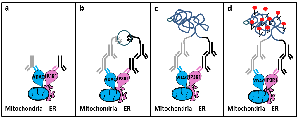
根据Szabadkai的发现的IP3受体/ GRP75 / VDAC络合物在MAM 11中 ,我们开发了一个定量的方法来分析ER-线粒体相互作用。我们使用原位接近ligati上测定来检测和量化VDAC1和IP3R1之间的相互作用,在固定单元12参与在MAM接口的钙 -channeling复杂二细胞器表面蛋白。简言之,我们在线粒体外膜(小鼠抗VDAC1初级抗体)与IP3R1在ER膜(兔抗IP3R1初级抗体)(图1,分图a)探测VDAC1。然后,根据该测定,我们增加既抗小鼠和抗兔IgG(小鼠和兔接近结扎分析探针),其偶联至互补寡核苷酸的扩展。如果两个目标蛋白质是在低于40纳米的距离,寡核苷酸可以与随后加入的接头的寡核苷酸杂交以允许环状DNA模板(图1,图b)的形成。此环形DNA分子连接并放大,产生共价连接到邻近探针之一的单链DNA产物(图1,分图c) 。由于在MAM接口的ER和线粒体之间的距离为10纳米的范围内,以25纳米6,接近结扎和扩增可以完成,从而导致后续检测由于德克萨斯红标记的寡核苷酸探针(图1,图d的杂交)。每个荧光点代表VDAC1 / IP3R1之间的相互作用,因而允许在单个细胞中原位 ER-线粒体相互作用的定量。

图1:由内质网线粒体相互作用检测的示意图原位接近结扎分析。 一)针对VDAC1和针对IP3R1可以在MAM接口绑定到其表位接近兔第一抗体的小鼠初级抗体, 二)除了一对接近结扎探针的针对小鼠和兔IgG。这些探针已附加的DNA链,可以形成用于连接寡核苷酸的结扎模板。 三)结扎后所形成的环状DNA链可以通过使用德克萨斯红标记的寡核苷酸扩增和d)通过显微镜观察作为荧光点。 请点击此处查看该图的放大版本。
原位接近连接测定实验相似可以与GRP75 / IP3R1对抗体,以及亲环D(CypD)来执行/ IP3R1抗体,考虑到CypD被证明与在MAM接口的IP3受体/ GRP75 / VDAC复合物相互作用12-14。
研究方案
1.溶液的制备
- 通过稀释5.5毫升37%甲醛的14.5毫升的PBS制备于PBS(低盐)10%的甲醛。制备1M甘氨酸,pH 8.0的,溶解在50ml的PBS将3.8g甘氨酸;稀释此溶液,得到在1×PBS中的100mM甘氨酸。
- 制备0.1%的Triton-X100在1×PBS中。通过使用3.0M的氯化钠和0.30柠檬酸三钠在25℃在去离子水缓冲液中制备具有pH为7.0制备20倍盐水柠檬酸钠(SSC)。这稀释缓冲1X和使用去离子水0.01X。
2.细胞固定
注意:我们使用在这项研究中的HuH7肝细胞癌细胞系,但这种方法也适用于其他的粘附细胞培养物。
- 板的HuH7细胞(在DMEM 1g / L的葡萄糖进行培养,补充有10%胎牛血清和0.01%青霉素 - 链霉素的库存)以每未涂覆35毫米玻璃底培养皿150,000个细胞。当原电池工作文化,用胶原蛋白涂层的菜肴。
- 次日,除去培养基。清洗细胞用1毫升PBS和吸出的。通过加入1ml的甲醛10%固定细胞,并孵育在搅拌下室温(RT)10分钟。
- 停止用1ml的1M甘氨酸使反应并通过旋转混合。除去停止反应溶液并通过加入1ml 1×PBS中洗细胞;通过旋转和吸搅动。 1毫升的100mM甘氨酸添加到细胞中,并孵育它们在室温下搅拌15分钟,然后吸出。
注:该协议可以在这里停了下来,下面的步骤可以推迟到另一天。在这种情况下,加入1ml的100mM甘氨酸和,保持在4℃,如果必要的。
3.细胞透
- 加入1ml 0.1%的Triton-X100的在1×PBS中,在室温下孵育在搅拌下15分钟,然后吸出。上一次电池文化性工作时20分钟 - 在该温育时间可以提高到15ES( 如初级小鼠肝细胞)。加入1 ml 1X PBS洗细胞;吸出。
4.封锁
- 添加40微升的封闭溶液以每个样品(由试剂盒提供);这个体积可以以覆盖样品增加。在湿度室中孵育30分钟的菜在37℃。
- 点击过的菜封闭液。试图获得平等的残液量为每张幻灯片,因为这会影响可重复性。不要让样品干!
5.一级抗体
- 稀释在1×PBS中的一抗,并添加溶液的培养皿(VDAC1小鼠抗体:1/100,IP3R1兔抗体:1/500)。可替代地,GRP75或CypD抗体(在1/500同时使用小鼠抗体)可以用来代替VDAC1。
- 在4℃的湿度室中孵育过夜。洗载玻片使用的Tris缓冲盐水两次含0.01%吐温(TBS-T)。
- 接近结扎分析探针由试剂盒。根据初级抗体的物种选择探针。
- 在抗体稀释液5:准备两个接近结扎分析探针1。使混合物静置在室温20分钟。添加稀释接近连接测定探头的解决方案。孵育在预加热的湿度室中的菜肴1小时,在37℃。用TBS-T洗碗两次。
7.结扎
- 使用在原位检测试剂德克萨斯红套件。
- 稀释5倍结扎股票(由试剂盒提供)1:5的高纯度水拌匀。稀释1倍连接液(由试剂盒提供)连接酶1:40和旋涡。等待添加连接酶,直到立即除了样品之前。
- 添加该溶液至每个样品(40微升为35毫米的玻璃底菜)孵育玻片在预热的湿度湛误码率在37℃,30分钟。用TBS-T洗载玻片两次。
8.放大
注意:小心,光敏感的试剂。
- 稀释5倍放大股票(由试剂盒提供)1:5的高纯度水。使用冷冻块中删除从冷冻聚合酶(-20℃)。稀释聚合酶(试剂盒提供)1:80在1倍放大的解决方案和旋涡。立即用混合前添加聚合酶。
- 添加该溶液至每个样品(40微升为35毫米的玻璃底菜)。孵育在预加热的湿度室100分钟的幻灯片在37℃。挖掘出幻灯片的扩增聚合酶溶液。
9.最后经洗涤
- 洗于1×SSC洗涤缓冲液将载玻片2分钟。洗在0.01X SSC洗涤缓冲液将载玻片2分钟。让菜在黑暗中在室温下干燥。
10.准备成像
- 使用含有DAPI(水相和不固化)安装介质的最小体积,以确保没有气泡得到盖玻片下抓装入滑动。指甲油可以用来密封边缘。 (:594纳米,发射:624纳米,放大倍数:63X激励)使用荧光或共聚焦显微镜分析前,等待约15分钟。
- 成像后,存放载玻片在黑暗中在-20℃。
结果
基于使用此协议我们的经验,我们可以放心地推荐为固定细胞ER-线粒体相互作用的可视化和量化这种方法。的在的HuH7肝癌细胞原位接近连接测定-可视ER-线粒体的相互作用,用几对抗体的代表性图像,被示出。如在图2中由荧光显微镜所示,每个红点代表两个目标蛋白质之间的相互作用,以及细胞核显示为蓝色。作为阴性对照,只使用一个一级抗?...
讨论
Collectively, our studies indicate that the in situ proximity ligation assay is truly a relevant strategy to follow and quantify endogenous ER-mitochondria interactions in fixed cells, without the need for using organelle-specific fluorophores or fluorescent proteins. The specific use of VDAC1/IP3R1 antibodies has been adapted to study ER-mitochondria interactions in HuH7 cells. However, alternative isoforms of VDAC and IP3R may be used, depending on the cell type. In this case, antibodies need to be validated b...
披露声明
The authors declare that they have no competing financial interests.
致谢
我们感谢所有在我们的实验室谁促成了优化和验证协议的人。这项工作是由INSERM和国家研究机构(ANR-09-JCJC-0116和ANR-11-BSV1-033-02)的支持。 ET博士学位由高等教育和研究的法国文化部的一个研究奖学金期间的支持。
材料
| Name | Company | Catalog Number | Comments |
| Formaldehyde | Sigma | F-8775 | |
| Glycine | Sigma | G-8898 | |
| Triton | Sigma | T8532 | |
| 35mm Glass bottom culture dishes | MatTeK corporation | P35G-0-14-C | |
| Blocking solution | Sigma | DUO-92004 or DUO-92002 | provided in the Duolink PLA probes, Sigma |
| VDAC1 antibody | Abcam | ab14734 | |
| IP3R1-H80 antibody | Santa Cruz | sc28614 | |
| CypD antibody | Abcam | ab110324 | |
| Grp75 antibody | Santa Cruz | sc13967 | |
| TBS 10X | euromedex | ET220 | Dilute to obtain 1X |
| Tween 100X | euromedex | 2001-B | dilute in TBS to obtain 0,01% |
| PLA Probes Mouse MINUS | Sigma | DUO-92004 | Duolink, Sigma |
| PLA Probes Rabbit PLUS | Sigma | DUO-92002 | Duolink, Sigma |
| Duolink detection reagents red | Sigma | DUO-92008 | Duolink, Sigma |
| Ligation solution | Sigma | DUO-92008 | Part of the Duolink detection reagents red, Sigma |
| Ligase | Sigma | DUO-92008 | Part of the Duolink detection reagents red, Sigma |
| Amplification solution | Sigma | DUO-92008 | Part of the Duolink detection reagents red, Sigma |
| Polymerase | Sigma | DUO-92008 | Part of the Duolink detection reagents red, Sigma |
| Duolink Mounting Medium | Sigma | DUO80102 | Duolink, Sigma |
| Softwares: | |||
| Blob-finder software | BlobFinder is a freely distributed software that can perform calculations on cells from fluorescence microscopy images. This software can be downloaded for free from The Centre for Image Analysis at Uppsala University who have developed the software and the work was supported by the EU FP6 Project ENLIGHT and Olink Bioscience. http://www.cb.uu.se/~amin/BlobFinder/index_files/Page430.htm | ||
| ImageJ software | Can be downloaded for free from: http://rsb.info.nih.gov/ij/download.html |
参考文献
- Bravo-Sagua, R., et al. Organelle communication: signaling crossroads between homeostasis and disease. The international journal of biochemistry & cell biology. 50, 55-59 (2014).
- Giorgi, C., et al. Mitochondria-associated membranes: composition, molecular mechanisms, and physiopathological implications. Antioxidants & redox signaling. 22, 995-1019 (2015).
- Phillips, M. J., Voeltz, G. K. Structure and function of ER membrane contact sites with other organelles. Nature reviews. Molecular cell biology. 17, 69-82 (2016).
- Cosson, P., et al. The RTM resistance to potyviruses in Arabidopsis thaliana: natural variation of the RTM genes and evidence for the implication of additional genes. PLoS One. 7, 39169 (2012).
- Mannella, C. A. Structure and dynamics of the mitochondrial inner membrane cristae. Biochim Biophys Acta. 1763, 542-548 (2006).
- Csordas, G., et al. Structural and functional features and significance of the physical linkage between ER and mitochondria. The Journal of cell biology. 174, 915-921 (2006).
- Mannella, C. A., Buttle, K., Rath, B. K., Marko, M. Electron microscopic tomography of rat-liver mitochondria and their interaction with the endoplasmic reticulum. Biofactors. 8, 225-228 (1998).
- Rizzuto, R., et al. Close contacts with the endoplasmic reticulum as determinants of mitochondrial Ca2+ responses. Science. 280, 1763-1766 (1998).
- Wieckowski, M. R., Giorgi, C., Lebiedzinska, M., Duszynski, J., Pinton, P. Isolation of mitochondria-associated membranes and mitochondria from animal tissues and cells. Nat Protoc. 4, 1582-1590 (2009).
- Csordas, G., et al. Imaging interorganelle contacts and local calcium dynamics at the ER-mitochondrial interface. Mol Cell. 39, 121-132 (2010).
- Szabadkai, G., et al. Chaperone-mediated coupling of endoplasmic reticulum and mitochondrial Ca2+ channels. J Cell Biol. 175, 901-911 (2006).
- Tubbs, E., et al. Mitochondria-associated endoplasmic reticulum membrane (MAM) integrity is required for insulin signaling and is implicated in hepatic insulin resistance. Diabetes. 63, 3279-3294 (2014).
- Paillard, M., et al. Depressing Mitochondria-Reticulum Interactions Protects Cardiomyocytes From Lethal Hypoxia-Reoxygenation Injury. Circulation. 128, 1555-1565 (2013).
- Rieusset, J., et al. Disruption of calcium transfer from ER to mitochondria links alterations of mitochondria-associated ER membrane integrity to hepatic insulin resistance. Diabetologia. 59, 614-623 (2016).
- Allalou, A., Wahlby, C. BlobFinder, a tool for fluorescence microscopy image cytometry. Computer methods and programs in biomedicine. 94, 58-65 (2009).
- Theurey, P., et al. Mitochondria-associated endoplasmic reticulum membranes allow adaptation of mitochondrial metabolism to glucose availability in the liver. Journal of molecular cell biology. , (2016).
- de Brito, O. M., Scorrano, L. Mitofusin 2 tethers endoplasmic reticulum to mitochondria. Nature. 456, 605-610 (2008).
- Soderberg, O., et al. Direct observation of individual endogenous protein complexes in situ by proximity ligation. Nature methods. 3, 995-1000 (2006).
- De Pinto, V., Messina, A., Lane, D. J., Lawen, A. Voltage-dependent anion-selective channel (VDAC) in the plasma membrane. FEBS letters. 584, 1793-1799 (2010).
- Kaul, S. C., Taira, K., Pereira-Smith, O. M., Wadhwa, R. Mortalin: present and prospective. Experimental gerontology. 37, 1157-1164 (2002).
转载和许可
请求许可使用此 JoVE 文章的文本或图形
请求许可探索更多文章
This article has been published
Video Coming Soon
版权所属 © 2025 MyJoVE 公司版权所有,本公司不涉及任何医疗业务和医疗服务。