Method Article
使用新型无泵流体系统维护和评估眼睛的各种组织和细胞类型
摘要
对活体组织进行实时分析可产生重要的功能和机理数据。本文描述了协议和关键变量,以确保通过一种新型的无泵多通道流体系统准确和可重复地生成数据,该系统可以维护和评估各种组织和细胞模型。
摘要
许多用于研究组织功能和细胞生物学的 体外 模型需要培养基流动,以提供足够的氧合和维持功能和活力所需的最佳细胞条件。为此,我们开发了一种多通道流式培养系统,以维持组织和细胞的培养状态,并通过在线传感器和/或收集流出部分来持续评估功能和活力。该系统将耗氧率的 8 通道连续光学传感与内置馏分收集器相结合,可同时测量代谢物的产生率和激素分泌。尽管它能够维持和评估广泛的组织和细胞模型,包括胰岛、肌肉和下丘脑,但在这里我们描述了它的工作原理和实验制剂/方案,我们用于研究分离的小鼠视网膜、小鼠视网膜色素上皮 (RPE)-脉络膜-巩膜和培养的人 RPE 细胞的生物能量调节。系统设计的创新,如无泵流体流动,大大简化了多通道流动系统的操作。视频和图像说明了如何组装、准备仪器进行实验,以及如何将不同的组织/细胞模型加载到围灌注室中。此外,还描述和讨论了为方案和组织特异性实验选择条件的指南,包括设置正确的流速与组织比率以获得一致和稳定的培养条件,以及准确测定消耗量和生产率。最佳组织维护和多个参数的实时评估相结合,产生了信息量很大的数据集,这些数据集将对眼睛生理学研究和治疗视力受损的药物发现具有很大的实用性。
引言
围灌注系统在生命科学领域有着悠久的历史。特别是,对于胰岛分泌功能的研究,它们已被用于表征胰岛素分泌响应促泌剂的动力学1。除了收集流出部分用于随后的激素和代谢物测定外,还集成了实时传感器,主要用于检测耗氧量2,3,4。由于缺乏生理学相关方法来评估眼睛各种分离成分(包括视网膜、视网膜色素上皮 (RPE)-脉络膜-巩膜和培养的 RPE 细胞)的代谢调节和失调,因此更好地理解介导眼部疾病机制的广泛努力受到限制。为培养细胞设计的静态系统已适用于组织5,但组织需要流动才能充分氧合。流动系统已成功准确且可重复地测量视网膜和 RPE-脉络膜-巩膜对耗氧率 (OCR) 的实时响应,并且组织保持代谢稳定超过 8 小时,从而允许涉及多种测试化合物的高度信息方案 4,6,7,8,9 .尽管如此,流体系统的操作历来需要定制的设备和训练有素的技术人员进行非标准化方法。在大多数实验室中,这种系统尚未被采用为标准方法。BaroFuse 是一种新开发的流体系统,它不依赖泵,而是依靠气体压力来驱动流经多个通道和组织室(图 1)。对每个通道进行OCR连续监测,并使用基于板的馏分收集器收集流出物,以便随后对内容物进行分析。重要的是,该仪器的组织围灌注室旨在容纳各种几何形状和大小的组织。
该仪器的核心是流体系统,其中流体从密封的加压储液罐通过小内径 (ID) 管(在流体回路中产生最显着的流动阻力)向上驱动,进入容纳组织的玻璃组织室。介质储液模块 (MRM) 的压力由连接到装有气体混合物(通常为 21% O 2、5% CO 2、平衡 N 2)的气瓶的低压和高压调节器提供,储液罐由容纳组织室组件 (TCA) 的围灌室模块 (PCM) 从顶部密封。流量由电阻管的长度和内径以及低压调节器的压力设置控制。连接到组织室顶部的流出管将流体输送到废物容器(连续称重以自动确定流速)或由馏分收集器控制的 96 孔板的孔中。O 2 检测系统可测量涂在组织下游每个玻璃组织室内部的 O2 敏感染料的寿命。然后,此信息用于连续计算 OCR。整个流体系统位于温控外壳中,储气罐、馏分收集器和计算机是仪器的主要组件(图 2A)。最后,运行仪器的软件用于控制其操作(包括进样测试化合物的制备和定时、流量测量系统和馏分收集器定时),以及处理和绘制 OCR 数据和其他补充测量。
在本文中,我们描述了使用流体系统围灌注和评估眼睛各种分离成分的 OCR 和乳酸生成率 (LPR) 的协议。LPR 是反映糖酵解速率的参数,与 OCR 高度互补,其中该对占细胞中碳水化合物产生能量的两个主要分支10。由于最好通过观看程序来了解组织制备并将其加载到组织室中,因此该视频将有助于说明在设置和操作过程中执行的几个关键步骤,这些步骤仅通过文本不容易传达。
协议的描述分为8个部分,对应于实验的不同阶段(图2B):1.实验前准备;2.围灌液的制备/平衡;3.仪器设置;4.组织平衡;5.实验方案;6、仪器故障;7. 数据处理;和 8.流出分数的测定。
研究方案
从大鼠和小鼠身上采集组织的所有程序均已获得华盛顿大学机构动物护理和使用委员会的批准。
1. 实验前准备
注意:以下任务至少在实验前一天完成。
- 设计实验方案
- 在通道中分配组织放置:选择要放置在 MRM 两侧 4 个通道中的 3 个中的组织或细胞模型。每侧各有一个组织室,无需组织即可进行基线校正。
- 使用两种典型设计之一排列样品 - 每侧使用不同的测试化合物方案(例如,MRM一侧的通道接收测试化合物,而另一侧的通道充当对照);MRM两侧的测试化合物注射方案相同,但与MRM两侧的对照组不同。
- 选择流速和组织量以获得最佳 OCR 测量:调整流速,直到寿命比乘以 100 的变化约为 3。
注: 表1 显示了眼睛成分的典型组织量和相应的流速,其中仪器在6-80μL/min/通道的流速下功能最佳。 - 所需培养基/缓冲液体积的计算:计算实验开始时要添加到每个MRM插入物中的培养基体积,如下
体积MRM = 30 mL + 实验方案持续时间(最小值)x 流速(mL/min)x 4 个通道(式 1)
例如,在 0.01 mL/min 时,60 mL 起始体积 MRM 将允许 12.5 小时方案(其中 30 mL 将耗尽,而剩余 30 mL),而在 0.04 mL/min 时,90 mL 起始体积MRM 将允许 6 小时方案(剩余 30 mL)。 - 测试化合物进样方案:选择要评估的测试化合物、要测试的浓度(通常选择以产生接近最大响应或作为浓度依赖性)和暴露持续时间。考虑溶解度,并在所需的溶剂(如水、DMSO或乙醇)中补足储备液。
- 选择进样和后续进样的时间,以便在添加后续代理之前使响应达到稳定状态。重复方案时,匹配注射时间,以便可以平均多个时间过程。
注:这里使用的化合物来自以前的线粒体(Mito)应激试验 11 ,寡霉素和羰基氰化物4-(三氟甲氧基)苯腙(FCCP)都需要DMSO在储备溶液中,以及最终的周融合物。 - 流出采样时间:选择所需的馏分收集间隔(1-60 分钟/样品),其中选择更快的采样率进行快速变化,并在接近稳态时选择更长的时间间隔。使用足够的孔体积(0.3 至 1.5 mL)以避免在采样间隔期间溢出(选择大于流速 x 时间间隔的体积)。
注意:采样时间会因方案的选择而异,但对于线粒体压力测试,我们通常在基线期间使用 5 分钟间隔,在注射期间使用 15 分钟间隔(-15、-10、-5、0、15、30、45、60、75、90、105,其中每次都是采样间隔的开始)。 - 在用户界面 (UI) 中输入上述测试化合物和馏分收集的选定值,该界面会生成此信息的图形表示。导出和传播文件以供小组评估和讨论(补充图1)。
- 列出配件和易损件
- 列出制造商提供和预包装的无菌用品,包括:TCA(8 件装)、流出管组件(8 管装)、测试化合物注射管 (2)、镊子、管夹 (3)、MRM、MRM 插件 (2)、搅拌棒 (2) 和吹扫管组件(放置在生物安全柜中)。
- 不要重复使用与液体接触的一次性部件,因为这会导致实验失败的增加。通过在实验之间清洁和高压灭菌来重复使用镊子和搅拌棒。
2.perifusate的制备和平衡(时间:30min,不包括孵育时间)
- 根据公式1的计算,在前一天制备培养基或Krebs-Ringer碳酸氢盐缓冲液(KRB),通常为200mL,然后在T225组织培养瓶中的39°C / 5%CO2 培养箱中孵育过夜,每个烧瓶中不超过90mL。
- 如果使用商业制备的KRB或培养基(加热至室温),则在实验当天早上准备perifustures,并置于5%CO2 培养箱中至少1小时。无菌制备所有溶液。
注意:所有液体和流体系统中与液体接触的部分在实验开始时都是无菌的。然而,系统的组装和组织的装载是在空气中进行的。
3. 平衡温度和溶解气体以设置仪器(时间:75 min)
- 将管路组件连接到 MRM
- 将 MRM 和液流套件放在仪器旁边的工作台上。确保管子夹 (3)、搅拌棒 (2) 和镊子已经在工具托盘上。
- 在MRM的每一侧放置一个未使用的MRM插入件,并带有搅拌棒(见 图3)。
- 将 TCA 连接到 MRM 两端的进样口,使管路的末端直接位于搅拌棒上方。确保两个测试化合物进样组件中较长的组件位于 MRM 的背面。
- 接下来,将进气管路和吹扫管组件分别连接到后部和前部空口(见 图 4A)。
- 将 MRM/管路组件放置在外壳中
- 将MRM(连接管路组件)放入MRM加热器中(图4B)。
- 将四个管道组件放入外壳底部的墙壁凹槽中(每侧两个),以便在中间外壳就位后,它们将突出到外壳的外部。
- 通过拧紧检测器支架上的两个轮子,将 MRM 固定在夹子之间。
- 将较长的测试化合物注射组件从外壳背面突出,穿过外壳侧面的两个管导轨,使管的开口朝前(图4C)。
- 夹紧每个封闭的测试化合物注射组件。
- 组装外壳并激活温度控制器
- 将为机柜内所有电气设备供电的接线板切换到 ON 位置。检测器支架上的风扇将打开,MRM 温度控制器应亮起,显示 38 °C 的设定值(图 5)。
- 使用 UI 将搅拌器打开至 70 rpm,以确保搅拌棒平稳旋转。观察到适当的搅拌后,关闭搅拌器。
- 将外壳的中间部分放在底座顶部。
- 将外壳中间部分的电缆连接到电箱的电缆,以接合环境温度控制器杠杆开关的电源,并为环境温度加热器供电。
- 将盖子盖在外壳上,上部温度控制器(环境温度控制器)的显示屏将亮起并读取 36 °C。 启动计时器 30 分钟,这是 MRM 加热器达到设定点温度所需的时间。
- 将 TCA 插入 PCM
- 使用 TCA 插入工具将 8 个 TCA 中的每一个插入 PCM 孔中,方法是用插入工具的表面用力按压适配器,直到缠绕在组织室周围的管套管的顶部接触 PCM 孔中的表面。
- 在插入下一个 TCA 之前,请完全插入一个 TCA。将部分组装好的 PCM 放在 PCM 支架和 6 个螺钉旁边。
注意: TCA 插入不完全会阻止顶部空间达到压力设定点,并且 perifusate 不会流动。
- 用预平衡的周灌液填充 MRM 中的两个插入片段
- 为此,在组装外壳且MRM达到温度30分钟后,使用50 mL移液管轻轻地将液体从侧面分配,将预平衡的周灌液转移到预热的MRM插入物中。
注意:这些步骤以及第 3.6 节中的步骤应立即执行,以避免气体在 MRM 中的 perifumate 和大气之间转移。
- 为此,在组装外壳且MRM达到温度30分钟后,使用50 mL移液管轻轻地将液体从侧面分配,将预平衡的周灌液转移到预热的MRM插入物中。
- 组装 MRM/PCM 以形成气密密封并定位 O2 探测器
- 通过将从 PCM 底部发出的 TCA 的电阻管插入 MRM 插入物(MRM 分频器每侧 4 个)中,将 PCM 置于 MRM 上。调整 PCM 的方向,使组织室在定位后可以靠在 O2 检测器上。
- 使用电动螺丝刀用 6 颗螺钉固定 PCM 和 PCM 支撑支架。
- 用弹性带将 TCA 固定在 PCM 支承鳍片内,方法是将其拉伸到橡胶垫圈的水平(图 6)。
- 将 O2 探测器放在探测器支架上,使其表面靠在 PCM 的翅片上。检查 LED/光电探测器对是否与组织室中的 O2 敏感染料对齐。如果需要,在松开 O2 探测器支架侧面的固定螺钉后调整 O2 探测器横向导轨。
- 将盖子放在外壳顶部。
- 用周灌液平衡MRM顶部空间中的气体
- 在高压阀完全固定并关闭的情况下,逆时针转动储气罐顶部的气瓶阀,打开储气罐阀门。
- 使用调节器上的旋钮将高压调节器调节到 10 psi 的压力。
- 通过将低压调节器设置为 1.0 psi 来对 MRM 加压(图 7A)。
- 松开吹扫管(图7B),使罐中的气体取代MRM顶空中的空气(测试化合物注射器保持夹紧)15分钟。通过将吹扫管的末端浸入烧杯中以观察气泡来确认气流。
- 确认流量后,启动 O2 检测器,如下文第 3.8 节所述。
- 15分钟后,以70rpm打开搅拌器,并在实验的其余部分保持运行。再过15分钟后,夹紧吹扫管组件(图7C)。
- 将低压调节器上的压力降低到达到所需液体流速的工作压力(如实验包规定的那样 - 通常在约 0.5-0.7 psi 之间)。如果流速高于 20 μL/min,请暂时将压力设置为 0.3 psi,以便有时间加载组织而不会使腔室溢出。如果在放置夹具后 15 分钟内加载组织,则不需要这样做。
注意:不要让缓冲液从组织室的外部流下,因为液体会干扰 O2 传感。
- 启动 O2 探测器
- 通过单击标有氧气检测仪的图标激活笔记本电脑上的 O2 检测器软件。
- 程序打开后(补充图 2),确认选择了正确的 COM 端口。如果需要,可以通过从计算机上拔下和插入 O2 检测器来识别 COM 端口,以便显示端口号。如果在应用程序运行时拔下 COM 端口,则必须在使用前关闭并重新打开应用程序。
- 单击 "开始 ",然后单击 "记录 "(并将数据保存在备份文件夹中)。接下来,单击 图形。
- 将生存期图左下角的平均值更改为 5(指示程序计算连续 5 个点的移动平均线)。一分钟后,第一个数据点显示在图形屏幕上,单击 "自动缩放"。
4.组织负荷和平衡期(时间:90分钟)
- 将筛板定位在组织室中
- 取下外壳的盖子和中间部分。
- 在组织室中的围灌液上升到预先定位的筛板顶部上方后,轻轻敲击筛板顶部,用筛板提示将筛板向下推,以去除筛板下方或内部形成的任何气泡。
- 将筛板放置在组织室底部上方约 0.25 英寸处。
- 将组织加载到组织室中
- 一旦介质水平距顶部 0.5 英寸,将组织装入腔室。
- 加载视网膜或 RPE-脉络膜-巩膜:如 6 所述收获视网膜或 RPE-脉络膜-巩膜。要加载组织,请使用细点镊子将组织轻轻地放入每个腔室中,注意不要折叠组织,同时使用纸巾擦拭以防止组织腔中的液体滴落到 O2 传感器上。观察组织朝向和沉入熔块。
注意:在收获组织和将组织装入腔室之间,确保避免对组织造成创伤,不要将组织留在基于碳酸氢盐的缓冲液/培养基中超过培养箱 10 分钟,并确保组织沐浴在足够的缓冲液/培养基(至少 1 mL/10 mg 组织)中,以防止组织缺氧并防止暴露在空气中。 - 将RPE细胞加载到transwell膜上:如前面 12 和 补充文件1所述制备RPE细胞。使用 0.25% 胰蛋白酶-EDTA 和聚对苯二甲酸乙二醇酯上的种子传代细胞,跟踪蚀刻过滤器(细胞培养插入物,孔径 0.4 mm),最小 2.0 x 10⁵ 个细胞/cm2。在实验当天,将膜切成三个宽度相等的条带,并用镊子加载到组织室中(见 图8A)。
- 将流出管组件连接到组织室
- 从包装中取出流出管组件,并将流出管分离器放在外壳中间部分的边缘上,使流出管适配器位于外壳内部(图 8B,C)。
- 注意不要用力推动TCA(否则它们会从MRM上松动),将流出管适配器连接到组织室TCA的顶部(图8D)。更换外壳中间并重新连接环境温度控制电缆。
- 在更换外壳盖之前,确认外壳内流体系统的组件(包括 O2 检测器、PCM、组织室、流出管、MRM 和加热器)均已正确放置, 如图 8E 所示。
- 装回机柜盖。将八个流出管送入馏分收集器导向臂。
- 激活馏分收集器
- 确保馏分收集器相对于外壳的右壁和流出管支架居中:馏分收集器底座的左侧支架应靠在外壳壁的边缘。
- 在笔记本电脑上,单击 UI 快捷方式,将打开实验信息页面(补充图 3 顶部)。
- 在实验信息页面上的相应框中填写信息(可以在实验开始之前完成),然后单击顶部的 "流量和馏分收集 器"页面(补充图3 ,底部)。
- 设置自动流量测量的参数
- 在顶部中间的样品采集时间下拉菜单中选择所需的积分时间,以平衡所需的精度(与积分时间成正比)和时间分辨率。
- 如果实验中没有收集流出分数,请单击"开始"并转到第 4.7 节。如果要收集流出分数,请执行第 4.6 节中的步骤。
- 收集流出分数
- 在 UI 软件中,选中 Collect fractions? "实验信息"页面或"流量和馏分收集器"页面上的框。然后单击 * 计算 FC 设置 ] 按钮。
- 当新窗口打开时,在方案中填写第一次进样的时间(定义为时间 = 0)和每个通道的流速,以及每个样品的时间间隔。然后,单击 "计算"。
- 验证集合间隔后,单击 "生成并启动"。
- 测量单个通道的流量(可选)
- 如果要测量单个通道的流速(在正常条件下仅相差百分之几),请称量八个(或更少)微量离心管,并记录其重量。
- 将装有预称重微量离心管的试管支架放在板架上。单击其他实用程序部分中的 手动测量流速 。
- 选择测量持续时间,然后单击 "生成模板"。关闭窗口,然后单击 "开始"。馏分收集器将在测量期间从流出管中收集流体,然后臂将返回其原位。
- 收集后称量微量离心管,并使用重量差除以测量持续时间来计算流速(其中1mg = 1μL)。
- 基线稳定
- 一旦组织和/或细胞被加载到组织室中,让系统平衡90分钟以建立O2 消耗的平坦基线,此时可以注射第一种测试化合物(被认为是时间= 0)。
- 在第一次进样前30分钟,在进样页面中输入每个通道的平均最后3个FR值,以制备测试化合物进样。
5.实验方案(时间:2-6 h)
注意:基线稳定开始后,接下来的任务是注入测试化合物并更换馏分收集器上的板(如果将使用多个)。
- 制备测试化合物注射液
- 在"进样"页面的UI表格中输入化合物的名称、所需浓度(最终溶液和储备溶液)和进样时间。确认程序中显示的信息,包括进样时MRM中剩余的体积以及进样量以达到所需浓度的储备溶液(补充图4)。
- 要计算进样所需的围纤毛液和储备液量,请填写进样表的白框,然后单击 计算。使用计算值,在注射前用围灌液稀释测试化合物储备溶液,使注射体积为注射后MRM中体积的5%。
- 通过混合储备溶液和周融合液来制备每种测试化合物。在注射时间前将注射器加载至少10分钟,并保持在CO2 培养箱中(保持在37-40°C之间的温度),直到准备注射。
- 注射测试化合物
- 将含有测试化合物的注射器连接到注射管(图9A)。松开通向进样管的软壁蜂管,以约3mL / min的速度缓慢进样测试化合物(图9B)。重新夹紧注射管上的管路,然后取出注射器(图9C);对 MRM 的另一侧重复上述步骤。
- 注射每种测试化合物后,根据UI注射页面准备要注射的每种后续测试化合物。
- 确定每个通道的流入 O2 信号
- 在每个实验结束时,注射呼吸抑制剂KCN(3mM)以确定每个通道的流入寿命信号,该信号用于校正基线传感器寿命和非线粒体氧气消耗的变化。
6. 结束实验并分解系统(时间:30 分钟)
- 保存氧气数据
- 点击氧气检测仪软件绘图窗口左上角的 保存 按钮;命名文件并将其存储在将保存该文件的文件夹中。点击 停止录制 主窗口上的按钮以保存备份文件。
- 保存 UI 实验性信息文件
- 单击UI常规页面左上角的 "保存配置文件 "按钮;命名文件并将其存储到将保存该文件的文件夹中。在 UI 的 fraction & flow 页面上,单击 "工具 "下拉菜单,然后单击 "保存"。保留生成的名称或选择另一个名称,并将文件存储在所需的位置。如有必要,可以访问和保存备份文件。
- 分解仪器
- 由于KCN是易挥发的,因此将流体组件丢弃在通风橱中;将 MRM 和 FC 废液盘中的培养基倒入废液容器中,然后用水冲洗 MRM 插入物并彻底搅拌棒。将废物容器(含KCN)的内容物倒入贴有标签的化学废物容器中,以便其后由化学安全部门处置。在下一次实验之前,彻底清洁并高压灭菌搅拌棒和镊子。
7. 数据处理(时间:15-45分钟)
- 在 Mac 或 PC 计算机上打开数据处理器应用程序。选择实验生成的 .csv 数据文件。如果此实验的实验方案与之前分析的实验类似,请选择该设置文件,然后单击 "下一步"。否则,只需单击 "下一步 "即可开始输入实验的设置。
- 填写实验的各种设置。在确定参考时间点时,选择 KCN 生效且生命周期值降低之前的时间点。使用滑块或在框中键入时间值来选择此点。
- 单击 "计算 ",根据公式 2 生成 OCR 图形:
OCR = ([O 2]in- [O 2]out) x FR = (217 nmol/mL - [O 2]out) x FR/组织基础(方程 2)
其中,在37°C下与21%O 2平衡时,KRB中的O2浓度为217 nmol/mL,FR是流速(以mL / min为单位),组织基础是加载到腔室中的组织量(即视网膜,RPE-脉络膜-巩膜或RPE细胞的数量)。 - 按"导出图形"按钮将 图形 另存为 .pdf 文件。将 OCR 图形化为绝对值,或通过选中与要设置为 1 的测试化合物相对应的框,将 OCR 作为稳态值的分数。
8. 流出分数的测定
- 如果实验后不能立即测定样品,则将板置于4°C(如果第二天测定),或冷冻(如果储存时间更长)。如果板被冷冻,则在4°C下解冻样品(使样品保持低温)。
- 对选定的流出馏分进行测定后,将数据列(每个通道一列)输入到.csv文件中,最左边的列中显示时间(以分钟为单位),右侧列为浓度(以nmol/mL或ng/mL为单位);按下数据处理程序中的链接,上传模板。
- 将此文件上传到数据处理器以计算和绘制数据。
结果
为了说明从眼睛的分离组件生成的数据的分辨率,按照常用的方案(线粒体压力测试10;图 10、图 11 和图 12)。每种组织的组织用量如表1所示。使用为流体系统开发的软件包对数据进行处理和绘制。视网膜和 RPE-脉络膜-巩膜的制备相对简单,每种组织类型只需不到 20 分钟。在注射测试化合物期间,OCR是恒定的,表明组织的健康和功能稳定,并支持该方法的有效性(图10)。一旦针对每种组织类型进行了验证,我们发现没有必要运行对照,因为每次实验都没有注射测试化合物。与使用更常规的围灌注方法获得的数据一致 6,8,13,对寡霉素的反应是 OCR 降低,而对 FCCP 的反应是 OCR 增加。LPR的变化与OCR的变化方向相反:寡霉素增加LPR,然后LPR下降(但仅略有下降)(图11)。为了比较每种顺序测试化合物效果的统计学意义,进行了 t 检验(由仪器随附的软件自动计算)。由于本文的目标是描述如何执行该方法,因此携带的重复次数并不总是高到足以产生统计学意义。然而,一般来说,当重复次数为 3 次或更多时,FCCP 和寡霉素对 OCR 和 LPR 的影响都显着。
RPE 细胞以前未使用流动系统进行过分析,但对 RPE-脉络膜-巩膜的反应类似(与大部分 OCR 是由 RPE 细胞引起的观点一致;图 11)。这些说明性示例突出了系统维持组织活力的能力,这反映在控制通道中 OCR 的稳定性,以及寡霉素和 FCCP 诱导的 OCR 变化的高信噪比,超过 100 比 1。此外,流出分数的测定可用于关联与细胞外液交换的各种化合物的摄取或产生速率,这些化合物与OCR(在本例中为LPR)互补。该仪器的这些特性允许准确量化并行执行的组织类型之间组织反应的特征差异。RPE-脉络膜-巩膜和RPE细胞的OCR对寡霉素的敏感度始终高于视网膜(图11和图12),尽管对于RPE-脉络膜-巩膜,暴露于FCCP的持续时间不足以达到稳定状态。使用DMSO作为溶剂时需要考虑的一点。在较高浓度下,(0.2%) DMSO对视网膜的OCR具有瞬时效应(可能反映了DMSO对膜通透性的影响引起的渗透压变化的影响)。
基于KCN通过直接作用于细胞色素c氧化酶完全抑制呼吸的假设,KCN暴露结束时的OCR设置为0,并且所有OCR值都是根据相对于KCN值的变化计算的。OCR 可独立于呼吸链和细胞色素 c 氧化酶发生。然而,这种对整体OCR的贡献程度通常不超过百分之几(数据未显示),并且组织暴露于KCN的时间延长,确保不属于电子传递链的氧化酶底物已经耗尽。
统计分析
如图所示,单个实验显示,但使用多个通道进行平均。然后将数据绘制为标准误差(SE)±平均值;计算为SD/√n)。
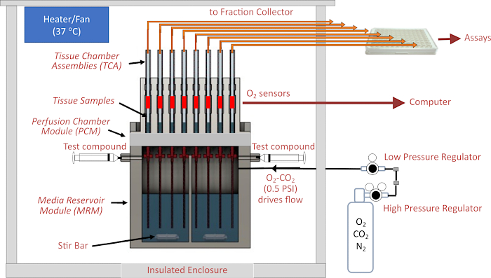
图 1.流体/评估系统示意图。 主要组件包括外壳、温度控制元件、流体和组织室系统、周围熔断液上方顶部空间的气体压力调节、馏分收集器/流速监测和 O2 检测器。缩写:MRM = 培养基储液器模块,PCM = 围灌注室模块,TCA = 组织室组件。 请点击这里查看此图的较大版本.
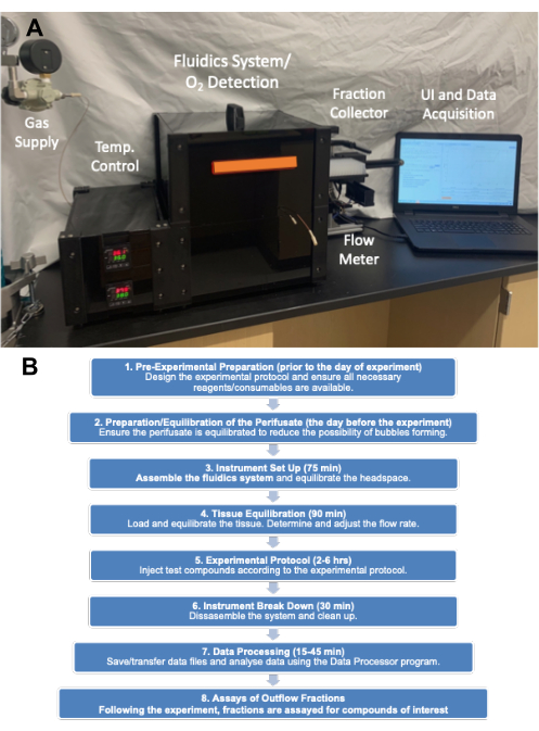
图2. (A) 仪器主要部件的图片。主要部件包括储气罐(压力调节器)、外壳、馏分收集器和计算机。(B) 实验流程图,显示主要步骤类别和完成这些步骤所需的时间。 请点击这里查看此图的较大版本.
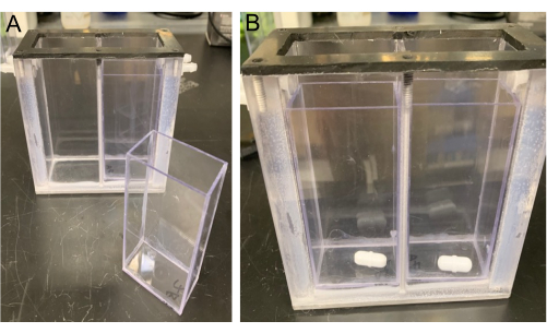
图3.MRM 视图。 MRM图中有一个MRM插件(左)和搅拌棒(右),放置在MRM插件的底部(位于MRM分频器的每一侧)。 请点击这里查看此图的较大版本.
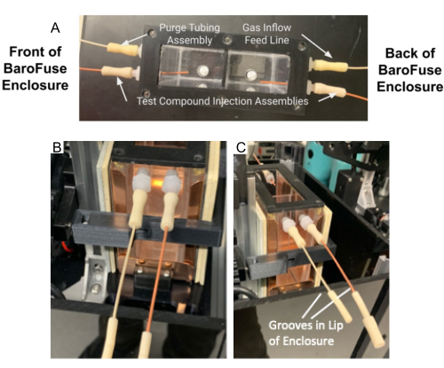
图4.MRM 中的管路组件和吹扫管路组件。(A) 测试化合物注入管组件和吹扫管组件连接到 MRM 上的端口。(B-C)测试化合物注入组件和吹扫管组件 (B) 放置在外壳 (C) 前部的凹槽中。请点击这里查看此图的较大版本.
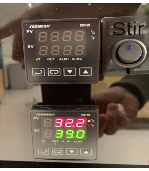
图5.接通MRM温度控制器的电源。请点击这里查看此图的较大版本.
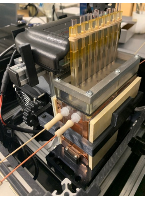
图6.组织室和储气罐。将 O2 检测器放置在检测器支架上(也支持 MRM 和 PCM),并将条带放置在 PCM 的翅片周围,以帮助将组织室固定到位。 请点击这里查看此图的较大版本.
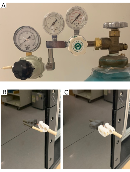
图 7. (A) 储气罐上的高压和低压调节器。(B-C)吹扫管。吹扫管允许MRM中的顶部空间清除空气,并充满来自供应罐的气体。图片显示打开的吹扫管 (B) 和关闭的吹扫管 (C)。测试化合物注射组件在吹扫过程中保持关闭状态。请点击这里查看此图的较大版本.
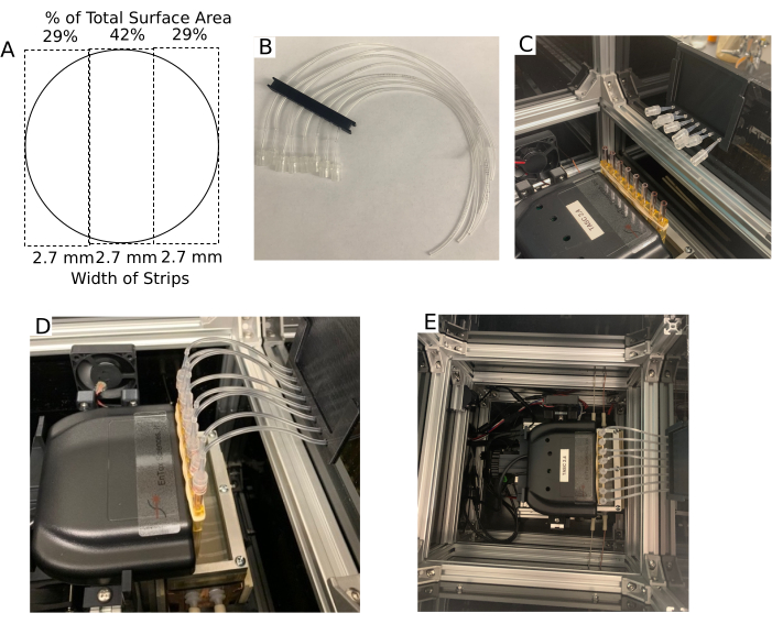
图8.组织室和流出装置。 (A) Transwell膜切成三条等宽的条带后的尺寸。(B)流出多管支撑。(C) 流出多管支架位于外壳的边缘,管路适配器靠近组织室。(D) 连接到组织室的流出管组件的图片。(E) 不带盖子的外壳鸟瞰图。 请点击这里查看此图的较大版本.
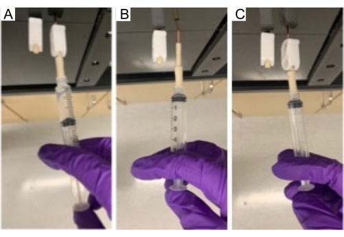
图 9.在MRM中注入化合物。 使用 5 mL 注射器通过进样口将测试化合物注入 MRM。 请点击这里查看此图的较大版本.
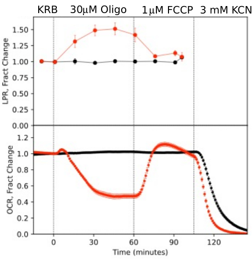
图 10.响应测试化合物的 OCR 和 LPR 曲线。 通过从小鼠(1 个视网膜/通道)中分离的视网膜进行 OCR 和 LPR,以响应指示的测试化合物的存在与否(对照)。每条曲线是单个实验中 6 次重复的平均值(误差线为 SE;p 值是通过执行配对 t 检验来计算的,该检验将每个测试代理的稳态值与前一个测试代理的稳态值进行比较)。 请点击这里查看此图的较大版本.
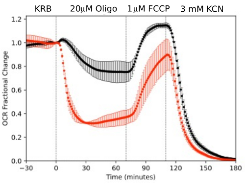
图 11.OCR 曲线。 通过从小鼠(1 个视网膜或 2 个 RPE-脉络膜-巩膜/通道)中分离的 RPE-脉络膜-巩膜和视网膜进行 OCR,并行测量以响应所示的测试化合物。数据是单个实验重复的平均值(RPE-脉络膜-巩膜和视网膜分别为 n = 2 和 4;p 值是通过执行配对 t 检验来计算的,将每个测试代理的稳态值与前一个测试代理的稳态值进行比较)。 请点击这里查看此图的较大版本.
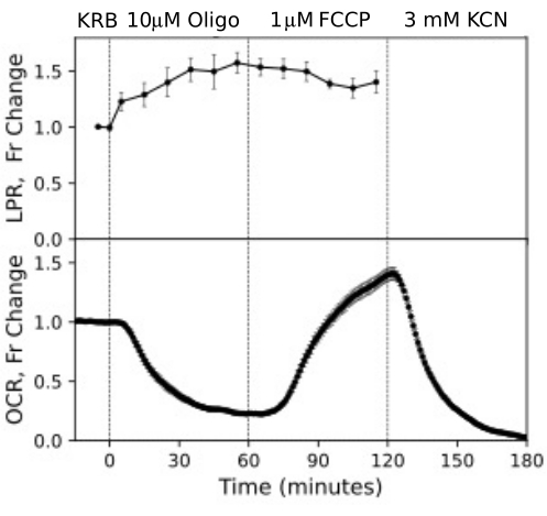
图 12.RPE 细胞的 OCR 和 LPR 曲线。 来自 RPE 细胞的 OCR 和 LPR,这些细胞附着在被切成条状并加载到围灌室中的跨孔膜上。数据是单个实验重复的平均值(n = 3,1.5 个膜/通道(360,000 个细胞/通道);p 值是通过执行配对 t 检验来计算的,将每种测试剂的稳态值与前一个测试剂的稳态值进行比较)。 请点击这里查看此图的较大版本.
| 组织/细胞 | 金额/渠道 | 流速:mL/MIN |
| 视网膜(鼠标) | 1 | 0.025 |
| RPE-脉络膜-巩膜(小鼠) | 2 | 0.02 |
| Transwell 膜上的 RPE 细胞 | 360,000 个细胞(4 x 1/3 滤条) | 0.016 |
表 1.针对不同组织的推荐操作规格。
补充图 1.实验设计的图形表示。 暴露于测试化合物的时间和组成,以及馏分收集的时间。浓度增量 (Conc Inc) 是要实施的浓度变化。 请点击这里下载此文件。
补充图2.启动时的用户界面。O 2 检测软件的启动窗口的 UI,该软件监控插入 PCM 的组织室中的 O2。请点击这里下载此文件。
补充图3。实验设置的用户界面。 用于输入实验信息(左)和选择收集流出分数的时间(右)的用户界面。 请点击这里下载此文件。
补充图4.注入页面的用户界面。 进样页面,根据所需测试化合物浓度和MRM中剩余体积计算进样体积。 请点击这里下载此文件。
补充文件1:组织样本制备方法。请点击这里下载此文件。
讨论
由于生物能量学在细胞功能和眼睛各种成分的维持的各个方面都很重要,因此迫切需要研究其调节的方法。特别是,神经视网膜和RPE依赖于代谢来产生能量以及细胞内和细胞间信号传导14,15,16,17。由于它们的高氧化能力,眼睛的分离组织在静态条件下不能很好地维持18,19,因此对眼睛的分离成分的研究需要能够维持和评估代谢过程的流动系统。流体系统旨在从各种组织类型生成 OCR 和 LPR 数据,在本文中,我们提出了详细的协议,这些协议被发现可以产生最佳结果。
使用流动系统生成可靠数据的主要决定因素包括在39°C下对基于CO2的介质/缓冲液进行预平衡(以确保周融合液不会被溶解气体过饱和,从而在实验过程中脱气)。特别是,储存在4°C的培养基或KRB缓冲液将相对于37°C过饱和,如果预平衡时间不足,则在实验过程中将脱气。此外,由于撕裂或组织分离不完全,或将组织暴露在少量碳酸氢盐缓冲液中太长时间,不得因组织过度隔离而对装入组织室的组织造成创伤。O2检测的温度控制、流量稳定性和可靠性变化不大,这些因素对故障率的影响不大。
该仪器有八个同时运行的流道/组织室,从两个储液器中供应外融合液,每个储液器有四个组织室。为了获得最准确的 OCR 时程,动力学曲线通过未加载组织的腔室进行基线校正。因此,典型的实验方案将涉及两组三个组织室。方案通常分为两类:一类是每侧不同的测试化合物方案(例如,MRM一侧的药物/载体,另一侧的载体);第二种是在MRM两侧使用相同的测试化合物注射方案,但在MRM的每一侧使用不同的组织或组织模型。在本文中,通过未暴露于任何测试化合物的组织将寡霉素和FCCP对视网膜的影响与OCR进行了比较,并在相同的方案和条件下同时评估了两个组织,以确定组织特异性行为。后者在本研究中通过显示在同一实验中 RPE-脉络膜-巩膜相对于视网膜的代谢率动态范围增加来说明。其他报告描述了更广泛的研究设计,包括测量不同的 O2 水平对 OCR 和 LPR 的影响,以及燃料、药物和毒素的浓度依赖性20,21。此外,尽管我们将流出分数的分析限制在乳酸的测量和LPR的计算上,但如果测定流出分数中的多种化合物和化合物类别,例如激素、神经递质、细胞信号和可以离开细胞的代谢物,则实验的信息内容会大大增加20,22,23.
离体视网膜或RPE-脉络膜-巩膜的加载很简单,一旦分离出,只需用镊子将这些组织放入组织室的顶部,然后让其下沉到熔块中。在滤器插件上培养的 RPE 细胞在培养 4-8 周后产生适当的极化和 RPE 成熟标志物。如果要保持 RPE 成熟度和极化,一旦附着在跨孔膜上,去除用于活细胞分析的 RPE 是不可行的24。围灌注室可以容纳用手术刀切割的transwell膜条,同时浸没在缓冲液中并快速插入组织室。尽管切割滤条已被置于静态系统24中,但没有其他流体方法来评估这些重要的细胞类型。RPE细胞的反应比视网膜或RPE-脉络膜-巩膜更快、更动态,部分原因可能是RPE细胞的顶端和基底都立即进入,这些细胞被配置为膜插入物上的单层。
确保数据具有最高信噪比的另一个因素是选择加载到围灌注室中的组织相对于流速的最佳比例。相对于流速而言,组织太少会导致流入和流出之间的溶解 O2 浓度差异非常小且难以可靠测量。相反,如果流动太慢,则 O2 的浓度会变得如此之低,以至于组织会受到缺氧的影响。尽管如此,气体压力驱动的液体流量可以保持在低至 5 mL/min 的流速下,只需少量组织即可进行准确的 OCR 和 LPR 测量。在此所示的实验中,使用约20 mL / min /通道,适用于一个视网膜,两个RPE脉络膜硬化或360,000个RPE细胞。为了最大限度地减少延迟和分散组织暴露于注射的测试化合物的系统效应,提供了多种尺寸的组织室,以便组织量(和流速)与适当的腔室尺寸相匹配。
本文中显示的分析数据以两种方式表示:相对于速率的绝对幅度,或相对于稳态或基线的分数变化。重点是说明对测试化合物的反应的测量。然而,流体系统非常适合在围灌注分析(如基因修饰)之前评估和比较组织治疗的效果。如果分析了处理对测试化合物归一化反应的影响,则测试处理是否与对照不同是最可靠的。如果分析需要绝对量级,则如果在同一围灌注实验中进行预处理标本的评估和对照,则预处理标本分析的统计功效将最大化。
除搅拌器外,所有与液体接触的部件均由制造商作为消耗品提供,并已消毒。这些部件不应重复使用,因为由于清洁不彻底和表面污染,实验偶尔会丢失。设置开始时的系统是无菌的。然而,将培养基添加到MRM中,并在非无菌条件下将组织加载到腔室中。我们已经测量了系统中的 OCR,该系统由无菌部件组装而成,但实验本身是在非无菌条件下进行的。细菌积累到具有可测量的OCR(未发表的结果)大约需要14小时。如果使用的方案少于 10 小时左右,则细菌的积累和由此引起的任何影响都可以忽略不计。
许多研究人员使用的仪器旨在测量单层细胞静态孵育下的 OCR,具有相对较高的通量25,26。相比之下,我们在本文中测试和描述的流体仪器通过确保足够的 O2 输送来维持组织,这对于组织标本中存在的更大扩散距离至关重要。此外,它能够收集分数,允许与 OCR 并行评估多个参数,这大大增强了研究它们之间关系的能力。最后,可以控制溶解气体浓度(如 O 2 和 CO 2),从而增加使用碳酸氢盐基介质和缓冲液进行实验的持续时间,使用户能够研究 O2 的影响。应该指出的是,这两种方法的局限性是无法研究测试化合物的洗脱,这是其他围灌注系统所具有的功能 4,27,28。在确定最佳分析模式时,另一个考虑因素是流体系统比静态系统使用更多的介质和测试化合物。尽管由于可以使用的低流速,当前的流体系统将额外费用降至最低。
总体而言,描述了使用新的流量/评估仪器进行实验的协议的详细说明。使用视网膜和RPE-脉络膜-巩膜生成的数据概括了以前使用更难使用(且不易获得)的系统获得的结果。研究还表明,该系统可以维持和评估附着在transwell膜上的RPE细胞,这是一个非常重要的细胞模型,由于细胞的脆弱性,以前没有用流动系统进行过分析。该协议的主要部分包括 75 分钟的设置时间,然后是 90 分钟的平衡期和实验协议,使其适合不专门从事流体系统操作的实验室的常规使用。虽然我们专注于测量组织对测试化合物的急性反应,但该系统非常适合比较来自各种来源的组织,例如经过基因改变或经过测试处理/条件的动物模型或细胞模型。此外,可以对流出部分进行的检测范围很广,包括代谢物、细胞信号分子和分泌的激素/神经递质,以及通过质谱对馏分和组织进行的多组分分析。
披露声明
I.R.S.、M.G. 和 K.B. 与 EnTox Sciences, Inc.(华盛顿州默瑟岛)有财务联系,该公司是本研究中描述的 BaroFuse 围灌注系统的制造商/分销商。所有其他作者均声明无利益冲突。
致谢
这项研究由美国国立卫生研究院 (R01 GM148741 IRS)、U01 EY034591、R01 EY034364、BrightFocus 基金会、预防失明研究 (J.R.C.) 和 R01 EY006641、R01 EY017863 和 R21 EY032597 (J.B.H.) 资助。
材料
| Name | Company | Catalog Number | Comments |
| BIOLOGICAL SAMPLES | |||
| C57BL/6J mice | Envigo Harlan (Indianapolis, IN) | N/A | |
| REAGENTS | |||
| FCCP | Sigma-Aldrich | C2920L9795 | |
| Glucose | Sigma-Aldrich | G8270G | |
| KCN | Sigma-Aldrich | 60178 | |
| Lactate | MilliporeSigma | L6661 | |
| Oliigomycin A | Sigma-Aldrich | 75351L9795 | |
| CELL CULTURE AND TISSUE HARVESTING | |||
| Beuthanasia-D | Schering-Plough Animal Health Corp., Union, NJ | N/A | |
| Bovine serum albumin | Sigma-Aldrich | A3059 | |
| Euthasol, 390 mg/ml sodium pentobarbital | Virbac | RXEUTHASOL | |
| Fetal bovine serum | Sigma-Aldrich | 12303C | |
| Hank’s Buffered Salt Solution | GIBCO | 14065056 | |
| Krebs Ringer Bicarbonate (KRB) | Thermo Fisher Scientific | J67795L9795 | |
| Matrigel | ThermoFisher | #CB-40230 | |
| Penicillin-streptomycin | ThermoFisher Scientific | 15140122 | |
| ROCKi | Selleck Chemicals | Y-27632 | |
| Trypsin-EDTA | ThermoFisher | #25-200-072 | |
| SUPPLIES | |||
| Gas Cylinders: 21% O2/5% CO2/balance N2 | Praxair Distribution, Inc | N/A | |
| Transwell filters | MilliporeSigma | 3470 | |
| COMMERCIAL ASSAYS | |||
| Amplex Red Glucose/Glucose Oxidase Assay Kit | ThermoFisher | A22189 | |
| Glucose Oxidase from Aerococcus viridans | Invitrogen (Carlsbad, CA) | A22189L9795 | |
| Lactate Oxidase | Sigma-Aldrich | L9795 | |
| EQUIPMENT | |||
| BaroFuse Multi-Channel Perifusion system | EnTox Sciences, Inc (Mercer Island, WA | Model 001-08 | |
| Synergy 4 Fluorometer | BioTek (Winooski, VT) | S4MLFPTA |
参考文献
- Lacy, P. E., Walker, M. M., Fink, C. J. Perifusion of isolated rat islets in vitro: Participation of the microtubular system in the biphasic release of insulin. Diabetes. 21 (10), 987-998 (1972).
- Doliba, N. M., et al. Metabolic and ionic coupling factors in amino acid-stimulated insulin release in pancreatic beta-HC9 cells. American Journal of Physiology. Endocrinology and Metabolism. 292 (6), E1507-E1519 (2007).
- Sweet, I. R., et al. Regulation of ATP/ADP in pancreatic islets. Diabetes. 53 (2), 401-409 (2004).
- Chertov, A. O., et al. Roles of glucose in photoreceptor survival. The Journal of Biological Chemistry. 286 (40), 34700-34711 (2011).
- Kooragayala, K., et al. Quantification of Oxygen Consumption in Retina Ex Vivo Demonstrates Limited Reserve Capacity of Photoreceptor Mitochondria. Investigative Ophthalmology & Visual Science. 56 (13), 8428-8436 (2015).
- Bisbach, C. M., et al. Succinate Can Shuttle Reducing Power from the Hypoxic Retina to the O2-Rich Pigment Epithelium. Cell Reports. 31 (5), 107606 (2020).
- Du, J., et al. Inhibition of mitochondrial pyruvate transport by zaprinast causes massive accumulation of aspartate at the expense of glutamate in the retina. The Journal of Biological Chemistry. 288 (50), 36129-36140 (2013).
- Hass, D. T., et al. Succinate metabolism in the retinal pigment epithelium uncouples respiration from ATP synthesis. Cell Reports. 39 (10), 110917 (2022).
- Kamat, V., et al. Fluidics system for resolving concentration-dependent effects of dissolved gases on tissue metabolism. Elife. 10, e66716 (2021).
- Stryer, L. . Biochemistry. , (1995).
- Gu, X., Ma, Y., Liu, Y., Wan, Q. Measurement of mitochondrial respiration in adherent cells by Seahorse XF96 Cell Mito Stress Test. STAR Protocols. 2 (1), 100245 (2021).
- Engel, A. L., et al. Extracellular matrix dysfunction in Sorsby patient-derived retinal pigment epithelium. Experimental Eye Research. 215, 108899 (2022).
- Zhang, R., et al. Inhibition of Mitochondrial Respiration Impairs Nutrient Consumption and Metabolite Transport in Human Retinal Pigment Epithelium. Journal of Proteome Research. 20 (1), 909-922 (2021).
- Hurley, J. B. Retina Metabolism and Metabolism in the Pigmented Epithelium: A Busy Intersection. Annual Review of Vision Science. 7, 665-692 (2021).
- Xiao, J., et al. Autophagy activation and photoreceptor survival in retinal detachment. Experimental Eye Research. 205, 108492 (2021).
- Okawa, H., Sampath, A. P., Laughlin, S. B., Fain, G. L. ATP consumption by mammalian rod photoreceptors in darkness and in light. Current Biology. 18 (24), 1917-1921 (2008).
- Lakkaraju, A., et al. The cell biology of the retinal pigment epithelium. Progress in Retinal and Eye Research. , 100846 (2020).
- Yu, J., et al. Emerging strategies of engineering retinal organoids and organoid-on-a-chip in modeling intraocular drug delivery: Current progress and future perspectives. Advanced Drug Delivery Reviews. 197, 114842 (2023).
- Arjamaa, O., Nikinmaa, M. Oxygen-dependent diseases in the retina: role of hypoxia-inducible factors. Experimental Eye Research. 83 (3), 473-483 (2006).
- Kamat, V., et al. A Versatile Multi-Channel Fluidics System for the Maintenance and Real-Time Metabolic and Functional Assessment of Tissue or Cells. Cell Reports Methods. In Press. , (2023).
- Neal, A., et al. Quantification of Low-Level Drug Effects Using Real-Time, in vitro Measurement of Oxygen Consumption Rate. Toxicological Sciences. 148 (2), 594-602 (2015).
- Jung, S. R., et al. Reduced cytochrome C is an essential regulator of sustained insulin secretion by pancreatic islets. The Journal of Biological Chemistry. 286 (20), 17422-17434 (2011).
- Rountree, A. M., et al. Control of insulin secretion by cytochrome C and calcium signaling in islets with impaired metabolism. The Journal of Biological Chemistry. 289 (27), 19110-19119 (2014).
- Calton, M. A., Beaulieu, M. O., Benchorin, G., Vollrath, D. Method for measuring extracellular flux from intact polarized epithelial monolayers. Molecular Vision. 24, 425-433 (2018).
- Jarrett, S. G., Rohrer, B., Perron, N. R., Beeson, C., Boulton, M. E. Assessment of mitochondrial damage in retinal cells and tissues using quantitative polymerase chain reaction for mitochondrial DNA damage and extracellular flux assay for mitochondrial respiration activity. Methods in Molecular Biology. 935, 227-243 (2013).
- Perron, N. R., Beeson, C., Rohrer, B. Early alterations in mitochondrial reserve capacity; a means to predict subsequent photoreceptor cell death. Journal of Bioenergetics and Biomembranes. 45 (1-2), 101-109 (2013).
- Cabrera, O., et al. high-throughput assays for evaluation of human pancreatic islet function. Cell Transplantation. 16 (10), 1039-1048 (2008).
- Doliba, N. M., Qin, W., Vinogradov, S. A., Wilson, D. F., Matschinsky, F. M. Palmitic acid acutely inhibits acetylcholine- but not GLP-1-stimulated insulin secretion in mouse pancreatic islets. American Journal of Physiology. Endocrinology and Metabolism. 299 (3), E475-E485 (2010).
转载和许可
请求许可使用此 JoVE 文章的文本或图形
请求许可探索更多文章
This article has been published
Video Coming Soon
版权所属 © 2025 MyJoVE 公司版权所有,本公司不涉及任何医疗业务和医疗服务。