
Method Article
単一細胞分泌物の時空間イメージング用ラベルフリー技術
要約
細胞間の通信は、細胞内外の様々な生理活性を制御するために重要です。本稿では、単一細胞分泌物の時空間的な性質を測定するためのプロトコルについて説明します。これを達成するためには、学際的なアプローチは、生細胞イメージングとラベルフリーnanoplasmonic検知を統合が使用されます。
要約
Inter-cellular communication is an integral part of a complex system that helps in maintaining basic cellular activities. As a result, the malfunctioning of such signaling can lead to many disorders. To understand cell-to-cell signaling, it is essential to study the spatial and temporal nature of the secreted molecules from the cell without disturbing the local environment. Various assays have been developed to study protein secretion, however, these methods are typically based on fluorescent probes which disrupt the relevant signaling pathways. To overcome this limitation, a label-free technique is required.
In this paper, we describe the fabrication and application of a label-free localized surface plasmon resonance imaging (LSPRi) technology capable of detecting protein secretions from a single cell. The plasmonic nanostructures are lithographically patterned onto a standard glass coverslip and can be excited using visible light on commercially available light microscopes. Only a small fraction of the coverslip is covered by the nanostructures and hence this technique is well suited for combining common techniques such as fluorescence and bright-field imaging.
A multidisciplinary approach is used in this protocol which incorporates sensor nanofabrication and subsequent biofunctionalization, binding kinetics characterization of ligand and analyte, the integration of the chip and live cells, and the analysis of the measured signal. As a whole, this technology enables a general label-free approach towards mapping cellular secretions and correlating them with the responses of nearby cells.
概要
細胞間の通信は、細胞の内側と外側の両方の多くの生理活性の調節のために不可欠です。タンパク質および小胞の様々なその後、このような分化、創傷治癒、免疫応答、遊走、および増殖などの複雑な細胞プロセスを誘発することが分泌され得る。細胞間シグナル伝達経路の1-5誤動作は、癌、アテローム性動脈硬化症を含む多くの疾患に関与しています、および糖尿病、いくつかの名前を付けます。
最適な細胞分泌アッセイは、空間的および時間的に関連するシグナル伝達経路に影響を与えることなく、目的の分泌タンパク質をマッピングすることができるべきです。このようにして、濃度プロファイルと受信細胞の応答との間の因果関係を推測することができます。残念ながら、最も一般的に使用される蛍光ベースの技術の多くは、これらの基準を満たしていません。蛍光融合タンパク質は、被検体Wをタグ付けするために使用することができセルをithinが、分泌経路を妨害することができ、又は分泌場合、定量化が困難である細胞外へ拡散グローをもたらします。蛍光immunosandwichベースのアッセイは、細胞分泌物を検出するために最も一般的に使用される技術であるが、典型的には個々の細胞の単離を必要とする。また6-11、検知抗体の導入は、典型的には、実験や抗体標識の大きさを停止または終了150kDaのIgGについては、下流のシグナル伝達に障害となっています。
これらの障害物のそれはラベルフリー技術は、画像タンパク質分泌および既存のラベルフリー技術の中で利用されるプラズモン共鳴(SPR)の表面と、局在表面プラズモン共鳴(LSPR)センサは、優れた候補であることが好ましいからである。12-17これらセンサー広くタンパク質、エキソソームおよび他のバイオマーカーの分析物結合の研究のために使用されている。LSPRの場合は18〜24、プラズモンnanostructuresガラスカバースリップ上にリソグラフィでパターン化し、標準的な広視野顕微鏡の構成を介して可視光を用いて励起することができます。それらのナノスケールのフットプリントに、ガラス基板の大部分は、25-28。例えば、生細胞の顕微鏡との統合に適した明視野でよく、これらのプローブを作製する蛍光顕微鏡のような一般的な画像化技術のために利用可能である我々は、リアルタイム測定を示しましたそれぞれ、225ミリ秒と10ミクロンの空間的および時間的解像度で官能金プラズモンナノ構造を用いて、ハイブリドーマ細胞からの抗体分泌物。基本的なチップ構成は 、図1に示されている。顕微鏡28の出力光路は、画像及びナノ構造の指定された配列の部分占有率の定量決意にファイバ光学的に結合分析装置( 図2に使用されるCCDカメラの間に分割され)。
protoc同時に、標準的な明視野顕微鏡を用いて細胞の応答を監視しながら、本論文では、オール単一細胞分泌物のリアルタイム測定のための実験計画について説明します。学際的なアプローチは、ナノ構造体の製造、分析物の高親和性結合のためのナノ構造体の官能化、商業表面プラズモン共鳴(SPR)測定器を使用して、両方の最小化、非特異的結合と特徴づける運動速度定数の表面の最適化、細胞株の統合が含ま基板と、画像やスペクトルデータの解析へ。私たちは、この技術は、細胞の分泌物と細胞を受けたとの因果関係の時空マッピングを可能にする技術であることを期待しています。
プロトコル
1.ナノ構造作製
- ナノファブリケーションのための基質として170ミクロン(1.5号)のおおよその厚さと直径25mmのカバーガラスを選択してください。
- 少なくとも6時間(硫酸と過酸化水素水の比1 3)ピラニア溶液中でカバースリップを浸します。超純18.2MΩ脱イオン蒸留水(DDW)のおびただしい量のピラニア浸したカバースリップを洗ってください。
注意:ピラニア酸は、有機物質と激しく反応し、細心の注意を払って取り扱わなければなりません。 - ナノ構造のパターニングおよび撮像中電荷効果を回避するために、電子ビーム蒸着法によりカバーガラス上に堆積10nmのクロム薄膜。
- 二重層の第1層が45秒間2000rpmで乳酸エチルメチルメタクリレート(MMA_EL6)共重合体からなるレジストをスピンし、次いで150℃で焼きます。その後、180℃で焼く45秒間3000rpmでポリメチルメタクリレート(950PMMA_A2)の第二の層をスピン。
- パターN二重層300μC/ cm 2の面積線量で25キロボルトの電子ビームリソグラフィ(EBL)を用いたレジスト。イソプロピルアルコール(IPA)/メチルイソブチルケトン(MIBK)で開発:2/1およびIPAですすぎます。
- 電子ビーム蒸発器を用いて基板上に堆積したTi(5 nm)の/金(80 nm)のフィルム。
- 金の堆積に続いて、共重合体の二重層が4時間アセトンに基板を浸漬することにより、レジストのリフトオフ。
- ナノ構造の形状及び大きさを確認するために、走査型電子顕微鏡(SEM)を用いて基板を検査し、RTで60秒間CR-7のエッチャントを用いたウェットエッチングによって基板から残りのクロムを除去した後、DDWですすぎます。
- アレイ間の細胞イメージングのための余地を残すために33ミクロンの中心の配列間隔を設計します。電子ビーム描画装置を用いて、300nmのピッチで配列ごとに20×20のパターンでパターンナノ構造を。各チップは、80±2.5 nmの高さと70±2.5 nmでディアメの典型的なナノ構造寸法の300の配列が含まれていますター。
- 大きさや均一性を検証するための原子間力顕微鏡(AFM)を用いて、配列のサブセットを検査します。
- UV硬化性エポキシを使用して、カバーガラスの背面に、サポートリング、典型的にはシリコンを取り付けます。
2.チップクリーニングと自己組織化単分子膜への応用
- 洗浄のため、同一条件下で5分間、チャンバをクリーニングした後、チップ、5%水素の300ミリトール混合物中の40 Wの電力でプラズマアッシュ、45秒間95%のアルゴンを再生します。
- 1 SH-の比率(CH 2)8 -EG 3 -OH(SPO)およびアミンのいずれかを有する成分:3からなる二成分エタノールチオール溶液にチップを浸漬することにより直ちにプラズマアッシング後の金ナノ構造を官能またはカルボキシル官能基、すなわち、SH-(CH 2)11 -EG 3 -NH 2(SPN)またはSH-(CH 2)11 -EG 3 -COOH(SPC)。
- チップIを残しますn個のチオール溶液をO / N自己組織化単分子膜(SAM)を形成します。
- 窒素ガスをエタノールとドライでチップを洗浄します。
- 必要に応じて、4℃で2週間まで官能チップを格納します。
- 使用可能な状態には、選択したリガンドに応じて化学反応を用いて、リガンドとSPNまたはSPCのコンポーネントを反応させた場合(下記参照)。
注意:チップは何十回も再生され、再官能化することができます。指定されたチップは、6ヶ月から一年間に及ぶ期間のために使用することができます。指定された配列で測定スペクトルは、確実に生体機能化に続いて、プラズマアッシングにより、繰り返し再生の後に再生されます。29
3.表面機能とキネティック特性評価
注:リガンドとアナライトとの間の動力学的速度定数を特徴付けるために、ならびに非特異的BにSAMの耐性を研究するために商業SPR機器で官能化チップを使用してinding。流量と効率的な表面機能を可能にするマイクロ流体設計の広い範囲があります。我々は、市販のSPRを持っているので、私たちはその推奨流量を中心に標準化しました。我々は、これらの流量はすべてのSPR機器の典型的なものであるので、制限するものではないことに注意してください。すべての機能化がLSPRチップ上で直接行うことができますが、それは多重化機器があるため、私たちのLSPRマイクロ流体セットアップがないのに対し、それが私たちの作業負荷を軽減なかったため、SPR機器は必要ありません。
- 第2節で説明したように、SAMを有する市販の裸の金チップを官能。
- 10 1-エチル-3-(3-ジメチルアミノプロピル)カルボジイミド(EDC)の133 mmでDDW 中のNヒドロキシスクシンイミドの33 mMの(NHS)の1:1混合物、SPCベースのSAMを使用して、1とカルボキシル基を活性化する場合分。
- 30μl/分の流速を使用して300秒間、目的の抗体/リガンドで活性化したカルボキシル基を結合。リガンドを準備中pH6のリン酸緩衝液は、典型的には、これは、分子に依存して変化し得ます。
- リガンドコンジュゲーションの後、30μL/分の速度で300秒間の不活性化工程として、リン酸緩衝生理食塩水(PBS)中の0.1 Mエタノールアミンを流します。エタノールアミンは、非特異的結合を最小限に抑えるのに役立ちます。
- 濃度の範囲を使用して、100μL/分の流速で、目的の分析物を導入し、反応速度分析ソフトウェアを用いて反応速度定数を算出します。
- 非特異的結合に問題がある場合は、SPCまたはSPNにSPOの割合を増加させます。
4. LSPR一般設定
- 顕微鏡の設定:
- サンプルを照らすケーラーに、100Wハロゲンランプを使用してください。共鳴シフト( 図2)に寄与しない波長を除去するために光路にロングパスフィルタ(通常は593 nmのカットオフ)を使用します。
- LSPRiデータ収集のために、40X油immersioで倒立顕微鏡を使用n個の対物レンズ(1.4 NA)と熱電は、16ビットのCCDカメラを冷却しました。
- 同時に画像とスペクトルを得るために、顕微鏡の出力ポートにビームスプリッタを配置します。
- 37に温度制御された顕微鏡ステージを設定します。 Cを°と4時間平衡化。
- それぞれ、5%および95%のCO 2濃度及び湿度を調節するために顕微鏡に追加の培養アセンブリを組み込みます。
- チップの準備と取り付け:
- SPR実験から決定された最適二成分のSAM比の第2節で説明したようにLSPRチップを官能。
- 次のようにカスタムメイドのマイクロ流体ホルダー内のチップをロードします。アルミボトムピースにチップを置きます。この底部片とシリコーンガスケットと透明なプラスチックトップピースとの間のチップを挟みます。アセンブリをクランプする4本のネジを使用してください。
- 1ミックス:典型的なSPCベースチオールアプリケーションでは、1μLのコート300をドロップSPCチオール成分のカルボキシル基を活性化するために、EDCの133ミリ、DDWでNHSの33 mMとチャー。
- 10分間待ってから、手動で10mMのPBSで表面を洗い流してください。
- リガンド溶液を300μlをコーティング降下によって、目的のリガンド(典型的には、抗体または抗体断片)で活性化されたカルボキシル基をコンジュゲート。
- 30分間待ってから、手動で10mMのPBSですすいでください。
- 非特異的結合を最小限に抑えるために、チップ上のコートを、PBS中の0.1Mエタノールアミンの300μLをドロップします。 10分間待ちます。
- PBSは、0.005%トゥイーン20(PBS-T20)を含有するエタノールを洗ってください。
- 更衣メニスカスに関連するデータの変動を低減するために、チップの上に水晶片を置きます。
- 顕微鏡に取り付けながら、PBS-T20緩衝液で濡れたチップを保管してください。
- 加熱されたステージの試料ホルダーにしっかりとLSPRチップアセンブリを配置し、マイクロ流体チューブを取り付けます。
- アセンブリにマイクロ流体チューブを取り付け、府の流れffer(または細胞研究のための無血清培地)定常状態に達するまで。
- アセンブリおよび顕微鏡は、少なくとも2時間平衡化することを許可します。
- 中央アレイは分光用光ファイバと整列するように、ジョイスティックを使用して、チップの位置を合わせます。分光分析データは、分光計およびスペクトル解析ソフトウェアを用いて行われます。
- ソフトウェアオートフォーカス、ツァイス明確なフォーカスまたは同等のオートフォーカス装置を用いて、実験を通じて、焦点の配列をしてください。
9E10ハイブリドーマ細胞から抗c-mycの分泌物の5 LSPRイメージング
注:この研究のために使用されるハイブリドーマ細胞株は、化学的トリガーを必要とせず、構成、従って抗c-myc抗体を発現します
- C-mycのペプチドでナノ構造体を官能。これは、クローン9E10ハイブリドーマ細胞によって分泌された抗c-mycの抗体について1.77 nMでのK D値を有します 。
- complet文化ハイブリドーマ細胞5%CO 2下、37℃でT75フラスコ中、10%ウシ胎児血清(FBS)および1%抗生物質を含む電子成長培地。 4×10 5細胞/ mlの細胞密度を維持します。
- 細胞分泌の研究のために、遠心分離によってT75フラスコからのペレット細胞は、分泌された抗体を除去し、4×10 6細胞/ mlの細胞密度を調整するためにRPMI-1640無血清培地(SFM)で2回洗浄します。
- LSPRチップにそれらを導入する前に、生存のために、細胞およびテストを収穫。
- 手動マイクロピペットでLSPRチップ上に細胞溶液50μlをご紹介します。数分後に25〜50の細胞は、LSPRチップの表面に付着します。
- マイクロ流体灌流システムを用いて、新鮮なSFMに溶液中に残った細胞を洗い流します。
- 10ミクロンの範囲内、接近しているイメージングのためのLSPRアレイを選択しますが、セルと重なっていません。
- 信号が分泌抗c-mycのantibに特異的であることを確実にするために、odiesは、抗体を用いたが、溶液中でのc-mycのペプチドの飽和濃度の存在によって遮断され、それらの結合部位を有する本だけでなく、抗体を用いてとせず、細胞培養培地をご紹介します。
- 抗c-mycの抗体(250 nM)を、飽和溶液を用いて各実行の最後にセンサーを調整します。これは、センサの応答を正規化するのに役立ちますし、各ランの生体機能化プロファイルに基づいて部分占有を決定します。
- 画像アラインメントソフトウェアを使用して、XとY方向のドリフトを修正してください。
結果
典型的な生細胞分泌研究で行われて、データ収集の複数のモードがあります。 図3は、正方形の配列を強調しLSPRi画像のオーバーレイ、および左下のセルを強調表示する透過光照らされた画像を示しています。データは、典型的には、以下に説明する正規化計算のための分析物の飽和溶液を導入し、続いて3時間の期間にわたって収集されます。蛍光画像は、フィルタキューブの自動切り替えにより、データ収集ルーチンに組み込むことができます。 図4の蛍光膜色素ローダミンDHPEで染色された細胞では葉状仮足のような拡張機能(矢印)を示します。そのような拡張は、配列と重複した場合、それらは、タンパク質の分泌のための偽陽性を与えるだろう。画像の複数のモードを持つことは、そのような発生を特定するのに役立ちます。
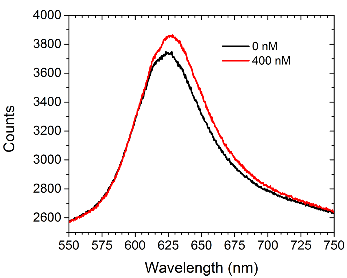
図5は、分析前のデータ及び後方を示し、ERのc-mycの官能基化アレイに市販抗c-mycの抗体の飽和溶液(400 nM)をの導入。いいえ細胞は、この実験中に存在しませんでした。スペクトルは赤方偏移と強度の増加の両方が表示されます。二つの曲線の下の面積の差は、CCDカメラでLSPRiモードで配列された画像強度の増加をもたらします。非線形最小二乗法データ分析手法は、スペクトルから表面結合リガンドの部分占有を推測するために開発された。30,31
実験の最後に、飽和強度値( すなわち、分数占有≈1)は次の式を使用して、各アレイについて正規化された応答を計算するために使用されます。
時刻tにおける時刻 tでの正規化された強度、実験、最終飽和強度の開始時の初期輝度、およびアレイの測定された強度である場合、それぞれ。
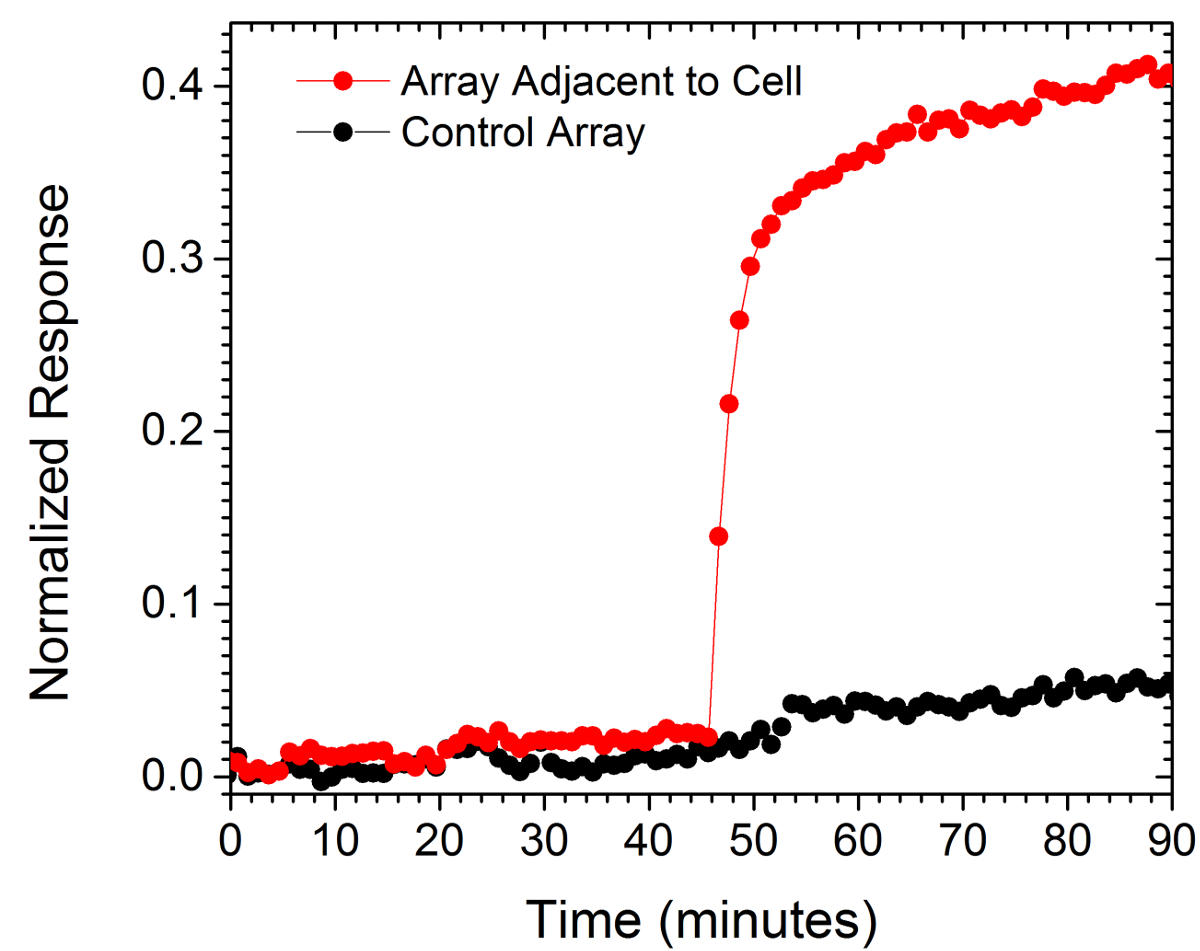
二つの配列からの正規化された値は 、図6に示されている。一つの配列は、コントロールとして使用し、他の一方で、調査中の細胞の10ミクロンの範囲内であった細胞から130ミクロンの距離でした。コントロール配列の平坦な応答に対する細胞の相対的に最も近い配列の正規化された応答の急激な増加は、分泌された抗体の局所的なバーストを示します。

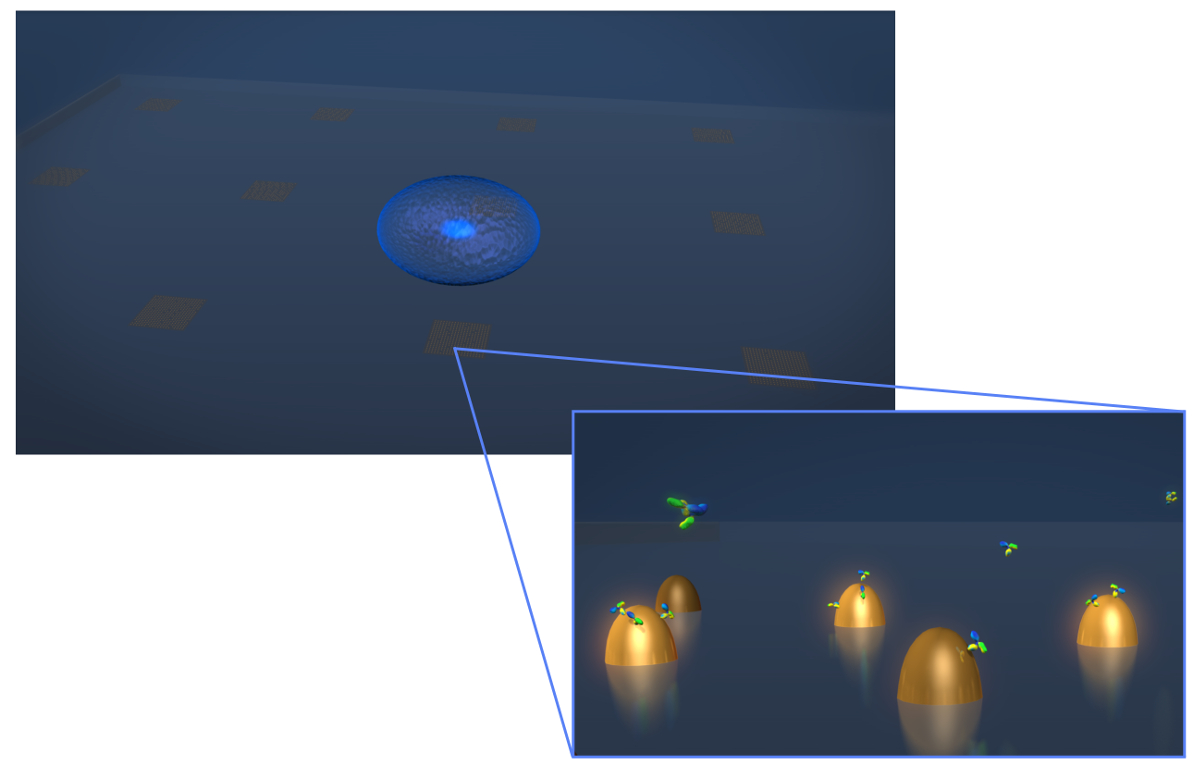
1.センサーの設計図。典型的な生細胞分泌実験のジオメトリを描いた図面。セル(青スフェロイド)がbiofunctionalized金ナノ構造体の配列が含まれているLSPRチップに上に堆積されます。ズームアップビューでは、目的の細胞分泌は、この場合には、抗体は、Y字型分子として示し、それらが表面に結合するように測定されます官能ナノ構造。 この図の拡大版をご覧になるにはこちらをクリックしてください。

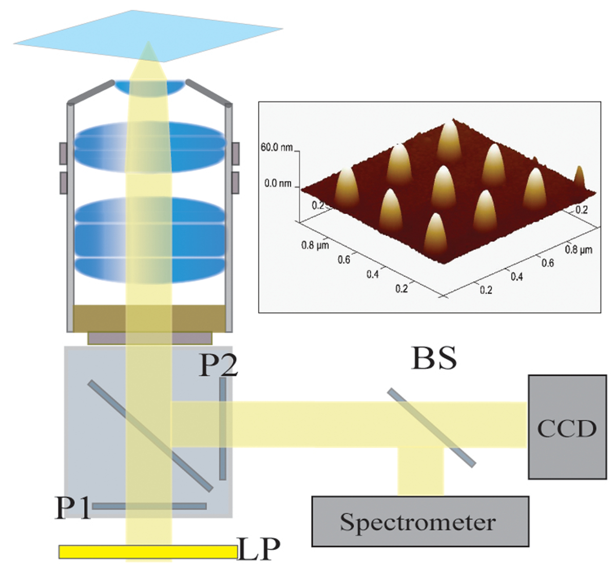
図2光学装置。ハロゲンランプから照射される光は、第一のロングパスフィルタ(LP)によってフィルタリングされます。光が直線的に(P1)偏光および40X / 1.4 NAの対物レンズを介して、サンプルを照明します。散乱光は、対物レンズによって収集され、交差偏光子(P2)を通過します。 50/50ビームスプリッター(BS)は、同時分光及び画像分析のために収集された光路に挿入されています。右上:300nmのピッチで区切られた9個々のナノ構造の原子間力顕微鏡画像はこちらをクリックしてくださいこの図の拡大版を表示します。

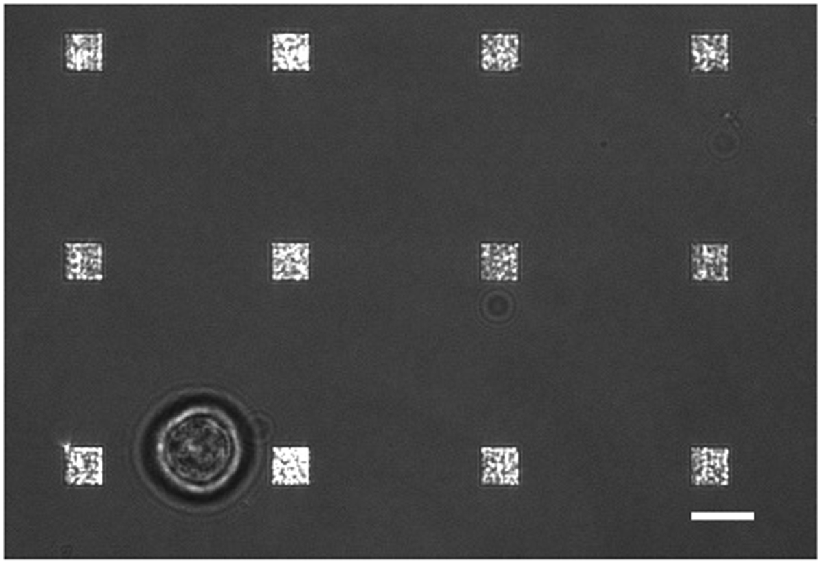
図3.生細胞LSPRi研究。Aが12の配列に囲まれた単一のハイブリドーマ細胞(左下)を示す透過光とLSPRi画像を統合しました。これは、コントラスト強調画像です。スケールバーは10μmである。 この図の拡大版をご覧になるにはこちらをクリックしてください。

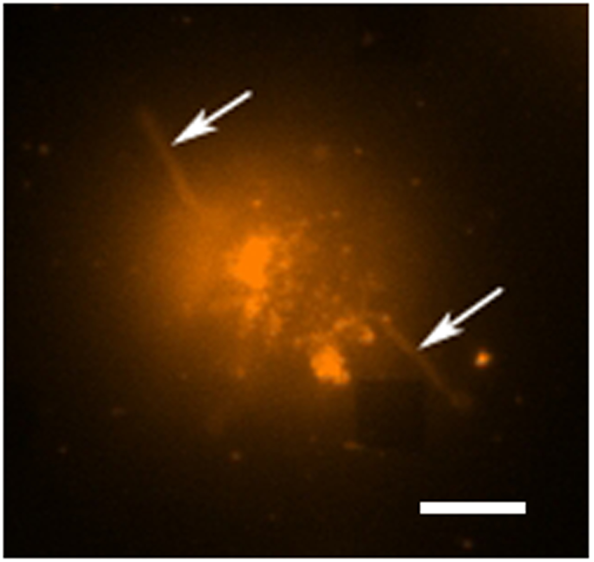
図4.ライブセル蛍光研究。膜色素であるローダミンDHPEで染色単一のハイブリドーマ細胞の蛍光偽カラー画像。配列は、一般的に表示されていない蛍光撮影モードでは、しかし、近くの配列はABとしてここに観察することができます右下の四角を欠いています。細胞は、細胞(矢印)から外側に拡張している触手のような拡張機能(おそらく糸状仮足や葉状仮足)が、配列から分離されることが分かります。スケールバーは10μmである。 この図の拡大版をご覧になるにはこちらをクリックしてください。

図5.スペクトルモダリティ。抗c-mycの抗体の400 nmのソリューションを導入する前と後のc-mycの官能化された配列から得られたスペクトル。いいえ細胞は、この研究では存在しなかった。 この図の拡大版をご覧になるにはこちらをクリックしてください。

図6.単一細胞分泌。15単セルのミクロンと130ミクロン離れて配置された1つ(コントロール)内に位置する配列の応答。スケールバーは10μmである。 この図の拡大版をご覧になるにはこちらをクリックしてください。
ディスカッション
The LSPR imaging technique described in this work has numerous advantages over more traditional methodologies for detecting cell secretions. First, the time resolution of our technique is on the order of seconds whereas the commercial alternative, an immunosandwhich assay known as EliSpot, has a typical time resolution of 2 to 3 days.7,32 As a result we were able to resolve sudden changes in the rate of protein secretion, such as that shown in Figure 6. Second, having arrays distributed over the chip allows for the secreted signal to be tracked in space and time which enables more rigorous comparisons to diffusion-based models of cell secretion. In addition, arrays like the control array shown in Figure 6 can be used to subtract out global changes in the image that typically arise from instrumental factors such as focus drift. Third, our technique requires no modification of the cells. If desired, the experiment can incorporate commonly used tags such as fluorescent proteins, but if there is concern that such tags may negatively affect cell viability or homeostasis the label-free nature of our approach does not require them. Fourth, using the spectroscopic data we have demonstrated that quantitative information regarding the fractional occupancy of surface bound ligands can be calculated.
There are numerous alternative methods to EBL for fabricating metallic nanoparticles. However, we have found that the EBL provides considerable flexibility for optimizing nanostructure and array dimensions to best suit the optics and the cells under investigation. Also critical is the fact that the chips can be readily regenerated by plasma ashing. In this way, a typical chip can be used dozens of times. Biofunctionalization details must be modified for the specific application. The protocol presented here conjugated the surface with relatively small c-myc peptide ligands. Larger ligands such as whole antibodies typically require more spacing and thus a higher SPO to SPN/SPC ratio. Regardless, a well formed SAM layer is essential for preventing non-specific binding in live-cell experiments. In general, larger molecular weight analytes are more readily detected by LSPR. Thus, in its single-cell manifestation, this technique may not be appropriate for detecting the secretion of small proteins, such as cytokines.
The current setup has been used for studying individual non-adherent cells. There are significant number of secreted signaling proteins and vesicles to which the results reported in this work are directly applicable. For example carcinoembryonic antigen (CEA) which for decades now has been a diagnostic marker for cancer. Colon cancer cells are known to secrete CEA at the rates of thousands of molecules/cell/hr and the molecular weight is 180 kDa which exceeds that of IgG antibodies. CEA is believed to be involved in autocrine and paracrine signaling pathways but the spatio-temporal nature of these secretions have never been measured. Our technique can directly address these signaling questions. An extension of this work will be to measure the spatio-temporal nature of CEA secretion from single cells.33 Future work will also focus on integrating LSPRi with two and three dimensional cell cultures of adherent cells. By incorporating multiplexed arrays capable of detecting a number of secreted proteins in parallel, this technique has the potential to open a new window into cell secretions and how they influence neighboring cells.
開示事項
We thank George Anderson for helpful comments and discussions. This work was supported by the Naval Research Laboratory’s Institute for Nanoscience and the National Research Council Research Associateship Award.
謝辞
The authors have nothing to disclose.
資料
| Name | Company | Catalog Number | Comments |
| 25 mm diameter glass coverslips | Bioscience Tools | CSHP-No1.5-25 | 170±5 µm is optimal |
| Poly-methyl methacrylate | Microchem | PMMA 950 A4 | |
| Ethyl lactate methyl metacrylate | Microchem | MMA EL6 | |
| Electron beam evaporator | Temescal | FC-2000 | |
| Electron beam lithography | Raith | Series 150 | |
| Ethanol | Sigma-Aldrich | 459836 | |
| Acetone | Sigma-Aldrich | 320110 | |
| CR-7 chromium etchant | Cyantek | CR-7 | |
| Scanning electron microscope | Zeiss | Ultra 55 | |
| Atomic force microscope | Veeco | Nanoscope III | |
| Plasma ashing system | Technics | Series 85 RIE | |
| SH-(CH2)8-EG3-OH (SPO) | Prochimia | TH 001-m8.n3-0.2 | |
| SH-(CH2)11-EG3-COOH (SPC) | Prochimia | TH 003m11n3-0.1 | |
| SH-(CH2)11-EG3-NH2 (SPN) | Prochimia | TH 002-m11.n3-0.2 | |
| Surface plasmon resonance system | Biorad | XPR36 | |
| Bare gold chip | Biorad | GLC chip | Plasma ashed to remove the monolayer |
| 1-Ethyl-3-(3-dimethylaminopropyl) carbodiimide | Thermo | 22980 | |
| N-hydroxysuccinimide (NHS) | Thermo | 24510 | |
| Pentylamine-Biotin | Thermo | 21345 | |
| Ethanolamine | Sigma-Aldrich | E9508 | |
| Neutraavidin | Thermo | 31000 | |
| Phosphate buffered saline | Thermo | 28374 | |
| Tween 20 | Sigma-Aldrich | P2287 | |
| Inverted microscope | Zeiss | Axio Observer | Microscope is equipped with 40X oil immersion objective; CO2 and humidity incubation from Pecon GmbH |
| CCD camera | Hamamatsu | Orca R2 | Thermoelectrically cooled (16 bit) |
| Spectrometer | Ocean Optics | QE65Pro | |
| Spectrasuite | Ocean Optics | version1.4 | |
| c-myc peptide HyNic Tag | Solulink | SP-E003 | |
| monoclonal anti-c-myc antibody | Sigma-Aldrich | M4439 | |
| Hybridoma cell line | ATCC | CRL-1729 | |
| Antibiotic Antimycotic Solution (100×) | Sigma-Aldrich | A5955 | |
| Serum free media RPMI 1640 | Invitrogen | 11835-030 | |
| Fetal bovine serum | ATCC | 30-2020 | |
| Rhodamine DHPE | Life Technologies | L-1392 |
参考文献
- Ludwig, A. -. K., Giebel, B. Exosomes: Small vesicles participating in intercellular communication. The International Journal of Biochemistry & Cell Biology. 44, 11-15 (2012).
- Friedl, P., Gilmour, D. Collective cell migration in morphogenesis, regeneration and cancer. Nature Reviews Molecular Cell Biology. 10, 445-457 (2009).
- Letterio, J. J., Roberts, A. B. Regulation of immune responses by TGF-beta. Annual Review of Immunology. 16, 137-161 (1998).
- Werner, S., Grose, R. Regulation of wound healing by growth factors and cytokines. Physiological Reviews. 83, 835-870 (2003).
- Werner, S., Krieg, T., Smola, H. Keratinocyte-fibroblast interactions in wound healing. Journal of Investigative Dermatology. 127, 998-1008 (2007).
- Bailey, R. C., Kwong, G. A., Radu, C. G., Witte, O. N., Heath, J. R. DNA-encoded antibody libraries: A unified platform for multiplexed cell sorting and detection of genes and proteins. Journal of the American Chemical Society. 129, 1959-1967 (2007).
- Gazagne, A., et al. A Fluorospot assay to detect single T lymphocytes simultaneously producing multiple cytokines. Journal of Immunological Methods. 283, 91-98 (2003).
- Han, Q., et al. Polyfunctional responses by human T cells result from sequential release of cytokines. Proceedings of the National Academy of Sciences of the United States of America. 109, 1607-1612 (2012).
- Han, Q., Bradshaw, E. M., Nilsson, B., Hafler, D. A., Love, J. C. Multidimensional analysis of the frequencies and rates of cytokine secretion from single cells by quantitative microengraving. Lab on a Chip. 10, 1391-1400 (2010).
- Ma, C., et al. A clinical microchip for evaluation of single immune cells reveals high functional heterogeneity in phenotypically similar T cells. Nature Medicine. 17, 738-743 (2011).
- Shirasaki, Y., et al. Real-time single-cell imaging of protein secretion. Scientific Reports. 4, (2014).
- Milgram, S., et al. On chip real time monitoring of B-cells hybridoma secretion of immunoglobulin. Biosensors and Bioelectronics. 26, 2728-2732 (2011).
- Abbas, A., Linman, M. J., Cheng, Q. A. New trends in instrumental design for surface plasmon resonance-based biosensors. Biosensors & Bioelectronics. 26, 1815-1824 (2011).
- Ermakova, A., et al. Detection of a Few Metallo-Protein Molecules Using Color Centers in Nanodiamonds. Nano Letters. 13, 3305-3309 (2013).
- Haes, A. J., Van Duyne, R. P. A nanoscale optical blosensor: Sensitivity and selectivity of an approach based on the localized surface plasmon resonance spectroscopy of triangular silver nanoparticles. Journal of the American Chemical Society. 124, 10596-10604 (2002).
- Horowitz, V. R., Aleman, B. J., Christle, D. J., Cleland, A. N., Awschalom, D. D. Electron spin resonance of nitrogen-vacancy centers in optically trapped nanodiamonds. Proceedings of the National Academy of Sciences of the United States of America. 109, 13493-13497 (2012).
- Sepulveda, B., Angelome, P. C., Lechuga, L. M., Liz-Marzan, L. M. LSPR-based nanobiosensors. Nano Today. 4, 244-251 (2009).
- Barbillon, G., et al. Biological and chemical gold nanosensors based on localized surface plasmon resonance. Gold Bulletin. 40, 240-244 (2007).
- Endo, T., et al. Multiple label-free detection of antigen-antibody reaction using localized surface plasmon resonance-based core-shell structured nanoparticle layer nanochip. Analytical Chemistry. 78, 6465-6475 (2006).
- Endo, T., Kerman, K., Nagatani, N., Takamura, Y., Tamiya, E. Label-free detection of peptide nucleic acid-DNA hybridization using localized surface plasmon resonance based optical biosensor. Analytical Chemistry. 77, 6976-6984 (2005).
- Haes, A. J., Hall, W. P., Chang, L., Klein, W. L., Van Duyne, R. P. A localized surface plasmon resonance biosensor: First steps toward an assay for Alzheimer's disease. Nano Letters. 4, 1029-1034 (2004).
- Jonsson, M. P., Jonsson, P., Dahlin, A. B., Hook, F. Supported lipid bilayer formation and lipid-membrane-mediated biorecognition reactions studied with a new nanoplasmonic sensor template. Nano Letters. 7, 3462-3468 (2007).
- Park, J. H., et al. A regeneratable, label-free, localized surface plasmon resonance (LSPR) aptasensor for the detection of ochratoxin A.. Biosensors & Bioelectronics. 59, 321-327 (2014).
- Mayer, K. M., Hao, F., Lee, S., Nordlander, P., Hafner, J. H. A single molecule immunoassay by localized surface plasmon resonance. Nanotechnology. 21, (2010).
- Endo, T., Yamamura, S., Kerman, K., Tamiya, E. Label-free cell-based assay using localized surface plasmon resonance biosensor. Analytica Chimica Acta. 614, 182-189 (2008).
- Huang, Y. X., Cai, D., Chen, P. Micro- and Nanotechnologies for Study of Cell Secretion. Analytical Chemistry. 83, 4393-4406 (2011).
- Oh, B. R., et al. Integrated Nanoplasmonic Sensing for Cellular Functional Immunoanalysis Using Human Blood. ACS Nano. 8, 2667-2676 (2014).
- Raphael, M. P., Christodoulides, J. A., Delehanty, J. B., Long, J. P., Byers, J. M. Quantitative Imaging of Protein Secretions from Single Cells in Real Time. Biophysical Journal. 105, 602-608 (2013).
- Raphael, M. P., et al. A New Methodology for Quantitative LSPR Biosensing and Imaging. Analytical Chemistry. 84, 1367-1373 (2011).
- Raphael, M. P., et al. Quantitative LSPR imaging for biosensing with single nanostructure resolution. Biophysical Journal. 104, 30-36 (2013).
- Raphael, M. P., et al. A new methodology for quantitative LSPR biosensing and imaging. Analytical Chemistry. 84, 1367-1373 (2012).
- Henn, A. D., et al. Modulation of single-cell IgG secretion frequency and rates in human memory B cells by CpG DNA, CD40L, IL-21, and cell division. Journal of Immunology. 183, 3177-3187 (2009).
- Bramswig, K. H., et al. Soluble Carcinoembryonic Antigen Activates Endothelial Cells and Tumor Angiogenesis. Cancer Research. 73, 6584-6596 (2013).
転載および許可
このJoVE論文のテキスト又は図を再利用するための許可を申請します
許可を申請さらに記事を探す
This article has been published
Video Coming Soon
Copyright © 2023 MyJoVE Corporation. All rights reserved