유세포 분석 및 형광 활성화 세포 분류 (FACS): 비장 B 림프구의 분리
Overview
출처: 퍼셰 티보1,2,3,뮤니에 실뱅1,2,3,소피 노볼트4,레이첼 골럽1,2,3
1 림프포포에이시스, 면역학학과, 파스퇴르 연구소, 파리, 프랑스
2 INSERM U1223, 파리, 프랑스
3 유니버시테 파리 디드로, 소르본 파리 시테, 셀룰레 파스퇴르, 파리, 프랑스
4 흐름 세포측정플리트에서, 세포측정및 바이오마커 UtechS, 번역 과학 센터, 파스퇴르 연구소, 파리, 프랑스
면역 계통의 전반적인 기능은 전염성 유기체 및 그밖 침략자로부터 바디를 방어하는 것입니다. 백혈구, 또는 백혈구는 면역 계통의 주요 한 선수입니다. 감염시, 그(것)들은 활성화되고 면역 반응을 시작합니다. 백혈구는 생물학적, 물리적 및/또는 기능적(예를 들어, 크기, 세분성 및 분비)이 될 수 있는 상이한 파라미터를 기반으로 다양한 하위 집단(예를 들어, 골수성 세포, 림프구, 수지상 세포)으로 나눌 수 있다. 백혈구를 특성화하는 한 가지 방법은 주로 수용체인 표면 단백질을 통해서입니다. 각 백혈구 인구는 인구 중 하위 집합을 정의할 수 있는 수용체(예를 들어, 세포독성, 활성화, 이주 수용체)의 특정 조합을 표현합니다. 면역 계통은 세포 인구의 넓은 범위를 포괄하기 때문에, 면역 반응에 있는 그들의 참여를 해독하기 위하여 그(것)들을 특성화하는 것이 필수적입니다.
유동 세포측정법(FC 또는 FCM)은 세포 표면 및 세포내 분자의 발현을 분석하여 이종성 세포 혼합물에서 상이한 세포 유형을 특성화하고 정의하는 데 널리 사용되는 방법입니다. 유동 세포계는 유체, 광학 및 전자 의 세 가지 주요 하위 시스템으로 구성됩니다. 유체 시스템은 레이저 앞에서 하나씩 통과할 수 있도록 스트림에서 세포를 수송합니다. 광학 시스템은 입자를 조명하는 광원(레이저), 생성된 빛을 지시하는 광학 필터, 적절한 검출기로 형광 신호로 구성됩니다. 마지막으로 전자 시스템은 감지된 광 신호를 컴퓨터에서 처리할 수 있는 전자 신호로 변환합니다. 개별 셀이 레이저 빔 앞에서 지나갈 때 빛이 흩어져 있습니다. 빔 앞의 검출기는 측면 분산(SC)을 측정하기 위해 전방 산란(FS) 및 여러 검출기를 측정합니다. FS는 세포 크기와 상관 관계가 있으며 SC는 세포의 세분성에 비례합니다. 이러한 방식으로, 세포 인구는 종종 혼자 그들의 크기와 세분성의 차이에 따라 구별 될 수 있습니다.
세포의 크기, 모양 및 복잡성을 분석하는 것 외에도, 유동 세포측정은 세포 표면 수용체(1)의 발현을 검출하는 데 널리 사용된다. 이것은 알려진 세포 특정 수용체에 결합하는 불소 크롬 표시 단클론 항체를 사용하여 달성됩니다. 이 결합된 불소크롬은 방출 파장이라고 불리는 특정 파장의 빛을 방출하여 검출및 득점할 수 있습니다. 형광 측정은 형광으로 표지된 세포 표면 수용체에 대한 정량적 및 정성적 데이터를 제공합니다. 혈액학자는 면역 세포 집단 (2)의 치료 후속을 위해 FC를 사용하기 위해 먼저 사용하였다. 지금, 면역 페노티핑, 세포 생존성, 유전자 발현, 세포 계수 및 GFP 분석과 같은 광범위한 응용 분야에 사용된다.
FACS (형광 활성 세포 선별기)는 형광 라벨링을 사용하여 세포의 인구를 하위 집단으로 분류하는 유동 세포 측정의 전문 유형입니다. 기존의 유동 세포측정과 마찬가지로 최초의 FS, SC 및 형광 데이터가 수집됩니다. 이어서, 기계는 전하(음수 또는 양성)를 적용하고 정전기 편향 시스템(전자석)은 세포를 포함하는 충전된 물방울의 수집을 적절한 튜브로 용이하게 한다.

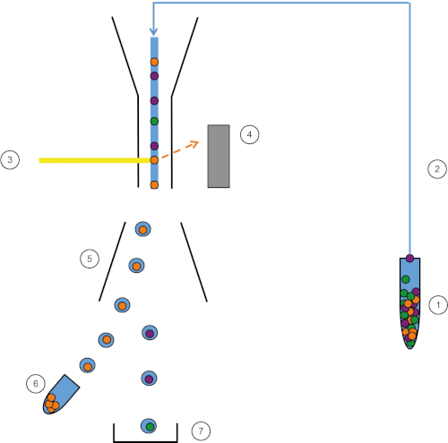
그림 1: FACS의 회로도 표현. 샘플(1)은 FACS(2)에서 흡인되어 레이저(3) 앞에 전달된다. 세포 형광은 형광 검출기 (4)에 의해 감지됩니다. 마지막으로, 세포는 액적에 통합되고 관심 있는 세포는 편향 플레이트(5)에 의해 편향되고 수집 관(6)에서 수집된다. 나머지 세포는 휴지통(7)으로 이동합니다. 이 그림의 더 큰 버전을 보려면 여기를 클릭하십시오.
FACS의 정렬 측면은 많은 장점을 제공합니다. 많은 시험은 RT-qPCR, 세포 주기, 또는 사이토카인 분비와 같은 유전자 발현의 분석과 같은 면역 계통에 있는 특정 세포의 역할을 이해하는 것을 도울 수 있습니다. 그러나 세포는 명확하고 구체적인 결과를 얻으려면 상류에서 정제되어야 합니다. 여기서 FACS는 유용하게 제공되며 원하는 셀은 매우 안정적이고 재현 가능한 결과를 산출하여 매우 순도로 정렬 할 수 있습니다. FACS는 또한 핵 또는 그밖 세포내 염색에 근거하여 그리고 표면 수용체의 존재, 부재 및 밀도에 따라 세포를 분류하기 위하여 이용될 수 있습니다. FACS는 이제 세포의 하위 집단의 정화를 위한 표준 기술이고 동시에 4개의 인구까지 분류하는 능력을 가지고 있습니다.
이 실험실 운동은 비장 백혈구를 분리하는 방법과 FACS를 사용하여 비장 백혈구 세포 혼합물에서 B 림프구 세포를 구체적으로 분류하는 방법을 보여줍니다.
Procedure
1. 준비
- 시작하기 전에 실험실 장갑과 적절한 보호 복을 착용하십시오.
- 먼저 세제로 모든 해부 도구를 살균한 다음 70%의 에탄올로 완전히 건조시하십시오.
- 2% 태아 종아리 혈청(FCS)을 함유한 행크의 균형 잡힌 소금 용액(HBSS)의 50mL를 준비한다.
2. 해부
- 이산화탄소 전달 시스템을 사용하여 저산소증으로 마우스를 안락사시합니다. supine 위치에 있는 해부 판에 안락사 마우스를 고정하고 가위와 집게를 사용하여 세로 복강경을 수행합니다.
- 집게를 사용하여 복부 오른쪽에 내장과 위를 움직여 위와 비장을 노출시십시오. 비장은 위장에 부착됩니다.
- 집게를 사용하여 위에서 비장을 조심스럽게 분리하고 HBSS 2 % FCS의 5mL가 들어있는 페트리 접시에 놓습니다.
.css-f1q1l5{display:-webkit-box;display:-webkit-flex;display:-ms-flexbox;display:flex;-webkit-align-items:flex-end;-webkit-box-align:flex-end;-ms-flex-align:flex-end;align-items:flex-end;background-image:linear-gradient(180deg, rgba(255, 255, 255, 0) 0%, rgba(255, 255, 255, 0.8) 40%, rgba(255, 255, 255, 1) 100%);width:100%;height:100%;position:absolute;bottom:0px;left:0px;font-size:var(--chakra-fontSizes-lg);color:#676B82;}
Results
이 프로토콜에서, 우리는 FACS 기술을 사용하여 비장 B 림프구를 정제. 우리는 먼저 비장에서 백혈구를 분리하고 얼룩지게했습니다. B 셀 표면 마커의 조합을 사용하여 이를 정렬하는 게이팅 전략을 만들었습니다(그림 2, 상단 패널). 실험의 끝에서 우리는 수집 관내의 세포가 "순도 테스트"를 통해 B 세포인지 확인했습니다. 우리는 동일한 게이팅 전략을 유지하고 세포의 98 %...
Application and Summary
유동 세포측정은 면역 세포 집단을 높은 순도로 특성화하고 분류하는 직접적인 기술입니다. 그것은 특정 세포 인구의 농축을 허용 하고 병원체에 면역 반응을 해독하는 것을 허용하기 때문에 연구 필드에 있는 원시 공구입니다. 사용 가능한 형광크롬 및 세포계의 수가 증가함에 따라 검출 가능한 매개 변수의 수가 크게 증가합니다. 그 결과, FACS 데이터의 생물학적 분석이 나타나기 시작했고 세포...
Tags
건너뛰기...
이 컬렉션의 비디오:

Now Playing
유세포 분석 및 형광 활성화 세포 분류 (FACS): 비장 B 림프구의 분리
Immunology
93.2K Views

자기 활성화 세포 분류 (MACS): 흉선 T 림프구 분리
Immunology
23.1K Views

ELISA 분석: 간접, 샌드위치 및 길항
Immunology
239.4K Views

ELISPOT 분석: IFN-γ 분비 비장세포 검출
Immunology
28.8K Views

면역 조직 화학 및 면역 세포 화학: 광학 현미경을 통한 조직 이미징
Immunology
79.2K Views

항체 생성: 융합 세포를 사용한 단일 클론 항체 생성
Immunology
43.7K Views

면역 형광 현미경 검사법: 파라핀이 내장 된 조직 절편의 면역 형광 염색
Immunology
54.0K Views

공 초점 형광 현미경: 쥐 섬유 아세포에서 단백질의 국소화를 결정하는 기술
Immunology
43.4K Views

면역침전반응 기반 기술: 아가로스 비즈를 사용한 내인성 단백질의 정제
Immunology
87.9K Views

세포주기 분석: 세포주기 CFSE 염색 및 유세포 분석을 사용한 자극 후 CD4 및 CD8 T 세포 증식 평가
Immunology
24.3K Views

입양 세포 전송: 기증자 쥐 비장 세포를 숙주 쥐에 도입 후 FACS를 통한 성공 평가
Immunology
22.6K Views

세포 사멸 분석: 세포 독성 능력의 크롬 방출 분석
Immunology
151.5K Views
Copyright © 2025 MyJoVE Corporation. 판권 소유
