A subscription to JoVE is required to view this content. Sign in or start your free trial.
Method Article
Using In Vitro Fluorescence Resonance Energy Transfer to Study the Dynamics Of Protein Complexes at a Millisecond Time Scale
In This Article
Summary
Protein-protein interactions are critical for biological systems, and studies of the binding kinetics provide insights into the dynamics and function of protein complexes. We describe a method that quantifies the kinetic parameters of a protein complex using fluorescence resonance energy transfer and the stopped-flow technique.
Abstract
Proteins are the primary operators of biological systems, and they usually interact with other macro- or small molecules to carry out their biological functions. Such interactions can be highly dynamic, meaning the interacting subunits are constantly associated and dissociated at certain rates. While measuring the binding affinity using techniques such as quantitative pull-down reveals the strength of the interaction, studying the binding kinetics provides insights on how fast the interaction occurs and how long each complex can exist. Furthermore, measuring the kinetics of an interaction in the presence of an additional factor, such as a protein exchange factor or a drug, helps reveal the mechanism by which the interaction is regulated by the other factor, providing important knowledge for the advancement of biological and medical research. Here, we describe a protocol for measuring the binding kinetics of a protein complex that has a high intrinsic association rate and can be dissociated quickly by another protein. The method uses fluorescence resonance energy transfer to report the formation of the protein complex in vitro, and it enables monitoring the fast association and dissociation of the complex in real time on a stopped-flow fluorimeter. Using this assay, the association and dissociation rate constants of the protein complex are quantified.
Introduction
Biological activities are ultimately carried out by proteins, most of which interact with others for proper biological functions. Using a computational approach, the total amount of protein-protein interactions in human is estimated to be ~650,0001, and disruption of these interactions often leads to diseases2. Due to their essential roles in controlling cellular and organismal processes, numerous methods have been developed to study protein-protein interactions, such as yeast-two-hybrid, bimolecular fluorescence complementation, split-luciferase complementation, and co-immunoprecipitation assay3. While these methods are good at discovering and confirming protein-protein interactions, they are usually non-quantitative and thus provide limited information about the affinity between the interacting protein partners. Quantitative pull-downs can be used to measure the binding affinity (e.g., the dissociation constant Kd), but it does not measure the kinetics of the binding, nor can it be applied when the Kd is very low due to an inadequate signal-to-noise ratio4. Surface plasmon resonance (SPR) spectroscopy quantifies the binding kinetics, but it requires a specific surface and immobilization of one reactant on the surface, which can potentially change the binding property of the reactant5. Moreover, it is difficult for SPR to measure fast association and dissociation rates5, and it is not appropriate to use SPR to characterize the event of exchanging protein subunits in a protein complex. Here, we describe a method that allows measuring rates of protein complex assembly and disassembly at a millisecond time scale. This method was essential for determining the role of Cullin-associated-Nedd8-dissociated protein 1 (Cand1) as the F-box protein exchange factor6,7.
Cand1 regulates the dynamics of Skp1•Cul1•F-box protein (SCF) E3 ligases, which belong to the large family of Cullin-RING ubiquitin ligases. SCFs consist of the cullin Cul1, which binds the RING domain protein Rbx1, and an interchangeable F-box protein, which recruits substrates and binds Cul1 through the adaptor protein Skp18. As an E3 ligase, SCF catalyzes the conjugation of ubiquitin to its substrate, and it is activated when the substrate is recruited by the F-box protein, and when Cul1 is modified by the ubiquitin-like protein Nedd89. Cand1 binds unmodified Cul1, and upon binding, it disrupts both the association of Skp1•F-box protein with Cul1 and the conjugation of Nedd8 to Cul110,11,12,13. As a result, Cand1 appeared to be an inhibitor of SCF activity in vitro, but Cand1 deficiency in organisms caused defects that suggests a positive role of Cand1 in regulating SCF activities in vivo14,15,16,17. This paradox was finally explained by a quantitative study that revealed the dynamic interactions among Cul1, Cand1, and Skp1•F-box protein. Using fluorescence resonance energy transfer (FRET) assays that detect the formation of the SCF and Cul1•Cand1 complexes, the association and dissociation rate constants (kon and koff, respectively) were measured individually. The measurements revealed that both Cand1 and Skp1•F-box protein form extremely tight complex with Cul1, but the koff of SCF is dramatically increased by Cand1 and the koff of Cul1•Cand1 is dramatically increased by Skp1•F-box protein6,7. These results provide the initial and critical support for defining the role of Cand1 as a protein exchange factor, which catalyzes the formation of new SCF complexes through recycling Cul1 from the old SCF complexes.
Here, we present the procedure of developing and using the FRET assay to study the dynamics of the Cul1•Cand1 complex7, and the same principle can be applied to study the dynamics of various biomolecules. FRET occurs when a donor is excited with the appropriate wavelength, and an acceptor with excitation spectrum overlapping the donor emission spectrum is present within a distance of 10-100 Å. The excited state is transferred to the acceptor, thereby decreasing the donor intensity and increasing the acceptor intensity18. The efficiency of FRET (E) depends on both the Förster radius (R0) and the distance between the donor and acceptor fluorophores (r), and is defined by: E = R06/(R06 + r6). The Förster radius (R0) depends on a few factors, including the dipole angular orientation, the spectral overlap of the donor-acceptor pair, and the solution used19. To apply the FRET assay on a stopped-flow fluorimeter, which monitors the change of the donor emission in real-time and enables measurements of fast kon and koff, it is necessary to establish efficient FRET that results in a significant reduction of donor emission. Therefore, designing efficient FRET by choosing the appropriate pair of fluorescent dyes and sites on the target proteins to attach the dyes is important and will be discussed in this protocol.
Protocol
1. Design the FRET assay.
- Download the structure file of the Cul1•Cand1 complex from the Protein Data Bank (file 1U6G).
- View the structure of the Cul1•Cand1 complex in PyMOL.
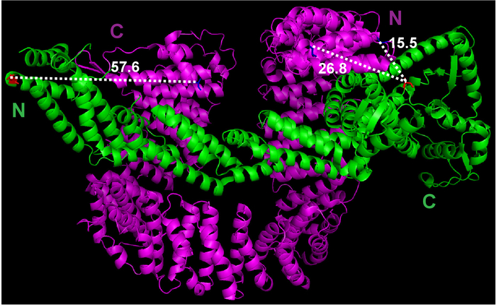
- Use the Measurement function under the Wizard menu of PyMOL to estimate the distance between the first amino acid of Cand1 and the last amino acid of Cul1 (Figure 1).
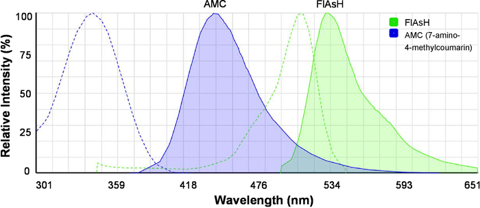
- Load the online spectra viewer (see Table of Materials) and view the excitation and emission spectra of 7-amino-4-methylcoumarin (AMC) and FlAsH simultaneously (Figure 2). Note that AMC is the FRET donor and FlAsH is the FRET acceptor.

Figure 1: The crystal structure of Cul1•Cand1 and measurement of the distance between potential labeling sites. The crystal structure file was downloaded from Protein Data Bank (File 1U6G), and viewed in PyMOL. Measurements between selected atoms were done by PyMOL. Please click here to view a larger version of this figure.

Figure 2: The excitation and emission spectra of the fluorescent dyes for FRET. Spectra of AMC (7-amino-4-methylcoumarin) and FlAsH are shown. Dashed lines indicate excitation spectra, and solid lines indicate emission spectra. The image was originally generated by the Fluorescence SpectraViewer and was modified for better clarity. Please click here to view a larger version of this figure.
2. Preparation of Cul1AMC•Rbx1, the FRET donor protein
- Construct plasmids for expressing human Cul1sortase•Rbx1 in E. coli cells. Note that the two plasmids co-expressing human Cul1•Rbx1 in E. coli cells are described in detail in a previous report20.
- Add a DNA sequence coding “LPETGGHHHHHH” (sortase-His6 tag) to the 3’ end of Cul1 coding sequence through standard PCR and cloning methods21,22.
- Sequence the new plasmid to confirm the gene insert is accurate.
- Co-express Cul1sortase•Rbx1 in E. coli cells. The method is derived from a previous report20.
- Mix 100 ng each of the two plasmids with BL21 (DE3) chemically competent cells for co-transformation using the heat shock method23. Grow cells on LB agar plate containing 100 µg/mL ampicillin and 34 µg/mL chloramphenicol at 37 °C overnight.
- Inoculate 50 mL of LB culture with freshly transformed colonies and grow overnight at 37 °C with 250 rpm shaking. This gives a starter culture.
- Inoculate 6 flasks, each with 1 L of LB medium, with 5 mL starter culture each and grow at 37 °C with 250 rpm shaking until the OD600 is ~1.0. Cool the culture to 16 °C and add isopropyl-β-D-thiogalactoside (IPTG) to 0.4 mM. Keep the culture at 16 °C overnight with 250 rpm shaking.
- Harvest the E. coli cells through centrifugation at 5,000 x g for 15 min and collect cell pellets in 50 mL conical tubes.
NOTE: The cell pellets can be processed for protein purification or be frozen at -80 °C before proceeding to the protein purification steps.
- Purification of the Cul1sortase•Rbx1 complex. This method is derived from a previous report20.
- Add 50 mL of lysis buffer (30 mM Tris-HCl, 200 mM NaCl, 5 mM DTT, 10% glycerol, 1 tablet of protease inhibitor cocktail, pH 7.6) to the pellet of E. coli cells expressing Cul1sortase•Rbx1.
- Lyse the cells on ice with sonication at 50% amplitude. Alternate between 1-second ON and 1-second OFF and run for 3 min.
- Repeat step 2.3.2 2-3x.
- Transfer the cell lysate into a 50-mL centrifugation tube and remove the cell debris by centrifugation at 25,000 x g for 45 min.
- Incubate the clear cell lysate with 5 mL of glutathione beads at 4 °C for 2 h.
- Centrifuge the beads-lysate mixture at 1,500 x g for 2 min at 4 °C. Remove the supernatant.
- Wash the beads with 5 mL of lysis buffer (with no protease inhibitors) and remove the supernatant after centrifugation at 1,500 x g for 2 min at 4 °C.
- Repeat step 2.3.7 2x.
- Add 3 mL of lysis buffer to the washed beads and transfer the bead slurry into an empty column.
- Add 5 mL of elution buffer (50 mM Tris-HCl, 200 mM NaCl, 10 mM reduced glutathione, pH 8.0) into the column. Incubate for 10 min and collect the eluate.
- Repeat step 2.3.10 3-4x.
- Add 200 µL of 5 mg/mL thrombin (see Table of Materials) to the eluate from the glutathione beads and incubate overnight at 4 °C.
NOTE: The protocol can be paused here. - Dilute the protein sample with Buffer A (25 mM HEPES, 1 mM DTT, 5% glycerol, pH 6.5) threefold.
- Equilibrate a cation exchange chromatography column (see Table of Materials) on an FPLC system with Buffer A.
- Load the protein sample to the equilibrated cation exchange chromatography column at a 0.5 mL/min flow rate.
- Elute the protein with a gradient of NaCl in 40 mL by mixing Buffer A and 0 to 50% Buffer B (25 mM HEPES, 1 M NaCl, 1 mM DTT, 5% glycerol, pH 6.5) at a 1 mL/min flow rate.
- Check the eluted protein in different fractions using SDS-PAGE24.
NOTE: The protocol can be paused here. - Pool the eluate fractions containing Cul1sortase•Rbx1.
- Concentrate the pooled Cul1sortase•Rbx1 sample to 2.5 mL by passing the buffer through an ultrafiltration membrane (30 kDa cutoff).
- Add AMC to the C-terminus of Cul1 through sortase-mediated transpeptidation21,22.
- Change the buffer in the Cul1sortase•Rbx1 sample to the sortase buffer (50 mM Tris-HCl, 150 mM NaCl, 10 mM CaCl2, pH 7.5) using a desalting column (see Table of Materials).
- Equilibrate a desalting column with 25 mL of sortase buffer.
- Load 2.5 mL of Cul1sortase•Rbx1 sample into the column. Discard the flow-through.
- Elute the sample with 3.5 mL of sortase buffer. Collect the flow-through.
- In 900 µL of the Cul1sortase•Rbx1 solution in the sortase buffer, add 100 µL of 600 µM purified sortase A solution and 10 µL of 25 mM GGGGAMC peptide. Incubate the reaction mixture at 30 °C in the dark overnight. Note that this step will generate Cul1AMC•Rbx1.
CAUTION: Fluorescent dyes are sensitive to light, so avoid exposing them to ambient light during the protein and sample preparation as much as possible.
NOTE: The protocol can be paused here. - Add 50 µL of Ni-NTA agarose beads to the reaction mixture and incubate at room temperature for 30 min.
- Pellet the Ni-NTA agarose beads by centrifugation at 5,000 x g for 2 min and collect the supernatant.
- Equilibrate a size exclusion chromatography column (see Table of Materials) with buffer (30 mM Tris-HCl, 100 mM NaCl, 1 mM DTT, 10% glycerol) on the FPLC system.
- Load all the Cul1AMC•Rbx1 sample on the size exclusion chromatography column. Elute with 1.5x column volume of buffer.
- Check the eluate fractions by SDS-PAGE24.
- Pool the eluate fractions containing Cul1AMC•Rbx1.
- Measure the protein concentration using its absorbance at 280 nm with a spectrophotometer.
- Aliquot the protein solution and store at -80 °C.
NOTE: The protocol can be paused here.
- Change the buffer in the Cul1sortase•Rbx1 sample to the sortase buffer (50 mM Tris-HCl, 150 mM NaCl, 10 mM CaCl2, pH 7.5) using a desalting column (see Table of Materials).
3. Preparation of FlAsHCand1, the FRET acceptor protein
NOTE: Most of steps in this part are the same as Step 2. Conditions that differ are described in detail below.
- Construct the plasmid for expressing human TetraCysCand1 in E. coli cells.
- Add DNA sequence coding “CCPGCCGSG” (tetracysteine/TetraCys tag) before the 15th amino acid of Cand1 through regular PCR25 (primer sequences: TGCTGTCCGGGCTGCTGCGGCAGCGGCATGACATCCAGCGACAAGGACTTTAG; CTAACTAGTGTCCATTGATTCCAAG).
- Insert the PCR product into a pGEX-4T-2 vector. Sequence the plasmid to confirm the gene insert is accurate and in frame.
- Express TetraCysCand1in E. coli cells in the same way as step 2.2, except that the plasmid is transformed into Rosetta competent cells.
- Purification of TetraCysCand1 from the E. coli cells.
- Lyse the E. coli cell pellets and extract TetraCysCand1using glutathione beads. These steps are the same as steps 2.3.1–2.3.12.
- Dilute the protein eluate from the glutathione beads with Buffer C (50 mM Tris-HCl, 1 mM DTT, 5% glycerol, pH 7.5) by three folds. Equilibrate an anion exchange chromatography column (see Table of Materials) on the FPLC system with Buffer C and load the diluted protein sample at a 0.5 mL/min flow rate.
- Elute the protein with a gradient of NaCl in 40 mL by mixing Buffer C and 0 to 50% Buffer D (50 mM Tris-HCl, 1 M NaCl, 1 mM DTT, 5% glycerol, pH 7.5) at a 1 mL/min flow rate. Check the eluted protein in different fractions using SDS-PAGE24, and pool the fractions containing TetraCysCand1. Note that TetraCysCand1 has a larger retention volume than free GST.
- Concentrate the pooled TetraCysCand1 sample by passing the buffer through an ultrafiltration membrane (30 kDa cutoff).
- Equilibrate the size exclusion chromatography column with labeling buffer (20 mM Tris-HCl, 100 mM NaCl, 2 mM TCEP, 1 mM EDTA, 5% glycerol) on the FPLC system. Load TetraCysCand1 sample (500 µL each time) and check the eluate fractions by SDS-PAGE24.
- Pool all the fractions containing TetraCysCand1 and concentrate the protein to ~40 µM by passing the buffer through an ultrafiltration membrane (30 kDa cutoff). Estimate the protein concentration using its absorbance at 280 nm. Store the protein as 50 µL aliquots at -80 °C.
NOTE: The protocol can be paused here.
- Preparation of FlAsHCand1.
- Add 1 µL of FlAsH solution (see Table of Materials) to 50 µL TetraCysCand1 solution.
- Mix well and incubate the mixture at room temperature in the dark for 1-2 h to get FlAsHCand1.
NOTE: The protocol can be paused here.
4. Preparation of Cand1, the FRET chase protein
NOTE: The protein preparation protocol is similar to Step 3, with the following modifications.
- Insert the coding sequence of full-length Cand1 into the pGEX-4T-2 vector.
- Change the buffer used in step 3.3.5 to a buffer containing 30 mM Tris-HCl, 100 mM NaCl, 1 mM DTT, 10% glycerol.
- Eliminate steps 3.3.7 and 3.3.8.
5. Test and confirm the FRET assay
- Prepare the FRET buffer containing 30 mM Tris-HCl, 100 mM NaCl, 0.5 mM DTT, 1 mg/mL ovalbumin, pH 7.6, and use at room temperature.
- Test the FRET between Cul1AMC•Rbx1 and FlAsHCand1 on a fluorimeter.
- In 300 µL of FRET buffer, add Cul1AMC•Rbx1 (FRET donor) to a final concentration of 70 nM. Transfer the solution into a cuvette.
- Place the cuvette in the sample holder of a fluorimeter. Excite the sample with 350 nm excitation light and scan the emission signals from 400 nm to 600 nm at 1 nm increments.
- Repeat step 5.2.1 but change Cul1AMC•Rbx1 to FlAsHCand1 (FRET acceptor). Scan the FlAsHCand1 sample using the same method as in step 5.2.2.
- (Optional) Excite the FlAsHCand1 with 510 nm excitation light, and scan the emission signal from 500 nm to 650 nm.
- In 300 µL FRET buffer, add both Cul1AMC•Rbx1 and FlAsHCand1 to a final concentration of 70 nM. Analyze the sample in the same way as in step 5.2.2.
- Confirm the FRET between Cul1AMC•Rbx1 and FlAsHCand1 by adding the chase (unlabeled Cand1) protein (Figure 3C).
- In 300 µL of FRET buffer, add 70 nM Cul1AMC•Rbx1 and 700 nM Cand1. Scan the sample emission as in step 5.2.2.
- In 300 µL of FRET buffer, add 70 nM FlAsHCand1 and 700 nM Cand1. Scan the sample emission as in step 5.2.2.
- In 300 µL of FRET buffer, add 70 nM Cul1AMC•Rbx1 and 70 nM FlAsHCand1, and incubate the sample at room temperature for 5 min. Then add 700 nM Cand1, and immediately after the addition, scan the sample emission as in step 5.2.2). Note that this step is similar to step 5.2.5.
- In 300 µL FRET buffer, sequentially add 70 nM Cul1AMC•Rbx1, 700 nM Cand1, and 70 nM FlAsHCand1. Incubate the sample at room temperature for 5 min, and scan the sample emission as in step 5.2.2. Note that this is the chase sample (green line in Figure 3C).
6. Measure the association rate constant (kon) of Cul1•Cand1
NOTE: Details of operating a stopped-flow fluorimeter has been described in a previous report26.
- Prepare the stopped-flow fluorimeter for measurement.
- Turn on the stopped-flow fluorimeter according to the manufacturer’s instruction.
- Set the excitation light at 350 nm, and use a band-pass filter that allows 450 nm emission light to pass and blocks 500-650 nm emission light.
- Keep the sample valves at the FILL position, and connect a 3 mL syringe filled with water. Wash the two sample syringes (A and B) with water by moving the sample syringe drive up and down several times. Discard all the water used in this step.
- Keep the sample valves at the FILL position, and connect a 3 mL syringe filled with FRET buffer. Wash the two sample syringes with the FRET buffer by moving the sample syringe drive up and down several time. Discard all the FRET buffer used in this step.
- Take a control measurement (Figure 4C).
- Connect a 3 mL syringe and load Syringe A with 100 nM Cul1AMC•Rbx1 in the FRET buffer. Turn the sample valve to the DRIVE position.
- Connect a 3 mL syringe and load Syringe B with the FRET buffer. Turn the sample valve to the DRIVE position.
- Use the Control Panel under Acquire in the software to take five shots (mix equal volume of samples from Syringe A and Syringe B) on the stopped-flow fluorimeter without recording the results.
- Open the Control Panel under Acquire in the software, and program to record the emission of Cul1AMC over 60 s. Then take a single shot.
- Repeat step 6.2.4 2x.
- Turn the sample valve to the FILL position. Empty Syringe B and wash with the FRET buffer.
- Measure observed association rate constants (kobs) of Cul1•Cand1 (Figure 4B).
- Keep the sample in Syringe A the same as in step 6.2.1.
- Connect a 3 mL syringe and load Syringe B with 100 nM FlAsHCand1 in the FRET buffer. Turn the sample valve to the DRIVE position.
- Use the Control Panel under Acquire in the software to take five shots without recording the results.
- Open the Control Panel under Acquire in the software, and program to record the emission of Cul1AMC over 60 s. Then take a single shot.
- Repeat step 6.3.4 2x.
- Empty Syringe B and wash with the FRET buffer.
- Repeat steps 6.3.1–6.3.6 several times with increasing concentrations of FlAsHCand1 in the FRET buffer.
- Fit the change (decrease) in fluorescent signals measured over time from each shot to a single exponential curve. This will give kobs in each measurement, and the unit is s-1. Note that the basis of this calculation has been well discussed in a previous report27.
- Calculate the average and standard deviation of kobs for each FlAsHCand1 concentration used. Plot the average kobs against Cand1 concentration (Figure 4D), and the slope of the line represents the kon of Cul1•Cand1, with a unit of M-1 s-1.
7. Measure the dissociation rate constant (koff) of Cul1•Cand1 in the presence of Skp1•F-box protein.
NOTE: This step is similar to Step 6, with the following modifications.
- In Syringe A, under the FILL position, load a solution of 100 nM Cul1AMC•Rbx1 and 100 nM FlAsHCand1 in the FRET buffer. Turn the sample valve to the “DRIVE” position.
- In Syringe B, under the FILL position, load a solution of Skp1•Skp2 (prepared following a previous report20). Turn the sample valve to the “DRIVE” position.
- Open the Control Panel under Acquire in the software, and program to record the emission of Cul1AMC over 30 s. Then take a single shot. The fluorescent signals increase over time after mixing solutions from Syringe A and Syringe B (Figure 5).
Results
To test the FRET between Cul1AMC and FlAsHCand1, we first determined the emission intensity of 70 nM Cul1AMC (the donor) and 70 nM FlAsHCand1 (the acceptor), respectively (Figure 3A-C, blue lines). In each analysis, only one emission peak was present, and the emission of FlAsHCand1 (the acceptor) was low. When 70 nM each of Cul1AMC and FlAsHCand1 were mixed to genera...
Discussion
FRET is a physical phenomenon that is of great interest for studying and understanding biological systems19. Here, we present a protocol for testing and using FRET to study the binding kinetics of two interacting proteins. When designing FRET, we considered three major factors: the spectral overlap between donor emission and acceptor excitation, the distance between the two fluorophores, and the dipole orientation of the fluorophores28. To choose the fluorophores for FRET, ...
Disclosures
The authors have nothing to disclose.
Acknowledgements
We thank Shu-Ou Shan (California Institute of Technology) for insightful discussion on the development of the FRET assay. M.G., Y.Z., and X.L. were funded by startup funds from Purdue University to Y.Z. and X.L.This work was supported in part by a seed grant from Purdue University Center for Plant Biology.
Materials
| Name | Company | Catalog Number | Comments |
| Anion exchange chromatography column | GE Healthcare | 17505301 | HiTrap Q FF anion exchange chromatography column |
| Benchtop refrigerated centrifuge | Eppendorf | 2231000511 | |
| BL21 (DE3) Competent Cells | ThermoFisher Scientific | C600003 | |
| Calcium Chloride | Fisher Scientific | C78-500 | |
| Cation exchange chromatography column | GE Healthcare | 17505401 | HiTrap SP Sepharose FF |
| Desalting Column | GE Healthcare | 17085101 | |
| Floor model centrifuge (high speed) | Beckman Coulter | J2-MC | |
| Floor model centrifuge (low speed) | Beckman Coulter | J6-MI | |
| Fluorescence SpectraViewer | ThermoFisher Scientific | https://www.thermofisher.com/us/en/home/life-science/cell-analysis/labeling-chemistry/fluorescence-spectraviewer.html | |
| FluoroMax fluorimeter | HORIBA | FluoroMax-3 | |
| FPLC | GE Healthcare | 29018224 | |
| GGGGAMC peptide | New England Peptide | custom synthesis | |
| Glutathione beads | GE Healthcare | 17075605 | |
| Glycerol | Fisher Scientific | G33-500 | |
| HEPES | Fisher Scientific | BP310-100 | |
| Isopropyl-β-D-thiogalactoside (IPTG) | Fisher Scientific | 15-529-019 | |
| LB Broth | Fisher Scientific | BP1426-500 | |
| Ni-NTA agarose | Qiagen | 30210 | |
| Ovalbumin | MilliporeSigma | A2512 | |
| pGEX-4T-2 vector | GE Healthcare | 28954550 | |
| Protease inhibitor cocktail | MilliporeSigma | 4693132001 | |
| Reduced glutathione | Fisher Scientific | BP25211 | |
| Refrigerated shaker | Eppendorf | M1282-0004 | |
| Rosetta Competent Cells | MilliporeSigma | 70953-3 | |
| Size exclusion chromatography column | GE Healthcare | 28990944 | Superdex 200 10/300 GL column |
| Sodium Chloride (NaCl) | Fisher Scientific | S271-500 | |
| Stopped-flow fluorimeter | Hi-Tech Scientific | SF-61 DX2 | |
| TCEP·HCl | Fisher Scientific | PI20490 | |
| Thrombin | MilliporeSigma | T4648 | |
| Tris Base | Fisher Scientific | BP152-500 | |
| Ultrafiltration membrane | MilliporeSigma | UFC903008 | Amicon Ultra-15 Centrifugal Filter Units, Ultra-15, 30,000 NMWL |
References
- Stumpf, M. P. H., et al. Estimating the size of the human interactome. Proceedings of the National Academy of Sciences of the United States of America. 105 (19), 6959-6964 (2008).
- Kuzmanov, U., Emili, A. Protein-protein interaction networks: probing disease mechanisms using model systems. Genome Medicine. 5 (4), 37 (2013).
- Titeca, K., Lemmens, I., Tavernier, J., Eyckerman, S. Discovering cellular protein-protein interactions: Technological strategies and opportunities. Mass Spectrometry Reviews. , 1-33 (2018).
- Lapetina, S., Gil-Henn, H. A guide to simple, direct, and quantitative in vitro binding assays. Journal of Biological Methods. 4 (1), 62 (2017).
- Zheng, X., Bi, C., Li, Z., Podariu, M., Hage, D. S. Analytical methods for kinetic studies of biological interactions: A review. Journal of Pharmaceutical and Biomedical Analysis. 113, 163-180 (2015).
- Pierce, N. W., et al. Cand1 promotes assembly of new SCF complexes through dynamic exchange of F box proteins. Cell. 153 (1), 206-215 (2013).
- Liu, X., Reitsma, J. M., Mamrosh, J. L., Zhang, Y., Straube, R., Deshaies, R. J. Cand1-Mediated Adaptive Exchange Mechanism Enables Variation in F-Box Protein Expression. Molecular Cell. 69 (5), 773-786 (2018).
- Zheng, N., et al. Structure of the Cul1-Rbx1-Skp1-F boxSkp2 SCF ubiquitin ligase complex. Nature. 416 (6882), 703-709 (2002).
- Kleiger, G., Deshaies, R. Tag Team Ubiquitin Ligases. Cell. 166 (5), 1080-1081 (2016).
- Zheng, J., et al. CAND1 binds to unneddylated CUL1 and regulates the formation of SCF ubiquitin E3 ligase complex. Molecular Cell. 10 (6), 1519-1526 (2002).
- Hwang, J. W., Min, K. W., Tamura, T. A., Yoon, J. B. TIP120A associates with unneddylated cullin 1 and regulates its neddylation. FEBS Letters. 541 (1-3), 102-108 (2003).
- Min, K. W., Hwang, J. W., Lee, J. S., Park, Y., Tamura, T., Yoon, J. B. TIP120A associates with cullins and modulates ubiquitin ligase activity. Journal of Biological Chemistry. 278 (18), 15905-15910 (2003).
- Goldenberg, S. J., et al. Structure of the Cand1-Cul1-Roc1 complex reveals regulatory mechanisms for the assembly of the multisubunit cullin-dependent ubiquitin ligases. Cell. 119 (4), 517-528 (2004).
- Chuang, H. W., Zhang, W., Gray, W. M. Arabidopsis ETA2, an apparent ortholog of the human cullin-interacting protein CAND1, is required for auxin responses mediated by the SCF(TIR1) ubiquitin ligase. Plant Cell. 16 (7), 1883-1897 (2004).
- Feng, S., et al. Arabidopsis CAND1, an Unmodified CUL1-Interacting Protein, Is Involved in Multiple Developmental Pathways Controlled by Ubiquitin/Proteasome-Mediated Protein Degradation. the Plant Cell Online. 16 (7), 1870-1882 (2004).
- Cheng, Y., Dai, X., Zhao, Y. AtCAND1, a HEAT-repeat protein that participates in auxin signaling in Arabidopsis. Plant Physiology. 135 (June), 1020-1026 (2004).
- Lo, S. -. C., Hannink, M. CAND1-Mediated Substrate Adaptor Recycling Is Required for Efficient Repression of Nrf2 by Keap1. Molecular and Cellular Biology. 26 (4), 1235-1244 (2006).
- Okamoto, K., Sako, Y. Recent advances in FRET for the study of protein interactions and dynamics. Current Opinion in Structural Biology. 46, 16-23 (2017).
- Hussain, S. A. An introduction to fluorescence resonance energy transfer (FRET). arXiv preprint. , (2009).
- Li, T., Pavletich, N. P., Schulman, B. A., Zheng, N. High-level expression and purification of recombinant SCF ubiquitin ligases. Methods in Enzymology. 398 (1996), 125-142 (2005).
- Popp, M. W., Antos, J. M., Grotenbreg, G. M., Spooner, E., Ploegh, H. L. Sortagging: A versatile method for protein labeling. Nature Chemical Biology. 3 (11), 707-708 (2007).
- Antos, J. M., Ingram, J., Fang, T., Pishesha, N., Truttmann, M. C., Ploegh, H. L. Site-Specific Protein Labeling via Sortase-Mediated Transpeptidation. Current Protocols in Protein Science. 89, (2017).
- Froger, A., Hall, J. E. Transformation of Plasmid DNA into E. coli Using the Heat Shock Method. Journal of Visualized Experiments. , (2007).
- Simpson, R. J. SDS-PAGE of Proteins. Cold Spring Harbor Protocols. 2006 (1), (2006).
- Kleiger, G., Saha, A., Lewis, S., Kuhlman, B., Deshaies, R. J. Rapid E2-E3 Assembly and Disassembly Enable Processive Ubiquitylation of Cullin-RING Ubiquitin Ligase Substrates. Cell. 139 (5), 957-968 (2009).
- Biro, F. N., Zhai, J., Doucette, C. W., Hingorani, M. M. Application of Stopped-flow Kinetics Methods to Investigate the Mechanism of Action of a DNA Repair Protein. Journal of Visualized Experiments. (37), 2-8 (2010).
- Patel, J. T., Belsham, H. R., Rathbone, A. J., Friel, C. T. Use of Stopped-Flow Fluorescence and Labeled Nucleotides to Analyze the ATP Turnover Cycle of Kinesins. Journal of Visualized Experiments. (92), 1-6 (2014).
- Bajar, B. T., Wang, E. S., Zhang, S., Lin, M. Z., Chu, J. A guide to fluorescent protein FRET pairs. Sensors (Switzerland). 16 (9), 1-24 (2016).
- Chen, A. K., Cheng, Z., Behlke, M. A., Tsourkas, A. Assessing the sensitivity of commercially available fluorophores to the intracellular environment. Analytical Chemistry. 80 (19), 7437-7444 (2008).
- Lin, C. T., Rorabacher, D. B. Mathematical approach for stopped-flow kinetics of fast second-order reactions involving inhomogeneity in the reaction cell. Journal of Physical Chemistry. 78 (3), 305-308 (1974).
- Toseland, C. P., Geeves, M. A. Rapid Reaction Kinetic Techniques. Fluorescent Methods for Molecular Motors. , 49-65 (2014).
- Adams, S. R., et al. New biarsenical ligands and tetracysteine motifs for protein labeling in vitro and in vivo: Synthesis and biological applications. Journal of the American Chemical Society. 124 (21), 6063-6076 (2002).
- Lin, C. W., Ting, A. Y. Transglutaminase-catalyzed site-specific conjugation of small-molecule probes to proteins in vitro and on the surface of living cells. Journal of the American Chemical Society. 128 (14), 4542-4543 (2006).
- Yin, J., et al. Genetically encoded short peptide tag for versatile protein labeling by Sfp phosphopantetheinyl transferase. Proceedings of the National Academy of Sciences of the United States of America. 102 (44), 15815-15820 (2005).
- Diaspro, A., Chirico, G., Usai, C., Ramoino, P., Dobrucki, J. Photobleaching. Handbook Of Biological Confocal Microscopy. , 690-702 (2006).
- Aoki, K., Kamioka, Y., Matsuda, M. Fluorescence resonance energy transfer imaging of cell signaling from in vitro to in vivo: Basis of biosensor construction, live imaging, and image processing. Development, Growth & Differentiation. 55 (4), 515-522 (2013).
- Kilic, S., et al. Single-molecule FRET reveals multiscale chromatin dynamics modulated by HP1α. Nature Communications. 9 (1), (2018).
- Shen, K., Arslan, S., Akopian, D., Ha, T., Shan, S. O. Activated GTPase movement on an RNA scaffold drives co-translational protein targeting. Nature. 492 (7428), 271-275 (2012).
- Bajar, B. T., et al. Improving brightness and photostability of green and red fluorescent proteins for live cell imaging and FRET reporting. Scientific Reports. 6 (February), 1-12 (2016).
Reprints and Permissions
Request permission to reuse the text or figures of this JoVE article
Request PermissionThis article has been published
Video Coming Soon
Copyright © 2025 MyJoVE Corporation. All rights reserved