
需要订阅 JoVE 才能查看此. 登录或开始免费试用。
Method Article
绵羊眼玻璃体内注射
摘要
在羊眼中进行玻璃体内注射,目的是将病毒介导的基因治疗传递到视网膜。
摘要
有几种方法可以将治疗剂输送到视网膜,包括玻璃体内 (IVT)、视网膜下、脉络膜上、眼周或局部给药。IVT药物输送涉及注射到眼睛的玻璃体中,玻璃体是一种充满眼睛后房并保持眼球形状的凝胶状物质。虽然IVT途径的特异性目标性不如视网膜下递送,但它的侵入性要小得多,并且在临床环境中广泛用于一系列眼部疾病。
我们之前证明了玻璃体内递送腺相关病毒(AAV)介导的基因治疗产物(AAV9.CLN5)在绵羊中具有天然存在的CLN5形式的神经元蜡样脂褐质沉着症(NCL)。受影响的绵羊一只眼睛接受IVT基因治疗,另一只未经治疗的眼睛作为内部对照。治疗后15个月内,接受治疗的眼睛保持视网膜结构和功能,而未经治疗的眼睛在尸检时表现出进行性下降和严重萎缩。根据绵羊研究,CLN5基因治疗产品于2021年9月被美国食品药品监督管理局批准为候选研究新药(IND)。本文详细介绍了IVT将治疗性病毒载体递送到绵羊眼的手术方案。
引言
有几种方法可用于将治疗剂输送到视网膜,包括玻璃体内 (IVT)、视网膜下、脉络膜上、眼周或局部给药。每种给药途径都涉及克服诸如血液 - 视网膜屏障或内部和外部限制膜之类的屏障,并且根据所输送的药物和特定的视网膜靶标1,2具有不同的疗效率。
IVT药物输送涉及将注射到眼睛的玻璃体中,玻璃体是一种占据眼睛后房的凝胶状物质。玻璃体的主要功能是维持眼球的形状并保持眼组织(如晶状体和视网膜)就位。玻璃体液主要由水组成,少量胶原蛋白、透明质酸和其他非胶原蛋白3。IVT注射是一种简单而常见的程序,常规用于治疗各种眼部疾病,包括年龄相关性黄斑变性,糖尿病黄斑水肿,糖尿病视网膜病变,视网膜静脉阻塞和几种遗传性视网膜营养不良4,5。
神经元蜡样脂褐素糖(NCL;巴顿病)是一组致命的溶酶体贮积病,会导致大脑和视网膜严重退化。目前有13种已知的NCL变异,由不同基因(CLN1-8,CLN10-14)的突变引起,主要影响儿童,但发病年龄和疾病严重程度为6。NCL具有共同的进行性症状,包括认知和运动衰退,癫痫发作和视力丧失。NCL无法治愈;然而,脑定向酶替代疗法目前正在针对CLN2疾病7,8进行临床试验,AAV介导的基因疗法在临床前研究中显示出巨大的前景,CLN5基因治疗的临床试验预计将于2022年开始9,10。
许多其他物种发展出天然存在的NCL形式,包括猫,狗,羊和牛。NCL的两种绵羊模型目前正在新西兰积极研究中:Borderdale绵羊的CLN5疾病模型和南汉普郡绵羊的CLN6疾病模型。受影响的绵羊表现出人类疾病的许多临床和病理特征,包括视网膜萎缩和视力丧失10,11。尽管在患有CLN5疾病的绵羊中进行脑定向CLN5基因治疗可以预防或阻止脑萎缩和临床衰退,但接受治疗的绵羊仍然会失去视力9。这突出了治疗视网膜以保护视力和保持更好生活质量的必要性,从而建立了绵羊眼部基因治疗方案。
羊眼代表了人眼的良好模型,因为它在眼球尺寸、玻璃体体积和视网膜结构10、12、13 方面相似。本文详细介绍了IVT将小体积(≤100μL)治疗性病毒载体输送到羊眼的手术方案。
研究方案
所有实验方案均由林肯大学动物伦理委员会批准,并符合美国国立卫生研究院关于在研究中照顾和使用动物的指南和新西兰动物福利法案(1999)。边境代尔绵羊在14 岁出生时被诊断出来,并在林肯大学的研究农场饲养。三只3个月大的纯合子(CLN5-/-)母羊接受了左眼单次IVT注射,未经治疗的右眼作为内部对照。将视网膜电图和病理学数据与历史健康和受影响的对照数据进行比较。本研究中使用的病毒载体是自互补的腺相关病毒血清型9,含有鸡β作用(CBh)启动子和密码子优化的绵羊 CLN5 (scAAV9 / CBh-oCLN5opt)。病毒载体由美国北卡罗来纳州北卡罗来纳大学载体核心提供。
1. 术前
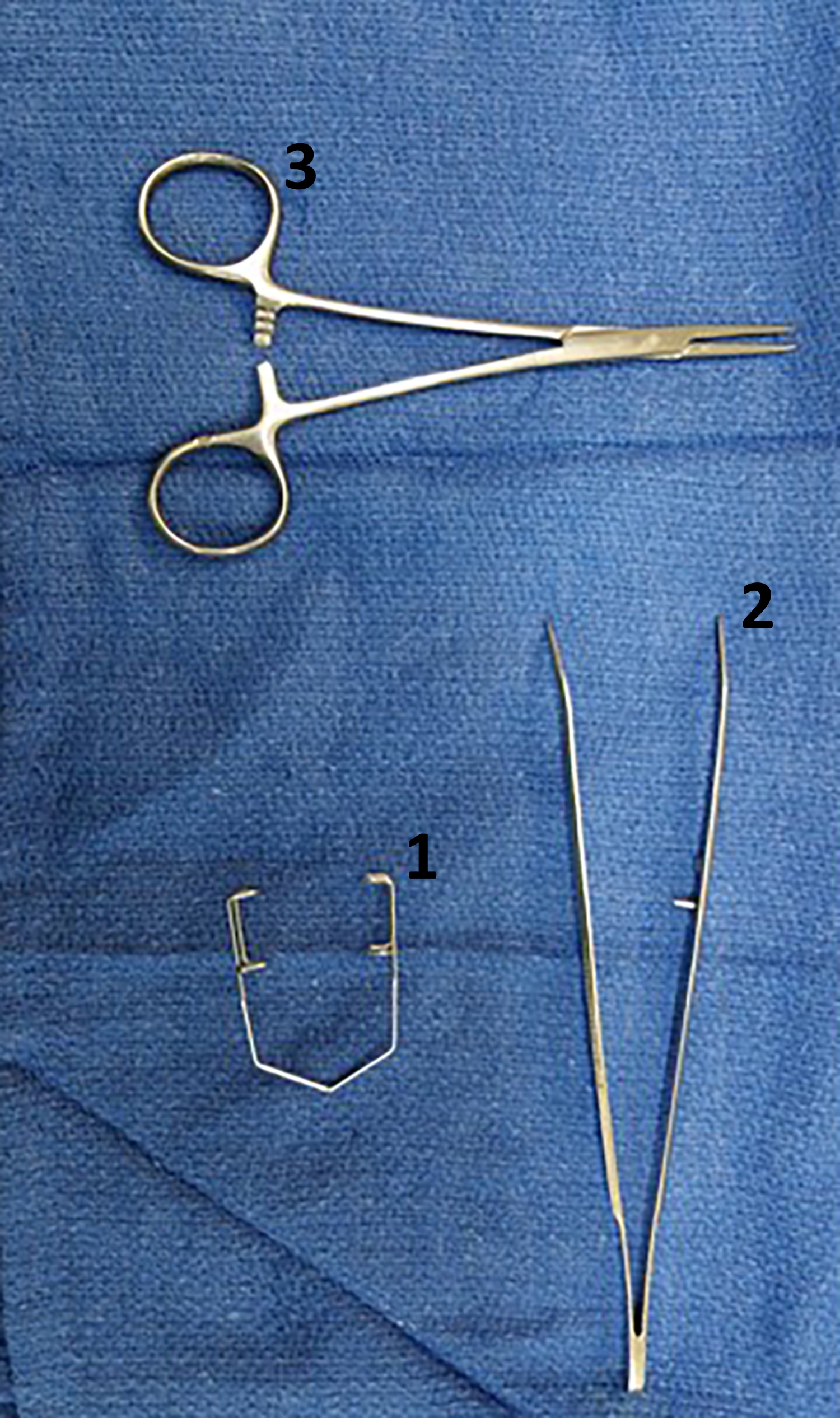
- 高压灭菌手术套件(图1)。
- 手术前禁食绵羊24小时。
- 手术前记录活体重。

图1:玻璃体内手术套件。 IVT手术所需的器械包括(1)保持眼睑张开的窥器,以及(2)一对弯曲的鼻钳,用于抓住延髓结膜并旋转眼睛。(3)还包括直鼻止血器作为替代仪器,用于抓住延髓结膜,并在眼睛回滚回眼眶时将眼睛固定到位。该套件在手术前进行高压灭菌。 请点击此处查看此图的大图。
2. 外科手术
- 约束动物,并使用电子剪刀从颈静脉的一侧剃掉颈部的羊毛。
- 通过在颈静脉沟底部施加压力来阻塞颈静脉,并观察凸起的静脉。
- 将适量的地西泮(0.3mg / kg)和氯胺酮(7.5mg / kg)抽入无菌注射器中,并连接无菌20 G针头。将针头插入颈静脉并轻轻拉回柱塞,以确保血液进入枢纽并且针头在静脉内。一旦确诊,通过静脉(颈)给药诱导。
- 诱导后,立即将动物置于背卧中,伸展颈部,将舌头向上和向前保持,使用喉镜观察喉部。当动物呼气时,通过在声带之间轻轻插入气管插管(大小 6.0-9.0,取决于绵羊的大小)来进行气管插管。立即给气管内袖带充气,并在下颌用系带固定插管。确认通过管子的气流。
- 将绵羊转移到手术台上,并将其放在侧面卧位。
- 立即将气管插管连接到麻醉机的软管上,以在100%氧气中输送异氟醚。最初从3%-4%异氟醚开始,然后减少到2%-3%进行维持治疗。观察羊的自发通风。
- 在整个手术过程中监测心脏(脉搏)频率、呼吸频率、血氧饱和度、潮气末 CO2 水平和直肠体温。有关麻醉绵羊中这些参数的生理值,请参见 表1 (可变,但用作指导)。
- 将一个大的无菌方形布放在外科手术车上,然后放置无菌器械。
- 将无菌开窗手术窗帘放在要注射的眼睛上。
- 使用无菌20毫升注射器无菌消毒眼睛,用1-5%聚维酮碘溶液冲洗眼睛。
- 将1-2滴Alcaine 0.5%W / V眼用溶液作为局部麻醉剂涂抹在眼睛上。
- 将Nopa Barraquer-Colibri眼窥器(10毫米)安装到眼睑上,以保持眼睛睁开。
- 用镊子抓住眼睛背外侧的延髓结膜,并向腹内侧旋转眼球。
| 意识 | 麻醉 | 推荐的关键干预点 | |
| 心率(心跳/分钟) | 50-80(静止)至 280(活动) | 50-80 | <50, >100 |
| 呼吸频率(呼吸/分钟) | 15-40(休息)至350(过热) | 10-30 | <8, >40 |
| 血氧饱和度(毫米汞柱) | 95-100 | 98-100 | <90 |
| 潮气末二氧化碳(毫米 汞柱) | 35-45 | 35-45 | >55 |
| 体温(°C) | 38.5-39.5 | 38.5-39.5 | <36, >40 |
表1:麻醉绵羊中要监测的参数的生理值。
3. 病毒制备
- 将AAV载体等分试样储存在-80°C直至使用。
- 在手术当天,解冻所需数量的小瓶,以便在冰上进行IVT输送。
- 在给药前,涡旋病毒载体等分试样并以400× g 离心10秒以收集内容物。
- 在无菌过滤的 1x 磷酸盐缓冲盐水 (PBS) 中将每个病毒载体等分试样稀释至所需剂量,最终体积为 100 μL。 使用无菌过滤移液器吸头在无菌 1.5 mL 低蛋白结合微量离心管中制备载体稀释液。将所有与病毒载体接触的消耗品丢弃在消毒液中(见 材料表)。
注意:在原始出版物15 中治疗剂(AAV9。CLN5)是1.9 x 10¹⁰ 病毒基因组。推荐剂量将根据所施用的治疗剂而有所不同;因此,此处介绍的标准方案中未包含剂量。 - 将全部 100 μL AAV 载体制剂吸入无菌、低死区 1 mL 注射器中,并永久连接的 28 G x 1/2 针头立即注射。确保从制备到注射的时间长度小于2分钟。
4. 病毒给药
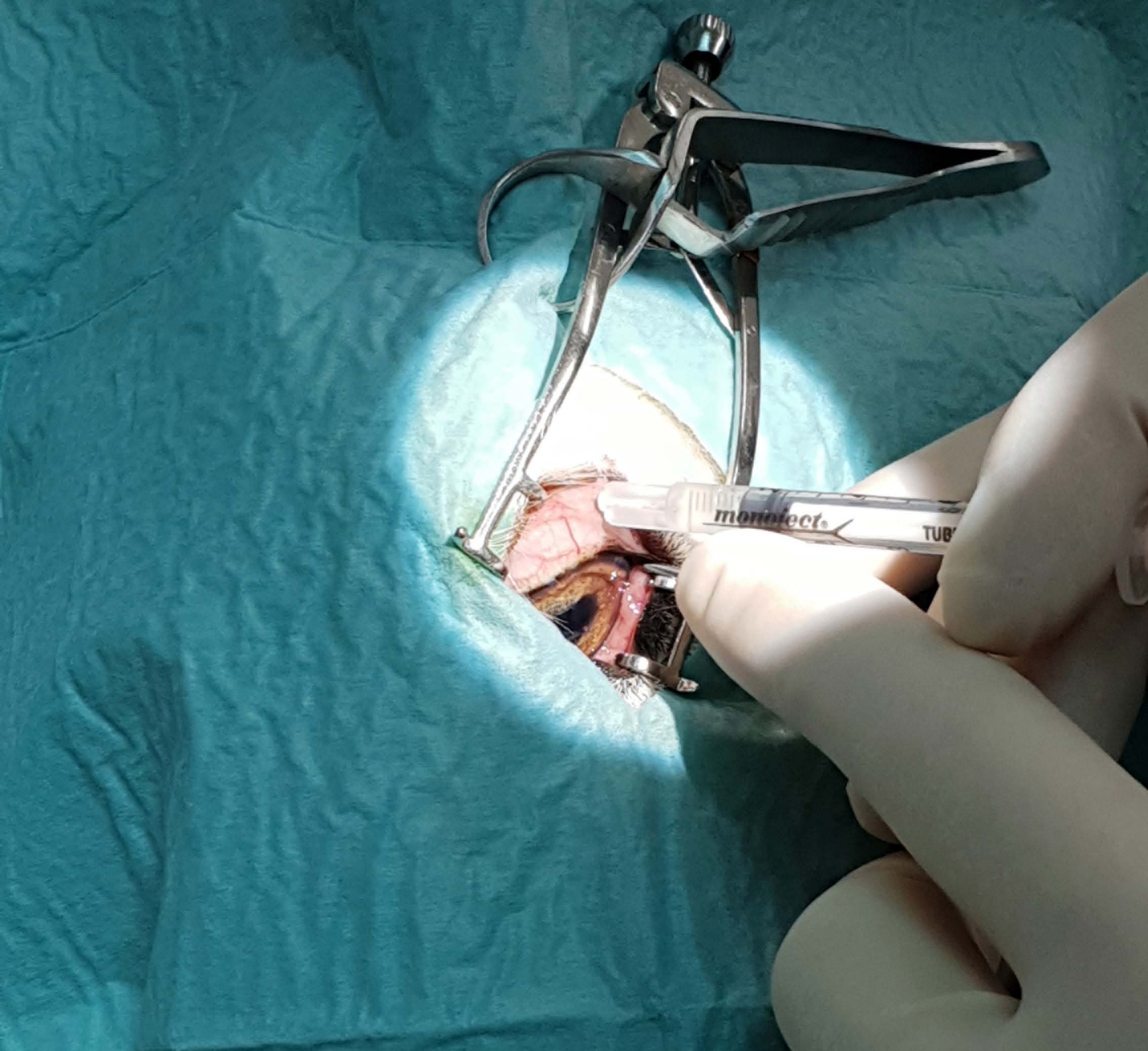
- 将针头插入眼睛外侧巩膜后方约7毫米处,并向后倾斜以避开晶状体(图2 和 图3)。在不干扰视网膜表面的情况下,单次注射 100 μL 作为推注。
- 用大约 10-15 mL 的 1-5% 聚维酮碘溶液冲洗眼睛,然后用 10 mL 盐水冲洗眼睛,然后取出窥器和悬垂物。
- 将羊翻过来,如果需要,用另一只眼睛重复。

图2:眼球的心内侧旋转 。 (A)用无齿镊子抓住延髓结膜和(B)腹内侧旋转(即向下和朝向鼻子)以暴露眼睛的背外侧表面进行注射。缩写:V = 腹侧,D = 背侧,M = 内侧,L = 侧侧。 请点击此处查看此图的大图。

图3:注射位置和深度。 将针头注射在眼球的背外侧,并将针杆的整个长度(0.5英寸/12.7毫米)插入眼中。注意针头朝向眼睛后部的角度,以避开晶状体并尽可能靠近视网膜注射。 请点击此处查看此图的大图。
5.术后管理
- 手术完成后,停止异氟醚气体吸入麻醉,用100%氧气冲洗管路,断开软管与气管插管的连接,然后将绵羊转移到恢复室。
- 将绵羊放在胸骨卧位,双腿藏在下面,并监测直至完全恢复。确保动物的嘴没有任何障碍物。
- 当观察到吞咽反射时,部分放气气管插管的袖带,轻轻地将管子从口腔中取出。
- 在后肢股二头肌内注射非甾体抗炎药,在颈部侧面或肩后皮下注射抗生素,将 0.5% 氯霉素滴眼液注射至眼球表面。
- 一旦绵羊可以独立站立,就提供水和食物(卢塞恩颗粒和谷壳)。
- 手术后 7 天,每天服用 0.5% 氯霉素滴眼液 2-3 次。
- 将绵羊留在室内过夜,然后在手术后约24小时返回室外围场。
- 每天记录直肠温度,持续 3 周。监测脉搏或呼吸频率、食物消耗、神经行为、体温、体重、姿势、眼睛健康和健康不良临床体征的任何变化。如果有任何不良事件的迹象,请寻求适当的兽医治疗。
6. 评估体内疗效
- 如果IVT注射的目标是保护视力,则通过迷宫测试或视网膜电图(ERG)等方法监测 体内 疗效以评估视网膜细胞功能或光学相干断层扫描(OCT)以评估视网膜结构。
注意:这些疗效措施在IVT基因治疗11,15,16之后得到了很好的描述。
7. 尸检组织分析
- 玻璃体内注射手术后,在适当的终点通过批准的方法对绵羊实施安乐死。
注意:建议的安乐死方法,例如静脉注射兽医安乐死药物或对颈椎进行穿透性固定螺栓,然后快速放血,详见其他专题15,16。 - 使用手术锋利/钝弯剪刀收获羊眼球。切开外眦和内眦以增加眼窝开口,然后系统地切开结膜褶皱、结缔组织、肌肉和视神经,将眼球从眼窝中解放出来。
- 将完整的去核眼球浸入10%福尔马林中2小时,然后在Bouin溶液中后固定4小时,在巩膜上切开一个小(0.5厘米)以允许足够的灌注。或者,将眼球浸入戴维森溶液中48小时。
- 通过常规石蜡包埋和切片在3-5μm处处理眼组织切片。
注意:苏木精和伊红(H&E)染色和免疫组织化学分析的染色程序已在前面描述过15,16。 - 通过视网膜总厚度、视网膜层厚度、外核层细胞行计数以及视网膜细胞类型、视网膜胶质细胞或目标蛋白质的免疫组织化学染色等措施评估死后组织中的疗效。
注意:有关这些分析的协议,请参阅以前的出版物15,16。
结果
该研究小组先前已证明 IVT 递送 CLN5 基因治疗载体在减轻 CLN5 NCL 绵羊视网膜功能障碍和变性的功效15.受影响的绵羊接受单次 100 μL IVT 注射包装在 AAV 血清型 9 (AAV9) 载体 (AAV9.CLN5)进入一只眼睛,对侧眼睛作为未经治疗的内部对照。从注射年龄(3个月)到终末期疾病(18个月)每月评估视力。对治疗和未治疗的眼睛以及年龄匹配的健康和受CLN5影响的对照组进行了视网膜组织学...
讨论
玻璃体内注射是人类眼科中最常见的外科手术之一,已被证明可有效将AAV介导的基因疗法输送到绵羊的视网膜。我们之前已经证明了AAV9的功效。CLN5基因治疗在玻璃体内使用CLN5 NCL15减轻绵羊视网膜功能障碍和变性。希望将这种给药途径转化为人类NCL患者也将证明是有益的。
小容量IVT注射到羊眼的方案相对简单和非侵入性,易于重现,并且易于非专家学习。?...
披露声明
作者没有利益冲突需要披露。
致谢
作者要感谢Steve Heap博士(BVSc,CertVOphthal)在建立该协议和执行Murray等人描述的注射方面的帮助15。作者还感谢来自CureKids New Zealand,Canterbury医学研究基金会,Neurogene Inc和Batten Disease Support and Research Association的资助。
材料
| Name | Company | Catalog Number | Comments |
| 1 mL low dead-space safety syringe with permanently attached 0.5 inch needle | Fisher Scientific, Auckland, New Zealand | 05-561-28 | Covidien Monoject Tuberculin Safety syringe or similar |
| 1.5 mL microcentrifuge tube | Sigma Aldrich | HS4323 | Autoclave tubes to sterilise prior to use |
| Anesthesia machine with gas bench and monitor | Hyvet Anesthesia, Christchurch, New Zealand | ||
| Antibiotic eye drops | Teva Pharma Ltd, Auckland, New Zealand | Commercial name: Chlorafast (0.5% chloramphenicol) | |
| BrightMount plus anti-fade mounting medium | Abcam, Cambridge, United Kingdom | ab103748 | |
| DAPI (4′ ,6-diamidino-2-phenylindole dihydrochloride) | Sigma Aldrich, St. Louis, Missouri, United States | 10236276001 | |
| Diazepam sedative | Ilium, Troy Laboratories Pty Ltd, Tauranga, New Zealand | 5 mg/mL | |
| Endotracheal tubes | Flexicare Medical Ltd, Mountain Ash, United Kingdom | Standard, cuffed. Sizes 7, 7.5, or 8 depending on sheep size | |
| Eye speculum | Capes Medical Ltd, Tauranga, New Zealand | KP151/14 | Nopa Barraquer-Colibri (10 mm) |
| Fenestrated surgical drape | Amtech Medical Ltd, Whanganui, New Zealand | DI583 | Or similar |
| Filter Tips | Interlab, Auckland, New Zealand | 10, 200, and 1,000 µL | |
| Formaldehyde solution (37%) | Fisher Scientific, Auckland, New Zealand | AJA809-2.5PL | Make up to 10% in distilled water with 0.9% NaCl |
| Goat anti-rabbit Alexa Fluor 594 | Invitrogen Carlsbad, CA, USA | A-11012 | Use at a dilution of 1:500 |
| Isoflurane anesthetic | Attane, Bayer Animal Health, Auckland, New Zealand | ||
| Ketamine HCl anesthetic/analgesic | PhoenixPharm Distributors Ltd, Auckland, New Zealand | 100 mg/mL | |
| Laryngoscope (veterinary) | KaWe Medical, Denmark | Miller C blade, size 2 | |
| Needles | Capes Medical Ltd, Tauranga, New Zealand | 302025 | BD Hypodermic Needles, or similar |
| Non-steroidal anti-inflammatory | Boehringer Ingelheim (NZ) Ltd, Auckland, New Zealand | 49402/008 | Commercial name: Metacam 20 (20 mg/mL meloxicam) |
| Non-toothed forceps | Capes Medical Ltd, Tauranga, New Zealand | AB864/16 | Or similar |
| Non-toothed hemostat | Capes Medical Ltd, Tauranga, New Zealand | AA150/12 | Or similar |
| Normal goat serum | Thermo Fisher Scientific, Christchurch, New Zealand | 16210072 | |
| Oxygen (medical) | BOC Gas, Christchurch, New Zealand | D2 cylinder, gas code 180 | |
| Phosphate buffered saline | Thermo Fisher Scientific, Christchurch, New Zealand | 10010023 | Sterile, filtered |
| Povidone-Iodine solution | Capes Medical Ltd, Tauranga, New Zealand | 005835 | Commercial name: Betadine (10% povidone-iodine) |
| Rabbit anti-cow glial fibrillary acidic protein (GFAP) | Dako, Glostrup, Denmark | Z0334 | Use at a dilution of 1:2,500 |
| Self-complementary adeno-associated virus serotype 9, containing the chicken beta action (CBh) promoter and codon-optimized ovine CLN5 | University of North Carolina Vector Core, NC, USA. | scAAV9/CBh-oCLN5opt | |
| Sodium Chloride 0.9% IV Solution | Capes Medical Ltd, Tauranga, New Zealand | AHB1322 | Commercial name: Saline solution |
| Subcutaneous antibiotics | Intervet Schering Plough Animal Health Ltd, Wellington, New Zealand | Commercial name: Duplocillin LA (150,000 IU/mL procaine penicillin and 115,000 IU/mL benzathine penicillin) | |
| Surgical sharp blunt curved scissors | Capes Medical Ltd, Tauranga, New Zealand | SSSHBLC130 | |
| Terumo Syringe Luer Lock | Amtech Medical Ltd, Whanganui, New Zealand | SH159/SH160 | Sterile syringes; 10 mL for drawing up induction drugs, 20 mL for drawing up saline |
| Virkon Disinfectant Powder | EBOS Group Ltd, Christchurch, NZ | 28461115 |
参考文献
- Himawan, E., et al. Drug delivery to retinal photoreceptors. Drug Discovery Today. 24 (8), 1637-1643 (2019).
- Murray, S. J., Mitchell, N. L. Ocular therapies for neuronal ceroid lipofuscinoses: More than meets the eye. Neural Regeneration Research. 17 (8), 1755-1756 (2022).
- Bishop, P. N. Structural macromolecules and supramolecular organisation of the vitreous gel. Progress in Retinal and Eye Research. 19 (3), 323-344 (2000).
- Grzybowski, A., et al. update on intravitreal injections: Euretina expert consensus recommendations. Ophthalmologica. 239 (4), 181-193 (2018).
- Pavlou, M., et al. Novel AAV capsids for intravitreal gene therapy of photoreceptor disorders. EMBO Molecular Medicine. 13 (4), 13392 (2021).
- Kousi, M., Lehesjoki, A. -. E., Mole, S. E. Update of the mutation spectrum and clinical correlations of over 360 mutations in eight genes that underlie the neuronal ceroid lipofuscinoses. Human Mutation. 33 (1), 42-63 (2012).
- Wibbeler, E., et al. Cerliponase alfa for the treatment of atypical phenotypes of CLN2 disease: A retrospective case series. Journal of Child Neurology. 36 (6), 468-474 (2021).
- Schulz, A., et al. Study of intraventricular cerliponase alfa for CLN2 disease. The New England Journal of Medicine. 378 (20), 1898-1907 (2018).
- Mitchell, N. L., et al. Longitudinal in vivo monitoring of the CNS demonstrates the efficacy of gene therapy in a sheep model of CLN5 Batten disease. Molecular Therapy. 26 (10), 2366-2378 (2018).
- Murray, S. J., Mitchell, N. L. Natural history of retinal degeneration in ovine models of CLN5 and CLN6 neuronal ceroid lipofuscinoses. Scientific Reports. 12 (1), 3670 (2022).
- Russell, K. N., Mitchell, N. L., Wellby, M. P., Barrell, G. K., Palmer, D. N. Electroretinography data from ovine models of CLN5 and CLN6 neuronal ceroid lipofuscinoses. Data in Brief. 37, 107188 (2021).
- Shafiee, A., McIntire, G. L., Sidebotham, L. C., Ward, K. W. Experimental determination and allometric prediction of vitreous volume, and retina and lens weights in Göttingen minipigs. Veterinary Ophthalmology. 11 (3), 193-196 (2008).
- Shinozaki, A., Hosaka, Y., Imagawa, T., Uehara, M. Topography of ganglion cells and photoreceptors in the sheep retina. The Journal of Comparative Neurology. 518 (12), 2305-2315 (2010).
- Frugier, T., et al. A new large animal model of CLN5 neuronal ceroid lipofuscinosis in Borderdale sheep is caused by a nucleotide substitution at a consensus splice site (c.571+1G>A) leading to excision of exon 3. Neurobiology of Disease. 29 (2), 306-315 (2008).
- Murray, S. J., et al. Intravitreal gene therapy protects against retinal dysfunction and degeneration in sheep with CLN5 Batten disease. Experimental Eye Research. 207, 108600 (2021).
- Ross, M., et al. Outer retinal transduction by AAV2-7m8 following intravitreal injection in a sheep model of CNGA3 achromatopsia. Gene Therapy. , (2021).
- Boyd, R. F., et al. Photoreceptor-targeted gene delivery using intravitreally administered AAV vectors in dogs. Gene Therapy. 23 (2), 223-230 (2016).
- Dalkara, D., et al. In vivo-directed evolution of a new adeno-associated virus for therapeutic outer retinal gene delivery from the vitreous. Science Translational Medicine. 5 (189), (2013).
- Gearhart, P. M., Gearhart, C., Thompson, D. A., Petersen-Jones, S. M. Improvement of visual performance with intravitreal administration of 9-cis-retinal in Rpe65-mutant dogs. Archives of Ophthalmology. 128 (11), 1442-1448 (2010).
- Ross, M., et al. Evaluation of photoreceptor transduction efficacy of capsid-modified adeno-associated viral vectors following intravitreal and subretinal delivery in sheep. Human Gene Therapy. 31 (13-14), 719-729 (2020).
- Kotterman, M. A., et al. Antibody neutralization poses a barrier to intravitreal adeno-associated viral vector gene delivery to non-human primates. Gene Therapy. 22 (2), 116-126 (2015).
- Whitehead, M., Osborne, A., Yu-Wai-Man, P., Martin, K. Humoral immune responses to AAV gene therapy in the ocular compartment. Biological Reviews. 96 (4), 1616-1644 (2021).
- Yun, C., Oh, J., Hwang, S. -. Y., Kim, S. -. W., Huh, K. Subconjunctival hemorrhage after intravitreal injection of anti-vascular endothelial growth factor. Graefe's Archive for Clinical and Experimental Ophthalmology. 253 (9), 1465-1470 (2015).
- Christensen, L., Cerda, A., Olson, J. L. Real-time measurement of needle forces and acute pressure changes during intravitreal injections. Clinical & Experimental Ophthalmology. 45 (8), 820-827 (2017).
- Allmendinger, A., Butt, Y. L., Mueller, C. Intraocular pressure and injection forces during intravitreal injection into enucleated porcine eyes. European Journal of Pharmaceutics and Biopharmaceutics. 166, 87-93 (2021).
- Ross, M., Ofri, R. The future of retinal gene therapy: Evolving from subretinal to intravitreal vector delivery. Neural Regeneration Research. 16 (9), 1751-1759 (2021).
- Henein, C., et al. Hydrodynamics of intravitreal injections into liquid vitreous substitutes. Pharmaceutics. 11 (8), 371 (2019).
- Park, I., Park, H. S., Kim, H. K., Chung, W. K., Kim, K. Real-time measurement of intraocular pressure variation during automatic intravitreal injections: An ex-vivo experimental study using porcine eyes. PloS One. 16 (8), 0256344 (2021).
- Willekens, K., et al. Intravitreally injected fluid dispersion: Importance of injection technique. Investigative Ophthalmology & Visual Science. 58 (3), 1434-1441 (2017).
- Peynshaert, K., Devoldere, J., De Smedt, S. C., Remaut, K. In vitro and ex vivo models to study drug delivery barriers in the posterior segment of the eye. Advanced Drug Delivery Reviews. 126, 44-57 (2018).
- Kiss, S. Vector Considerations for Ocular Gene Therapy. Adeno-associated virus vectors offer a safe and effective tool for gene delivery. Retinal Physician. 17, 40-45 (2020).
- Kleine Holthaus, S. -. M., et al. Gene therapy targeting the inner retina rescues the retinal phenotype in a mouse model of CLN3 Batten disease. Human Gene Therapy. 31 (13-14), 709-718 (2020).
- Kleine Holthaus, S. -. M., et al. Neonatal brain-directed gene therapy rescues a mouse model of neurodegenerative CLN6 Batten disease. Human Molecular Genetics. 28 (23), 3867-3879 (2019).
转载和许可
请求许可使用此 JoVE 文章的文本或图形
请求许可探索更多文章
This article has been published
Video Coming Soon
版权所属 © 2025 MyJoVE 公司版权所有,本公司不涉及任何医疗业务和医疗服务。