Method Article
通过侧尾静脉注射在成年小鼠中持续递送腺相关病毒
摘要
在这里,我们详细介绍了小鼠侧尾静脉注射的优化方案,以在成年小鼠中全身施用腺相关病毒 (AAV)。此外,我们描述了评估 AAV 转导的常用检测方案。
摘要
许多疾病影响多个器官或涉及身体的不同区域,因此系统性地提供治疗以靶向位于不同部位的受影响细胞至关重要。静脉注射是临床前研究中广泛使用的全身给药途径,这些研究旨在评估全身给药的治疗方法。在成年小鼠中,它涉及将治疗剂静脉注射到小鼠的侧尾静脉中。掌握后,尾静脉注射既安全又快速,只需要简单且常用的工具。然而,尾静脉注射在技术上具有挑战性,需要广泛的培训和持续实践,以确保准确输送预期剂量。
在这里,我们描述了一个详细的、优化的、侧尾静脉注射方案,该方案是我们根据我们的经验和其他小组之前报告的建议制定的。除了鼠标限制器和胰岛素注射器外,该方案只需要大多数实验室中容易获得的试剂和设备。我们发现,遵循该方案可始终成功地将腺相关病毒 (AAV) 静脉内输送到未镇静的 7-9 周龄小鼠的尾静脉中。此外,我们描述了用于组织学检测荧光报告蛋白和每个二倍体基因组 (vg/dg) 定量载体基因组的优化方案,用于评估 AAV 转导和生物分布。该协议的目标是帮助实验者轻松成功且一致地进行尾静脉注射,这可以减少掌握该技术所需的练习时间。
引言
单基因疾病占罕见病的 80%,共影响全球 3 亿人 1,2。目前还没有批准的治愈性疗法治疗大多数这些严重衰弱的罕见疾病 1,2,3。然而,单基因疾病是基因疗法的理想候选者,可以替代、补充、纠正或沉默功能失调的基因 4,5。目前,正在开发多种载体并将其用于向特定细胞类型递送基因疗法 4,6。其中一种载体是腺相关病毒 (AAV)。AAV 是一种非致病性细小病毒,越来越多地被用作基因治疗载体7。与其他病毒载体相比,AAV 具有较低的免疫原性、整合到宿主基因组中的可能性较低,并且能够有效转导各种组织中的分裂和非分裂细胞 7,8。此外,已经开发了多种方法来设计和鉴定具有理想特性的 AAV,例如特异性组织嗜性或进一步降低的免疫原性,这大大增强了 AAV 作为不同适应症的病毒载体的多功能性9。这些因素使 AAV 成为广泛研究的基因治疗载体,并导致多种 FDA 批准的基于 AAV 的基因疗法的开发10。
小鼠模型通常用于在体内测试潜在的基因疗法,并更好地了解单基因疾病的病理机制。这是由于小鼠模型对不同病症的病理学的概括,它们的基因组与人类基因组的相似性,以及小鼠处理、维护和生成相对容易 11,12,13。在研究影响身体多个系统或区域的疾病(例如肌营养不良)时,体内检测尤为重要。对于这些疾病,体外测试可能不足以全面评估全身给药后旨在到达不同身体区域的治疗的安全性、有效性、药代动力学和药效学14。
各种全身给药途径可用于给药。每种途径都有其优点、缺点以及与所研究的动物模型和药物的兼容性程度15。静脉内 (IV) 侧尾静脉注射是小鼠全身输送 AAV 的常用途径16。侧尾静脉注射允许快速、直接地将注射液注射到小鼠血液中,确保药物在体循环中的高生物利用度17。它们还需要执行相对简单且常用的工具。然而,主要是由于尾静脉直径小且难以定位静脉,外侧尾静脉注射在技术上具有挑战性,需要高度的技能和不断的练习,以避免注射尝试失败或剂量输送不完全 16,17,18,19。这些可能导致昂贵试剂的损失或结果不准确,尤其是在执行注射时未识别不完全注射的情况下。我们在这里总结的经验基于我们有据可查的文章中报道的协议,我们已针对我们的使用进行了调整,优化了外侧尾静脉注射程序的各个步骤,以确保始终如一地成功注射20、21、22、23、24、25、26、27。
在这里,我们描述了这种详细的优化侧尾静脉注射方案,以使用简单且常用的工具将 AAV 输送到未镇静的 7-9 周龄小鼠中。此外,我们还提供了用于评估 AAV 递送和生物分布的方法的方案。这些方案涵盖注射后组织收集、组织固定、DNA 提取和每个二倍体基因组 (vg/dg) 定量的数字聚合酶链反应 (dPCR) 载体基因组。此处提供的 IV 注射方案和指南旨在提高成功进行侧尾静脉注射的便利性。这可能有助于减少掌握注射技能所需的时间,同时提高注射的准确性和一致性。
研究方案
所有动物处理和注射程序均已获得 NINDS 动物护理委员会的批准。所有动物程序均按照 NINDS 动物护理和使用指南进行。
1. 注射前准备
- AAV 剂量制备
- 确定将要注射的小鼠的平均体重。
- 根据机构的动物护理指南计算最大允许注射液量,如等式 (1) 所示。最大进样体积通常为体积 (μL)/小鼠体重 (g) 值(例如,10 μL/g)。
最大进样液体积 (μL)/小鼠 = (最大进样液体积 (μL/g)) × (小鼠平均体重 (g)) (1)
注:样品计算:最大进样液体积/小鼠 = 10 μL/g × 20 g/小鼠 = 200 μL/小鼠 - 设置每只小鼠要输送的 AAV 载体基因组 (vg) 剂量。
注意:在不同小鼠之间,这可能是相同的绝对值(例如,无论每只小鼠的体重如何,所有小鼠都接受 1.5 ×10 12 vg)。或者剂量可以以 vg/kg 为单位,因此必须根据每只小鼠在注射日的体重计算每只小鼠注射的总 vg。- 如果剂量以 vg/kg 为单位,则在剂量制备前的注射日称量每只小鼠。
- 使用等式 (2) 根据每只小鼠的重量计算要递送的载体基因组:
在特定小鼠中递送的载体基因组 (vg) = 预先指定的 vg/kg 值 (vg/kg) × (2)
(2)
注意:在某些临床前研究中,使用 vg/kg 作为剂量单位而不是 vg/小鼠可能更合适,以确保注射剂量之间的有效比较。这是由于同龄的雄性和雌性小鼠之间或可能相同性别的小鼠之间的体重差异。 - 使用最大注射液体积和 AAV (vg) 剂量计算制备所需剂量所需的储备 AAV 和无菌磷酸盐缓冲盐水 (PBS) 的体积(参见方程 (3-6))。确保要进样的体积等于或小于允许的最大进样体积。始终准备比进样体积至少大 15 μL 的进样液体积,以解决移液错误和注射器死腔问题。
注射液浓度 (vg/μL) = (3)
(3)
制备注射液时要添加的 AAV 载体总基因组 (vg) = 注射液浓度 (vg/μL) ×制备体积 (μL) (4)
为制备注射液而添加的 AAV 原液体积) (μL) = (5)
(5)
制备注射液时要添加的 PBS 体积 (μL) = 要制备的体积 (μL) - 准备注射液时要添加的 AAV 储备液体积 (μL) (6)
注意:示例计算:
在重 25 g 的小鼠中以 200 μL/小鼠(要注射的体积)输送 6 ×10 13 vg/kg(剂量)。AAV 原液滴度为 3.0 ×10 13 (vg/mL)
该特定小鼠中要递送的载体基因组 (vg) = 6 × 10¹³ (vg/kg) × = 1.5 × 10¹² vg 对于该小鼠
注射液浓度 = = 7.5 × 10⁹ (vg/μL)
制备进样液时要添加的 AAV 载体总基因组 = 7.5 × 10⁹ (vg/μL) × (200 (μL) + 15 (μL)) = 1.6125 × 10¹² (vg)
为制备注射液而添加的 AAV 原液体积 = = 53.75 (μL)
制备进样液时要添加的 PBS 体积 = 215 (μL) - 53.75 (μL) = 161.25 (μL)
- AAV 剂量制备程序
注:按照该机构处理 AAV 的生物安全和 PPE 指南,在高压灭菌的无菌无 RNase 和无 DNase 的 1.7 mL 微量离心管中,根据步骤 1.1.3.3 中的计算,使用 AAV 原液和无菌 PBS 制备 AAV 注射液。始终将库存 AAV 和 AAV 注射液放在冰上。使用干净的微量移液器和新的微量移液器吸头盒以确保无菌。根据机构的废物管理指南丢弃受 AAV 污染的微量移液器吸头。- 在冰上解冻库存 AAV。
注意:避免解冻和重新冷冻 AAV 原液。订购或制备 100-200 μL 等分试样的 AAV 原液,以避免在剂量制备后出现过量的 AAV,从而需要重新冷冻。
- 在冰上解冻库存 AAV。
- 注射站准备
- 清洗
- 用 70% 乙醇 (EtOH) 清洁工作区域。
- 使用杀菌剂、杀真菌剂和杀病毒剂对工作区域进行消毒。
- 用水和肥皂清洁鼠标管限制器。
- 工作站工具设置
- 将干净的空 15 mL 锥形管放入试管架/管架中。
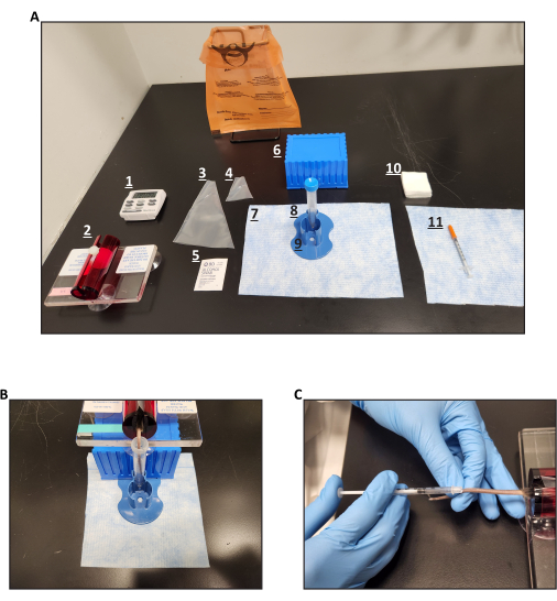
- 设置一个高架平台,将放置小鼠管限制器(图 1A、B)。
- 将干净的鼠标管限制器放在工作区域。
- 如果注射的小鼠比可用的小鼠管限制器小得多,请使用塑料啮齿动物限制锥制作限制套管。请参阅步骤 2.1.3-4。
- 将用于注射的注射器放在工作区域。使用 0.3 mL 胰岛素注射器和 29 G 针头。
- 将废液容器尽可能靠近注射站放置,以便立即处理受 AAV 污染的工具。
- 多次拉动和推动每个注射器的柱塞,以确保柱塞平稳移动,从而在注射过程中不会产生注射器引起的阻力。如果柱塞移动不顺畅,请丢弃此注射器并更换新注射器。
- 在进样区域旁边放置小鼠秤和微型离心机。
- 在注射区域准备好冰上准备好的 AAV。
- 准备温水 (38-40 °C)。注意水温不要超过 40 °C,以免烫伤小鼠尾巴。
- 清洗
2. 注射程序
- 鼠标约束
- 如果需要,称量每只小鼠以计算以 vg/kg 为单位的剂量。
- 确保鼠标完全被束缚且无法移动。
注意:如果鼠标没有完全约束,它可能会在静脉注射期间移动,从而导致针头移位。这可能会导致针头从静脉中伸出和/或伤害老鼠。 - 对于管式限制器:
- 在小鼠之间用温水和肥皂清洗过滤器,以清洁和加热支架。
- 握住鼠标的尾巴。将鼠标的尾巴插入管的顶部开口;然后,慢慢地将鼠标拉入管式限制器中。如果管式限制器的尺寸适合鼠标的尺寸,请将插头放在鼠标前面,以防止鼠标逸出。
注意: 插头应离鼠标足够近,以防止鼠标在管内移动或旋转,但插头不应阻塞鼠标的鼻子,以便让鼠标自由呼吸。如果鼠标小于试管限制器尺寸,则除了试管限制器外,还应使用柔性一次性限制锥,如下文步骤 2.1.4 中所述。
- 如果需要,对于灵活的一次性限制锥(为较小的小鼠制作限制套):
- 在圆锥体的鼻端切开,以确保鼠标的鼻子没有被阻塞,并且老鼠有足够的呼吸空间。
- 切开锥体的背面,使锥体的长度几乎与鼠标相同(这样锥体将适合管内)(图 1A、B)。
- 如上文步骤 2.1.3 中所述,将鼠标放入管式限制器中。握住鼠标尾巴,先将开口侧较宽的限紧锥插入管中。
- 握住鼠标的尾巴,让鼠标走进圆锥体;然后,将锥体的其余部分滑入管中。确保鼠标的尾巴完全从管子的后部伸出,并且鼠标在锥体内有呼吸的空间。
- 将管塞紧紧地固定在锥体的鼻子开口前,同时确保鼠标完全束缚并有足够的呼吸空间。
- 注入鼠标
- 用温水填充 15 mL 锥形管。
- 将装有鼠标的管限制器放在高架平台上(图 1B)。
- 将受束缚的小鼠尾巴尽可能多地浸入温水中至少 1 分钟,直到侧静脉明显扩张和可见(图 1B)。
- 在尾部加热步骤中,将 AAV 剂量装入注射器中。
- 将未加帽的含 AAV 的 1.7 mL 微量离心管放入试管架中。用惯用手将针头垂直插入管中。针头进入管内后,用非惯用手握住管子。
注意: 针头的垂直插入可防止因接触管壁而对针头造成损坏。可以使用其他替代的注射器加载方法,但应确保实验者和注射小鼠的安全。如果在握住管子时插入针头,用非惯用手握住管子可以防止意外穿刺针头。 - 将针头放在管内,同时将管子和注射器抬高到与眼睛齐平,同时确保针头不接触管壁。将双臂放在桌子上以稳定它们。慢慢将剂量拉入注射器中。
注意:缓慢吸液可防止细小气泡附着在点胶针筒侧面。 - 排出注射器中的气泡。如果注射 AAV,请确保气泡通过一次性吸水垫从注射器中排出,该吸水垫将被丢弃在生物危害箱中。
- 保持注射液至少 40 秒以使其预热。
注意:注射前始终确保注射液是温暖的。如果给予冷注射液,除了最初的几 μL 外,注射液可能不会流经静脉。 - 每分钟检查一次尾脉。
注意:静脉必须一直到注射部位都非常明显(增加暖尾时间,并根据需要更换新鲜的温水,直到静脉清晰可见,但不要超过机构动物处理协议允许的小鼠约束时间)。 - 确保静脉清晰可见后,从高架平台顶部取下管式限制器,将管式限制器直接放在桌子上。将鼠标放在限制器中,脚向下,不要放在一边,以便于处理尾巴。
注意: 鼠标不应侧放或背靠在限制器内。鼠标应完全被束缚,不能在束缚器内移动或旋转,也不能移动/拉动其尾巴。 - 用纱布快速擦拭尾巴,使尾巴干燥;用酒精棉签擦拭尾巴,然后用干纱布擦干。
注意:使用干纱布使尾巴足够干燥,以便牢固地抓住尾巴,但不要完全干燥。当尾巴完全干燥时,可能更难看到静脉。 - 将尾巴向左或向右旋转约 90°,使两条侧静脉中的一条朝上。在尾部的中间三分之一内找到合适的注射部位。如果由于尝试失败或实验设计需要额外注射,则从远端(更靠近尾尖)开始初始注射,并向近端移动。
注意: 不要尝试注射到先前注射部位的远端,因为注射液可能会从先前的注射部位泄漏出来。可选:使用非惯用手的拇指和食指对注射部位近端(上游/更靠近小鼠身体)施加压力 10 秒。手指充当止血带,进一步扩张注射部位的静脉。止血带 10 秒后,立即松开止血带手指,确保两条侧静脉中的一条朝上且清晰可见。 - 用非惯用手握住注射部位远端的拇指和食指。将尾巴折叠在食指上,使注射部位平放在食指上。向后拉尾部,使尾部被拉伸,注射部位完全水平(0°)(平行于水平台)(图 1C)。
- 用惯用手的食指和中指握住注射器针筒法兰的两侧,同时保持拇指在柱塞处。
注意:一旦针头进入静脉内,这将更容易不移动拇指或针头(图 1C)。 - 将双手放在桌子上以稳定它们,然后将针头直接放在并平行于尾部和静脉上,斜面朝上。保持注射部位靠近握住尾部的食指,以提高注射部位的控制和稳定性。
注意: 管式限制器在桌子中应足够深,以便双手都支撑在桌子上。 - 在保持针头与尾静脉平行并向下按压针头的同时,将针头向前滑入静脉。
注意: 向下的压力应足以将针头以正确的角度插入静脉。静脉非常浅,因此在尝试进入静脉时,针头应尽可能平坦。 - 慢慢地将溶液注入静脉中。给药后,慢慢抽出针头并立即用纱布在注射部位施加压力至少 10 秒以止血。
注:根据需要施加压力,直到出血完全停止,以避免注射试剂的潜在损失。拔出针头后通常会出现血滴,表明针头已穿透静脉。有时,即使注射成功,血滴也不会出现。血滴并不表示注射成功;它仅表明针头穿透了静脉。可靠的注射成功指标是注射过程中完全没有柱塞阻力。如果针头在静脉内,则注射液注射过程中针柱塞处不应有阻力,并且注射部位附近的静脉会暂时显得颜色略浅(变白)(在某些小鼠品系中,静脉变白可能不是很清楚)。如果有阻力和/或注射部位开始出现凸起,则针头未正确放置在静脉内。如果发生这种情况,请从尾部完全取出针头,并尝试在靠近失败注射部位(更靠近小鼠身体)的新注射部位注射静脉。 - 根据该机构的废物管理指南丢弃受 AAV 污染的注射器和试管。
- 将鼠标从限制器中解脱出来,并将其放回与未注射的小鼠分开的新笼子中。监测小鼠 10 分钟以确保注射后活性水平正常。
注意:如果施用传染性药物,这可以避免注射剂可能传播给未注射的小鼠。 - 使用杀菌剂、杀真菌剂和杀病毒剂以及 70% 乙醇对工作区域进行消毒。用水和肥皂清洁鼠标管限制器。
- 将未加帽的含 AAV 的 1.7 mL 微量离心管放入试管架中。用惯用手将针头垂直插入管中。针头进入管内后,用非惯用手握住管子。
3. 解剖和组织采集固定27
- 组织采集站准备
- 根据制造商的说明,用 DNA 降解试剂清洁工作站,以降解工作区域中可能存在的污染 DNA。
- 将甲基丁烷放入金属容器中。将甲基丁烷金属容器放入聚苯乙烯泡沫塑料盒中;然后,用干冰包围金属容器,使容器周围的干冰液位高于容器内的甲基丁烷液位。
- 标记并将空的 2 mL 微量离心机组织储存管放在干冰上。在开始冷冻组织之前,将甲基丁烷和组织储存管在干冰上冷却至少 20 分钟。将组织转移钳放在干冰上。
- 用新鲜的 4% 多聚甲醛 (PFA) 标记并填充另一组 2 mL 微量离心机组织储存管,并保持在室温下。向每个试管中加入足够的 4% PFA,以完全浸没将要放入试管中的组织。
- 组织采集和固定
- 根据该机构的动物护理指南对小鼠实施安乐死。
注意:在这里,使用颈椎脱位对小鼠实施安乐死。 - 用 70% EtOH 充分喷洒鼠标。
- 收集所需的组织。
注意:此处描述的收集和固定方案已在骨骼肌和肝脏上进行了测试。 - 对于将用于 DNA 提取的组织:
- 将组织放入干冰上预冷的甲基丁烷中,并将组织置于甲基丁烷中至少 1 分钟。使用预冷的转移镊子将冷冻组织从甲基丁烷转移到预冷的空 2 mL 微量离心机组织储存管中。将组织储存在-80°C。
注:可选:组织可切成 20 mg 小块,然后滴入甲基丁烷,以准备在步骤 4.1.4 中使用。
- 将组织放入干冰上预冷的甲基丁烷中,并将组织置于甲基丁烷中至少 1 分钟。使用预冷的转移镊子将冷冻组织从甲基丁烷转移到预冷的空 2 mL 微量离心机组织储存管中。将组织储存在-80°C。
- 对于将用于组织学分析和保留报告蛋白荧光的组织:
- 使用保存在室温下的 PFA 指定镊子,将组织放入各自含有 4% PFA(保持在室温下)的微量离心管中,同时确保组织完全浸没在 4% PFA 溶液中。
注:PFA 污染会对不同的下游分子检测产生负面影响。处理 PFA 时,仅使用 PFA 指定的镊子,以防止 PFA 污染其他组织或工具。 - 将微量离心管放在架子上,并用铝箔纸盖住架子,使管子保持黑暗。将带盖的架子在 4 °C 下在摇床上轻轻摇动孵育过夜。
- 孵育过夜后,通过剧烈摇动将 5.0 g 蔗糖溶解在 70 mL 的 1x PBS 中,在 1x PBS 中制备 5% 蔗糖 (% w/v)。加入足够的 1x PBS 至最终总体积为 100 mL,以获得 5% 蔗糖溶液 (% w/v)。
- 使用 0.22 μm 注射器过滤器对 5% 蔗糖溶液进行灭菌。标记并用新鲜制备的 5% 蔗糖填充 2.0 mL 微量离心管。
- 将组织从 4% PFA 转移到各自的含有 5% 蔗糖(保持在室温下)的微量离心管中,同时确保组织完全浸没在 5% 蔗糖溶液中。
- 将微量离心管放在架子上,并用铝箔纸盖住架子,使管子保持黑暗。将带盖的架子在 4 °C 下在摇床上轻轻摇动孵育过夜。
- 孵育过夜后,通过剧烈摇动将 20.0 g 蔗糖溶解在 70 mL 的 1x PBS 中,在 1x PBS 中制备 20% 蔗糖 (% w/v)。加入足够的 1x PBS 至最终总体积为 100 mL,以获得 20% 蔗糖溶液 (% w/v)。
- 使用 0.22 μm 注射器过滤器对 20% 蔗糖溶液进行灭菌。标记并用新鲜制备的 20% 蔗糖填充 2.0 mL 微量离心管。
- 将组织从 5% 蔗糖转移到各自的含有 20% 蔗糖(保持在室温下)的微量离心管中,同时确保组织完全浸没在 20% 蔗糖溶液中。
- 将微量离心管放在架子上,并用铝箔纸盖住架子,使管子保持黑暗。将带盖的架子在 4 °C 下在摇床上轻轻摇动孵育过夜。
- 孵育过夜后,将甲基丁烷放入金属容器中,并将甲基丁烷金属容器放入聚苯乙烯泡沫塑料盒中。用干冰包围金属容器,使容器周围的干冰液位高于容器内的甲基丁烷液位。
- 标记并将空的 2 mL 微量离心机组织储存管放在干冰上。在开始冷冻组织之前,将甲基丁烷和组织储存管在干冰上冷却至少 20 分钟。将转移钳放在干冰上。
- 使用精密湿巾快速吸干组织,以去除任何多余的 20% 蔗糖。将组织放入在干冰上预冷的甲基丁烷中。将组织留在甲基丁烷中至少 1 分钟。
- 使用预冷的转移镊子将冷冻组织从甲基丁烷转移到预冷的空 2 mL 微量离心机组织储存管中。将组织储存在-80°C。
- 使用保存在室温下的 PFA 指定镊子,将组织放入各自含有 4% PFA(保持在室温下)的微量离心管中,同时确保组织完全浸没在 4% PFA 溶液中。
- 根据该机构的动物护理指南对小鼠实施安乐死。
4. 用于 vg/dg 定量的 dPCR
- 从组织中提取 DNA 和初始 RNA 消化
注:使用 材料表 中列出的 DNA 提取试剂盒手册得出该 DNA 提取方案。始终将装有冷冻组织块的试管放在干冰上。- 准备一桶冰块。
- 对于每个 DNA 样品,标记一个 1.5 mL 裂解珠管和两个空的无 RNase 和无 DNase 的 1.7 mL 微量离心管。
- 向每个微珠管中加入 180 μL 第一种 DNA 提取试剂盒缓冲液。去皮第一个含有缓冲液的珠管。
注意:如果在步骤 3.2.4.1 中未预先切割组织,请使用在干冰上预冷的剃须刀片将组织切成 20 毫克的小块。此步骤必须在 -20 °C 或更冷的干净低温恒温器内完成。 - 将一块组织加入试管中;称重并记录组织的重量(为 ~20 毫克)。
- 立即将装有组织的裂解珠管放在冰上。缓冲器可能会结晶。
- 对每个组织样品重复前面的步骤。
- 将试管转移到裂解珠管搅拌机中,并在 4 °C 下以最大速度(速度 10)运行 1 分钟。
- 将样品放在冰上,将其转移到离心机中。在 4 °C 下以 20,000 × g 离心 1 分钟。
- 在离心步骤中,将 20 μL 蛋白酶 K 添加到第一批 1.7 mL 微量离心管中。离心步骤后,将匀浆的上清液转移到含有蛋白酶 K 的 1.7 mL 试管中并充分混合。在 56 °C 下孵育 15 分钟,以 500 RPM 混合。
- 通过使用微型离心机离心管 1-2 秒,从管壁和盖子收集液滴。在室温下孵育 2 分钟。
- 加入 4 μL RNase A,通过短暂的脉冲涡旋混合。在室温下孵育 2 分钟。脉冲涡旋 15 秒。
- 通过使用微型离心机离心管 1-2 秒,从管壁和盖子收集液滴。加入 200 μL 第二种 DNA 提取试剂盒缓冲液。脉冲涡旋 15 秒。
- 通过使用微型离心机离心管 1-2 秒,从管壁和盖子收集液滴。加入 200 μL 100% 乙醇。脉冲涡旋 15 秒。
- 通过使用微型离心机离心管 1-2 秒,从管壁和盖子收集液滴。将裂解物转移到 DNA 提取离心柱中。以 6,000 × g 离心 1 分钟。
- 将离心柱放入新的收集管中。向离心柱中加入 500 μL 第三种 DNA 提取试剂盒缓冲液。以 6,000 × g 离心 1 分钟。
- 将离心柱放入新的收集管中。向离心柱中加入 500 μL 第四种 DNA 提取试剂盒缓冲液。以 20,000 × g 离心 3 分钟。
- 将离心柱放入新的 1.7 mL 微量离心管中。向离心柱中加入 100 μL 分子级水。在室温下孵育 1 分钟。在室温下以 6,000 × g 离心 1 分钟。
- 如果需要,测量 DNA 浓度。短期储存在 4 °C 或长期储存在 -20 °C 下。
- 从 FACS 分选的细胞中提取 DNA
注:使用 材料表 中列出的 DNA 提取试剂盒手册得出该 DNA 提取方案。- 分选细胞后,将样品以 300 × g 离心 5 秒,以收集侧面和盖子上的所有液滴。确保收集所有掉落物。
注:如果样品体积小于 1.5 mL,请直接进行下一步。如果样品体积大于 1.5 mL,则使用微量移液器小心去除并丢弃上清液的顶部,留下 1-1.5 mL 样品。 - 通过上下多次吹打混合样品,然后将样品转移到 1.7 mL 微量离心管中。在室温下以 515 × g 离心 1 分钟。
- 弃去除最后 50 μL 的上清液。将沉淀重悬于 50 μL 第一种 DNA 提取试剂盒缓冲液中,最终体积为 100 μL。
- 按照制造商的方案从小体积血液中分离基因组 DNA(参见 材料表)。
- 加入 10 μL 蛋白酶 K 和 100 μL 第二种 DNA 提取试剂盒缓冲液;通过脉冲涡旋混合 15 秒。将样品在 56 °C 下以 300 RPM 混合孵育 10 分钟。在此孵育期间,通过轻轻倒置将样品混合两次。
- 通过使用微型离心机离心管 1-2 秒,从管壁和盖子收集液滴。加入 50 μL 100% 乙醇,并通过脉冲涡旋混合 15 秒。将样品在室温下孵育 5 分钟。
- 通过使用微型离心机离心管 1-2 秒,从管壁和盖子收集液滴。将样品转移到 DNA 提取柱(柱位于 2 mL 收集管中),不要弄湿边缘。以 6,000 × g 离心 1 分钟。
- 将色谱柱放入干净的 2 mL 收集管中后,丢弃含有流出液的收集管。向柱中加入 500 μL 第三种 DNA 提取试剂盒缓冲液,不润湿边缘,并以 6,000 × g 离心 1 分钟。
- 同样,将色谱柱放入干净的 2 mL 收集管中后,丢弃含有流出液的收集管。向柱中加入 500 μL 第四种 DNA 提取试剂盒缓冲液,不要润湿边缘,并以 6,000 × g 离心 1 分钟。
- 将色谱柱放入干净的 2 mL 收集管中,丢弃含有流通液的收集管。以 20,000 × g 离心 3 分钟。
- 将色谱柱放入干净的 1.7 mL 微量离心管中,丢弃含流通液的收集管。向柱膜中心添加 20 μL 分子级水进行洗脱;盖上盖子,将样品与分子级水在室温下孵育 5 分钟。
- 以 20,000 × g 离心 1 分钟。将洗脱的 DNA 储存在 4 °C 下进行短期储存,或在 -20 °C 下进行长期储存。
- 分选细胞后,将样品以 300 × g 离心 5 秒,以收集侧面和盖子上的所有液滴。确保收集所有掉落物。
- RNA 消化和纯化
注:材料 表 中列出的 DNA 提取试剂盒手册用于得出该 DNA 纯化方案。根据 dPCR 条件、试剂以及引物和探针设计,在进行 dPCR vg/dg 定量之前,可能需要确保 DNA 样品中完全不存在 RNA。在某些 dPCR 条件下,RNA 污染可能导致不同程度的 vg/dg 值不准确。- 在 0.2 mL PCR 管或 1.7 mL 微量离心管中,向每个 DNA 样品中最多添加 20 μL 提取的 DNA 样品和 1.5 μL 不含 DNase 的 RNase。如果 DNA/RNase 混合物体积小于 21.5 μL,则添加足够的分子级水至最终体积为 21.5 μL,并通过倒置试管混合 25 次。在 37 °C 下孵育 30 分钟,每倒置试管每 10 分钟定期混合一次。
注:添加到试管中的核酸总量应在 175 ng 至 700 ng 之间。如果 DNA 样品的体积或核酸量超出此范围,或者 DNA 样品的分离方式不同,则可能需要进行修饰。 - 置于冰上 2 分钟。向每种 DNA/RNase 混合物中加入足够的分子级水,最终体积为 100 μL。
注:建议使用此处列出的 RNase,因为它可以消化污染的 RNA,而不会对靶 DNA 或下游 PCR 检测产生负面影响。 - 按照制造商的方案纯化基因组 DNA(参见 材料表)。
- 加入 10 μL 第一种 DNA 提取试剂盒缓冲液和 250 μL 第二种 DNA 提取试剂盒缓冲液。通过脉冲涡旋混合 10 秒。
- 将样品转移到 2 mL 收集管中的 DNA 提取柱中,不要弄湿边缘。以 6,000 × g 离心 1 分钟。
- 将色谱柱放入干净的 2 mL 收集管中后,丢弃含有流出液的收集管。向柱中加入 500 μL 第二种 DNA 提取试剂盒缓冲液,不要润湿边缘。以 6,000 × g 离心 1 分钟。
- 将色谱柱放入干净的 2 mL 收集管中,丢弃含有流通液的收集管。以 20,000 × g 离心 6 分钟。
- 将色谱柱放入干净的 1.7 mL 微量离心管中,丢弃含有流出液的收集管。在柱膜中心加入 20 μL 分子级水进行洗脱,盖上盖子,将样品与分子级水在室温下孵育 5 分钟。
- 以 20,000 × g 离心 1 分钟。短期储存在 4 °C 或长期储存在 -20 °C 下。
- 使用 PCR 引物对,通过终点 PCR 或 dPCR 或定量 PCR (qPCR) 确认 RNase 消化样品中不存在 RNA 污染,该引物对将扩增跨越多个外显子而不仅仅是单个外显子的 mRNA 区域。
注意:mRNA 靶标应为在靶组织/细胞类型中高度表达的基因,以确保如果存在 mRNA 污染,则能够正确识别。跨越多个外显子的扩增子区分污染 mRNA 和基因组 DNA 产生的条带。始终使用非 RNase 消化的 DNA 样品作为 PCR 反应的阳性对照,以确保检测到 mRNA 污染(如果存在)。
- 在 0.2 mL PCR 管或 1.7 mL 微量离心管中,向每个 DNA 样品中最多添加 20 μL 提取的 DNA 样品和 1.5 μL 不含 DNase 的 RNase。如果 DNA/RNase 混合物体积小于 21.5 μL,则添加足够的分子级水至最终体积为 21.5 μL,并通过倒置试管混合 25 次。在 37 °C 下孵育 30 分钟,每倒置试管每 10 分钟定期混合一次。
- 数字 PCR (dPCR)
- 终点 PCR,用于检查引物的特异性和最佳 PCR 条件(可选)
- 为载体基因组设计 dPCR 引物对和探针,为小鼠参考基因设计 dPCR 引物对和探针,用于定量样品中的二倍体基因组。
注意:扩增子大小的目标是在 60 bp 和 150 bp 之间。小鼠参考基因应该是每个二倍体基因组具有恒定基因拷贝数的基因。对于此处列出的计算,参考基因 (Polr2a) 每个二倍体基因组有两个拷贝。 - 对于 10 μL 终点 PCR 反应,使用试剂和终浓度制备 PCR 混合物,以便稍后用于 dPCR 反应。将 RNase 消化的模板 DNA 样品(核酸量范围 56-223 ng)加入终浓度为 1x dPCR 预混液中,该预混液含有 DNA 聚合酶和 dNTP、0.8 μM 每个正向引物、0.8 μM 每个反向引物、0.4 μM 每个探针和 0.025 U/μL 限制性内切酶(限制性内切酶的最终浓度取决于所使用的限制性内切酶和品牌)。加入分子级水,使最终体积达到 10 μL。
注:PCR 混合物中应至少有两个引物对和两个探针:一个引物对和一个探针用于检测载体基因组,一个引物对和一个探针用于检测小鼠基因组。 - PCR 热循环条件:在 95 °C 下进行初始热活化步骤,持续 2 分钟,然后在 95 °C 下进行 35-45 次变性步骤循环,持续 25 秒,在 58-62 °C 下进行联合退火/延伸步骤,持续 1 分钟。
注:必须确定每个扩增子和引物对的最佳退火温度。循环次数可以根据样品中模板 DNA 的量进行调整。 - 使用凝胶电泳在琼脂糖凝胶上观察 PCR 产物,以确定是否存在靶扩增子条带和任何可能的非特异性扩增条带。
- 在确认引物对和循环条件导致靶序列的特异性扩增后,继续进行下一个 dPCR 步骤。
- 为载体基因组设计 dPCR 引物对和探针,为小鼠参考基因设计 dPCR 引物对和探针,用于定量样品中的二倍体基因组。
- dPCR 反应
- 对于 40 μL dPCR 反应,加入至 4 μL RNase 消化的模板 DNA 样品(核酸量范围 50-330 ng)至终浓度为 1x dPCR 预混液中,该预混液含有 DNA 聚合酶和 dNTP、0.8 μM 每种正向引物、0.8 μM 每种反向引物、0.4 μM 每种探针, 和 0.025 U/μL 限制性内切酶(限制性内切酶的最终浓度取决于所使用的限制性内切内切酶和品牌)。加入分子级水,使最终体积达到 40 μL。
- dPCR 热循环条件:在 95 °C 下进行初始热活化步骤,持续 2 分钟,然后在 95 °C 下进行 40-50 次变性步骤,持续 25 秒,然后在 58-62 °C 下进行联合退火/延伸步骤,持续 1 分钟。
注:必须确定每个扩增子和引物对的最佳退火温度。循环次数可以根据样品中模板 DNA 的量进行调整。此处列出的体积、浓度和条件针对 材料表中列出的 dPCR 板、试剂和设备进行了优化。这些条件降低了任何可能降低反应准确性的潜在 dPCR 抑制剂的作用。 - 运行 dPCR 反应并获得载体基因组和小鼠参考基因的绝对值后,使用公式 (7-8) 计算样品中的 vg/dg。
对于具有两个基因拷贝/二倍体基因组的参考基因:
二倍体基因组的绝对值 (dg) = (7)
(7)
VG/DG = (8)
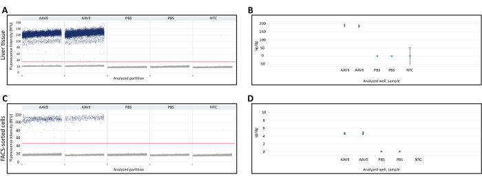
(8) - 检查 dPCR 反应的 1D 散点图以确认测定和定量的有效性(图 3A、C)。要使分析有效,请确认 1D 散点图满足以下所有标准:存在正分区和负分区;正负分区之间明确分离,以便准确确定阈值;以及正负分配之间存在少量液滴(也称为雨),这会降低 dPCR 定量的准确性。
- 终点 PCR,用于检查引物的特异性和最佳 PCR 条件(可选)
结果
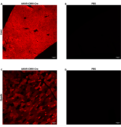
7 至 9 周龄雄性小鼠通过侧尾静脉注射以 1.5 ×10 12 vg/小鼠注射 AAV,注射液量为 150-200 μL。这里使用的 ssDNA AAV 递送了由 CMV 启动子驱动的 Cre 重组酶转基因。注射的小鼠是 Cre 报告基因 Ai14 等位基因的纯合子。当暴露于 Cre 重组酶时,含有 Ai14 等位基因的细胞表达荧光 tdTomato 蛋白。由于 tdTomato 表达是由 Cre 诱导的基因组重组引起的,因此表达 tdTomato 的细胞表示由 AAV 直接转导的细胞或转导细胞的后代细胞。此处显示的数据是以 1.5 × 10¹² vg/小鼠注射 AAV9-CMV-Cre 的小鼠,以 160 μL (5.8-5.9 × 10¹³ vg/kg) 给药。注射后 28 天处死小鼠,如上所述收集组织。消化少量骨骼肌和肝叶,并使用 FACS 收集其细胞。立即使用预冷的甲基丁烷冷冻几个肝叶进行核酸提取。将一些骨骼肌和肝叶固定冷冻用于荧光 tdTomato 的组织学成像。tdTomato 在整个肝脏(图 2A)和股四头肌(图 2C)中弥漫表达,表明 AAV9 广泛到达并转导了两个组织的不同区域。
从新鲜冷冻肝脏和 FACS 分选细胞中提取的 DNA 用于通过 dPCR 定量 vg/dg。Vg/dg 定量可用于评估分析样品中 AAV 的进样一致性和转导效率。使用来自新鲜冷冻肝组织样品和 FACS 分选细胞的 1D 液滴散点图来确保测定的有效性(图 3A、C)。散点图显示存在阳性和阴性分区,阳性和阴性分区之间的明确分离,可以准确确定检测阈值,以及阳性和阴性分区之间存在少量液滴,这可能会降低 dPCR 检测的准确性。满足所有这些标准表明 dPCR 检测结果是有效的。定量每个样品中 Polr2a 基因拷贝数以确定小鼠二倍体基因组(2 个 Polr2a 基因拷贝/小鼠二倍体基因组)的数量,并使用针对 Cre 重组酶转基因序列的引物/探针来定量病毒基因组(1 个转基因拷贝/病毒基因组, 表 1)。对新鲜冷冻肝组织样品和 FACS 分选细胞的 vg/dg 值进行量化,并显示每个样品中分别存在 187.7 vg/dg 和 4.7 vg/dg(图 3B、D)。来自 PBS 注射小鼠的样品和不含核酸的非模板对照用作阴性对照。

图 1:静脉注射站概述。 (A) 进行 IV 注射所需的工具。此处显示的是 (1) 计时器、(2) 小鼠管限制器、(3) 未切割和 (4) 切割的塑料限制锥、(5) 酒精棉签、(6) 用作升高小鼠管限制器平台的空移液器吸头盒、(7) 一次性吸收垫、(8) 15 mL 温水锥形管、(9) 15 mL 管架、(10) 纱布和 (11) 胰岛素注射器。(B) 首先将鼠标放入管式限制器内。然后,如果鼠标太小而无法仅由管式限制器约束,则插入切割的限制锥体以在鼠标周围形成一个限制套。确保鼠标的呼吸不受限制器的阻碍。管式限制器放置在高架平台的顶部,以便将小鼠尾巴放入温水中。(C) 注射前的小鼠尾巴位置和针头保持角度。向后拉尾部,使尾部伸展,注射部位完全水平。针平行于尾部和静脉,斜面朝上。 请单击此处查看此图的较大版本。

图 2:IV 注射后荧光报告蛋白的检测。 携带 Cre 报告基因 Ai14 等位基因的 7 至 9 周龄雄性小鼠静脉注射 1.5 ×10 12 vg/小鼠的 AAV9-CMV-Cre,以 160 μL(5.8-5.9 ×10 13 vg/kg)或 PBS 给药。AAV9 输送 Cre IV 注射后小鼠 (A) 肝脏或 (C) 股四头肌切片的代表性荧光图像。(B) 对注射 PBS 的小鼠的肝脏或 (D) 股四头肌切片进行成像,作为阴性对照。收集组织并在 IV 注射后 28 天固定冷冻。Cre 暴露后,荧光 tdTomato 蛋白在转导细胞和转导细胞的后代细胞中表达。以 10 倍放大倍率对 10 μm 厚的切片进行成像。比例尺 = 100 μm。 请点击此处查看此图的较大版本。

图 3:每个二倍体基因组 (vg/dg) 定量的载体基因组。 从注射 AAV9-CMV-Cre 或 PBS 的小鼠中收集的 (A) 肝组织或 (C) FACS 分选细胞中 dPCR 载体基因组定量的 1D 散点图。散点图显示了阳性和阴性 dPCR 分配,以及样品上水平线指示的检测阈值。(B,D) 定量 (B) 肝组织或 (D) FACS 分选细胞样品中的小鼠二倍体基因组和载体基因组后,进行 vg/dg 定量。此处显示的结果来自一只注射 AAV9 的小鼠和一只注射 PBS 的小鼠,每只小鼠都有一个技术 dPCR 副本。误差线表示每个样本的 95% 置信区间。缩写: NTC= 非模板对照;dPCR = 数字 PCR;FACS = 荧光激活细胞分选。 请单击此处查看此图的较大版本。
| 底漆 | 序列 |
| Cre 正向引物 | CTGACGGTGGGAGAATGTTAAT |
| Cre 反向引物 | CATCGCTCGACCAGTTTAGTT |
| Cre 探针 | /56-FAM/CGCAGGTGT/ZEN/AGAGAAGGCACTTAGC/3IABkFQ/ |
| Polr2a 正向引物 | GACTCCTTCACTCACTGTCTTC |
| Polr2a 反向引物 | TCTTGCTAGGCAGTCCATTATC |
| Polr2a 探针 | /5HEX/ACGAGATGC/ZEN/TGAAAGAGCCAAGGT/3IABkFQ/ |
表 1:用于 vg/dg 定量的引物和探针序列。 Cre 引物和探针用于定量载体基因组。Polr2a 引物和探针用于定量小鼠二倍体基因组。
讨论
由于 AAV 作为基因治疗载体的多功能性,基于 AAV 的疗法对单基因疾病具有巨大潜力,这使得定制 AAV 以满足不同疾病的各种递送需求成为可能 4,5,7,9。在临床前小鼠模型中,AAV 通常通过静脉注射给药,以测试潜在疗法的安全性和有效性16。由于不同的 AAV 注射剂量会导致实验结果的显着差异,因此实验者能够始终如一地注射预期的 AAV 剂量以确保生成的体内数据的有效性和稳健性至关重要28。静脉注射被广泛使用,但它们在技术上具有挑战性,需要广泛的培训和持续的实践来发展和保持确保持续成功注射的技能水平 16,17,18,19。除了正确注射 AAV 外,通常还需要使用检测来评估注射的 AAV 向目标组织或细胞的生物分布和递送效率29,30。
该方案旨在通过全面描述在 7-9 周龄未镇静小鼠中管理 AAV 的优化 IV 注射方案的细节,帮助实验者轻松、成功和一致地进行 IV 注射。重要的是要注意,由于静脉可见度降低或与该方法中使用的限制器不相容,在此处使用的年龄范围内明显小于或大于野生型小鼠的小鼠可能会带来更大的挑战。先前已经报道,由于血管尺寸小,尾部静脉注射不适合在 6 周龄以下的小鼠中静脉注射试剂31。尽管有可能,但可能很难持续注射体重低于 22.0 克的小鼠。使用非典型大小小鼠的研究人员可能需要适应该程序。该方案还概述了可用于评估 AAV 生物分布和转导效率的几种测定方法。
在遵循此协议时,需要牢记一些关键点。在注射过程中,如果针头不在静脉内,则 29 G 针头会提供更大的阻力。这减少了注射尝试失败期间因意外血管周围注射溶液而损失的体积。胰岛素注射器的死体积比普通注射器小。如果使用与此处列出的注射器和/或针头不同的注射器和/或针头,则可能需要在方案步骤 1.1.3.3 中准备额外的注射液体积,以解决更大的死腔体积(例如,在预期剂量中加入 30 μL 而不是 15 μL)。
如果在将 AAV 剂量吸入注射器时在注射器侧面形成细小的吸入引起的气泡,请慢慢地将注射液进一步拉到注射器上。这将去除大多数小气泡。将至少 10-15 μL AAV 额外加载到要注射的预期体积中。此额外体积用于说明在排出气泡或可能失败的进样尝试期间可能丢失的任何体积。(例如,如果要注射的目标体积为 150 μL,则将 165 μL 加载到注射器中(注射器刻度上 160 μL 和 170 μL 标记之间的中间位置)。如果针头正确放置在静脉内,并且在成功进样尝试前注射器中的体积为 165 μL,则输送试剂直到注射器中剩余 15 μL(介于 10 μL 和 20 μL 标记之间),从而输送 150 μL (165 μL - 150 μL = 15 μL))。将斜面管腔(斜面朝上)与注射器刻度对齐,可以在注射过程中跟踪输送的体积。
一些实验者可能更喜欢将鼠标侧放,这样与脚上的鼠标相比,它的一条静脉是笔直的且易于接近的。但是,在注射不同大小的小鼠时,根据鼠标的大小,小鼠侧面的尾巴会以不同的角度倾斜,需要调整注射角度。这可能会对手术成功的一致性产生负面影响。在最初的练习尝试中,实验者可以尝试两种鼠标约束方向,以确定他们喜欢的方法。让鼠标脚着可以快速轻松地进入两侧侧尾静脉。这减少了在多次注射尝试失败的情况下需要进入两条静脉时的抑制时间。
如果注射靠近尾部基部(更靠近小鼠身体)的侧静脉(特别是对于体重 >30 克的小鼠),请将注射角度从平行于静脉调整到与静脉 5°-10°,因为尾部底部的静脉比远端略深。
此处列出的 RNase 消化和 RNA 污染检查方案在从新鲜冷冻肝组织中分离的 DNA 样品上进行了验证,这些样品在 20 μL 中含有总共 175-700 ng 的核酸。还对从新鲜冷冻肝组织和 FACS 分选细胞中分离的 DNA 样品测试了 RNase 消化方案,以确认 RNase 消化后载体基因组和小鼠基因组的存在。使用目标扩增子的终点 PCR 扩增的琼脂糖凝胶电泳对结果进行可视化。
遵循所描述的方法可以减少掌握 IV 注射所需的培训和练习时间,并提高注射成功率,从而节省试剂。该协议使用简单且常用的工具,无需可能不容易获得的高级设备或设置。此外,此处列出的 IV 注射步骤可应用于需要静脉注射的各种注射液,例如反义寡核苷酸 (ASO),并根据注射液对注射液制备步骤进行适当修改。
披露声明
作者没有与本文中发表的工作相关的任何披露。
致谢
作者要感谢 NINDS 动物护理机构工作人员的支持。这项工作得到了 NIH 校内研究部 NINDS 的支持(年度报告编号 1ZIANS003129)。内容完全由作者负责,并不一定代表美国国立卫生研究院的官方观点。
材料
| Name | Company | Catalog Number | Comments |
| 0.22 µm syringe filter | Millipore | SLGVM33RS | |
| 0.3 mL insulin syringes with 29G needle | BD Biosciences | 324702 | |
| 1.7 mL microcentrifuge tube | Crystalgen | 23-2051 | |
| 10 mL syringe | BD Biosciences | 302995 | |
| 100% EtOH | The Warner Graham Company | 201096 | |
| 10x phosphate-buffered saline (PBS) | Corning | 46-013-CM | Used to prepare 1x PBS for tissue fixation |
| 15 mL conical tube | Corning | 430766 | |
| 15 mL conical tube holder | Multiple sources | N/A | |
| 190 proof ethyl alcohol | The Warner Graham Company | 6810-01-113-7320 | Used to prepare 70% ethanol |
| 1x sterile PBS | Gibco | 10010023 | |
| 2 mL microcentrifuge tissue storage tubes | Eppendorf | 022363344 | |
| 4% paraformaldehyde (PFA) | Electron Microscopy Sciences | 157-4 | |
| Adeno-associated virus (AAV) | Charles River | N/A | Single-stranded DNA (ssDNA) AAV was packaged to deliver Cre recombinase as the transgene driven by CMV promoter |
| Alcohol swab | BD Biosciences | 326895 | |
| Bead lysis tube | Next Advance | GREENE5 | |
| BsuRI (HaeIII) restriction enzyme | Thermo Fisher Scientific | ER0151 | |
| Bullet blender | Next Advance | BBX24B | |
| Ai14-derived mice from JAX 007914 strain (genetic background: C57BL/6J) | N/A | N/A | Mice containing Ai14 Cre-reporter allele were purchased from JAX (catalog number: 007914) |
| Disposable absorbent pads | Fisherbrand | 1420662 | |
| Dissection forceps | Fine Science Tools (F.S.T) | 11251-35 | |
| Dissection scissors | Fine Science Tools (F.S.T) | 14085-08 | |
| DNA degradation reagent (DNAZap) | Invitrogen | AM9890 | |
| DNA-Extraction RNase A | Qiagen | 19101 | For RNA digestion during nucleic acid extraction |
| DNase-free RNase for DNA cleanup | F. Hoffmann-La Roche | 11119915001 | For RNA digestion after nucleic acid extraction |
| dPCR Probe PCR Kit | Qiagen | 250102 | |
| dPCR software | Qiagen | N/A | QIAcuity Software Suite |
| Elevated platform | Multiple sources | N/A | An empty pipette tips box was used to elevate the mouse restrainer during tail warming up |
| Fluorescence microscope | Multiple sources | N/A | Model used here: Nikon Eclipse Ti |
| Fluorescence microscope software | Multiple sources | N/A | Software used here: NIS-Elements |
| Gauze | Covidien | 9022 | |
| Heat block | Eppendorf | Thermomixer 5350 | |
| High-speed centrifuge | Eppendorf | 22620689 | |
| Metal container | Vollrath | 80125 | |
| Methylbutane | J.T. Baker | Q223-08 | |
| Molecular grade water | Quality Biological | 351-029-131 | |
| Mouse tube restrainer | Braintree Scientific | TV-RED-150-STD | |
| Myfuge mini centrifuge | Benchmark Scientific | C1012 | |
| Polymerase chain reaction thermal cycler | Bio-Rad Laboratories | 1851148 | Model: C1000 Touch |
| Precision wipes | Kimberly-Clark Professional | 7552 | |
| Proteinase K | Qiagen | 19131 | |
| QIAcuity dPCR Nanoplate 26k 24-well | Qiagen | 250001 | |
| QIAcuity One dPCR system | Qiagen | 911020 | |
| Qiagen DNeasy Blood & Tissue Kit | Qiagen | 69504 | Used for DNA extraction from tissues |
| Qiagen QIAamp DNA Micro Kit | Qiagen | 56304 | Used for cleanup of genomic DNA, and the isolation of DNA from small volumes of blood prtocotol was used for DNA extraction from FACS-sorted cells |
| Rodent restrainer cone | Braintree Scientific | MDC-200 | |
| Scale | Ohaus | 72212663 | |
| Styrofoam box | Multiple sources | N/A | |
| Sucrose | Sigma-Aldrich | S9378-1kg | |
| Surface cleaner and disinfectant | Peroxigard | 29101 | |
| Timer | Multiple sources | N/A | |
| Transfer forceps | Fine Science Tools (F.S.T) | 91113-10 | |
| Vortex | Daigger & Company | 22220A | Model: Daigger Vortex Genie 2 |
参考文献
- Tisdale, A., et al. The ideas initiative: Pilot study to assess the impact of rare diseases on patients and healthcare systems. Orphanet J Rare Dis. 16 (1), 429 (2021).
- Condo, I. Rare monogenic diseases: Molecular pathophysiology and novel therapies. Int J Mol Sci. 23 (12), 6525 (2022).
- Vaisitti, T., et al. The frequency of rare and monogenic diseases in pediatric organ transplant recipients in italy. Orphanet J Rare Dis. 16 (1), 374 (2021).
- Bulcha, J. T., Wang, Y., Ma, H., Tai, P. W. L., Gao, G. Viral vector platforms within the gene therapy landscape. Signal Transduct Target Ther. 6 (1), 53 (2021).
- Lundstrom, K. Viral vectors in gene therapy: Where do we stand in 2023. Viruses. 15 (3), 698 (2023).
- Wang, C., et al. Emerging non-viral vectors for gene delivery. J Nanobiotechnology. 21 (1), 272 (2023).
- Naso, M. F., Tomkowicz, B., Perry, W. L., Strohl, W. R. Adeno-associated virus (aav) as a vector for gene therapy. BioDrugs. 31 (4), 317-334 (2017).
- Ghosh, S., Brown, A. M., Jenkins, C., Campbell, K. Viral vector systems for gene therapy: A comprehensive literature review of progress and biosafety challenges. Appl Biosaf. 25 (1), 7-18 (2020).
- Wang, D., Tai, P. W. L., Gao, G. Adeno-associated virus vector as a platform for gene therapy delivery. Nat Rev Drug Discov. 18 (5), 358-378 (2019).
- Srivastava, A. Rationale and strategies for the development of safe and effective optimized aav vectors for human gene therapy. Mol Ther Nucleic Acids. 32, 949-959 (2023).
- Mukherjee, P., Roy, S., Ghosh, D., Nandi, S. K. Role of animal models in biomedical research: A review. Lab Anim Res. 38 (1), 18 (2022).
- Jucker, M. The benefits and limitations of animal models for translational research in neurodegenerative diseases. Nat Med. 16 (11), 1210-1214 (2010).
- Vandamme, T. F. Use of rodents as models of human diseases. J Pharm Bioallied Sci. 6 (1), 2-9 (2014).
- Anders, H. J., Vielhauer, V. Identifying and validating novel targets with in vivo disease models: Guidelines for study design. Drug Discov Today. 12 (11-12), 446-451 (2007).
- Turner, P. V., Brabb, T., Pekow, C., Vasbinder, M. A. Administration of substances to laboratory animals: Routes of administration and factors to consider. J Am Assoc Lab Anim Sci. 50 (5), 600-613 (2011).
- Prabhakar, S., Lule, S., Da Hora, C. C., Breakefield, X. O., Cheah, P. S. Aav9 transduction mediated by systemic delivery of vector via retro-orbital injection in newborn, neonatal and juvenile mice. Exp Anim. 70 (4), 450-458 (2021).
- Hauff, P., Nebendahl, K., Kiessling, F., Pichler, B. J., Hauff, P. Drug administration. Small Animal Imaging: Basics and Practical. , 127-152 (2017).
- Saleem, M., et al. A new best practice for validating tail vein injections in rat with near-infrared-labeled agents. J Vis Exp. (146), (2019).
- Resch, M., Neels, T., Tichy, A., Palme, R., Rulicke, T. Impact assessment of tail-vein injection in mice using a modified anaesthesia induction chamber versus a common restrainer without anaesthesia. Lab Anim. 53 (2), 190-201 (2019).
- Glascock, J. J., et al. Delivery of therapeutic agents through intracerebroventricular (icv) and intravenous (iv) injection in mice. J Vis Exp. (56), 2968 (2011).
- Grames, M. S., Jackson, K. L., Dayton, R. D., Stanford, J. A., Klein, R. L. Methods and tips for intravenous administration of adeno-associated virus to rats and evaluation of central nervous system transduction. J Vis Exp. (126), 55994 (2017).
- Yano, J., Lilly, E. A., Noverr, M. C., Fidel, P. L. A contemporary warming/restraining device for efficient tail vein injections in a murine fungal sepsis model. J Vis Exp. (165), 61961 (2020).
- Warren, J. S. A., Feustel, P. J., Lamar, J. M. Combined use of tail vein metastasis assays and real-time in vivo imaging to quantify breast cancer metastatic colonization and burden in the lungs. J Vis Exp. (154), 60687 (2019).
- JoVE Science Education Database. . Lab Animal Research. Compound administration I. , (2023).
- UCSF Office of Research, Institutional Animal Care and Use Program. Lateral tail vein injection in mice and rats (preferred technique for vascular access in mice Available from: https://iacuc.ucsf.edu/sites/g/files/tkssra751/f/wysiwyg/STD%20PROCEDURE%20-%20Misc%20Rodent%20Procedures%20-%20Lateral%20Tail%20Vein%20Injection%20in%20Mice%20and%20Rats.pdf (2023)
- Jones, K. Tail vein injections in the mouse and rat sop. UBC Animal Care Guidelines SOP: ACC-2012-Tech03. , (2012).
- Liadaki, K., Luth, E. S., Kunkell, L. M. Co-detection of gfp and dystrophin in skeletal muscle tissue sections. Biotechniques. 42 (6), 699-700 (2007).
- Burr, A., et al. Allometric-like scaling of aav gene therapy for systemic protein delivery. Mol Ther Methods Clin Dev. 27, 368-379 (2022).
- Lang, J. F., Toulmin, S. A., Brida, K. L., Eisenlohr, L. C., Davidson, B. L. Standard screening methods underreport aav-mediated transduction and gene editing. Nat Commun. 10 (1), 3415 (2019).
- Rodriguez-Estevez, L., Asokan, P., Borras, T. Transduction optimization of aav vectors for human gene therapy of glaucoma and their reversed cell entry characteristics. Gene Ther. 27 (3-4), 127-142 (2020).
- Hatakeyama, S., Yamamoto, H., Ohyama, C. Tumor formation assays. Methods Enzymol. 479, 397-411 (2010).
转载和许可
请求许可使用此 JoVE 文章的文本或图形
请求许可探索更多文章
This article has been published
Video Coming Soon
版权所属 © 2025 MyJoVE 公司版权所有,本公司不涉及任何医疗业务和医疗服务。