
A subscription to JoVE is required to view this content. Sign in or start your free trial.
Method Article
Synthesis of Protein Bioconjugates via Cysteine-maleimide Chemistry
In This Article
Summary
This protocol details the important steps required for the bioconjugation of a cysteine containing protein to a maleimide, including reagent purification, reaction conditions, bioconjugate purification and bioconjugate characterization.
Abstract
The chemical linking or bioconjugation of proteins to fluorescent dyes, drugs, polymers and other proteins has a broad range of applications, such as the development of antibody drug conjugates (ADCs) and nanomedicine, fluorescent microscopy and systems chemistry. For many of these applications, specificity of the bioconjugation method used is of prime concern. The Michael addition of maleimides with cysteine(s) on the target proteins is highly selective and proceeds rapidly under mild conditions, making it one of the most popular methods for protein bioconjugation.
We demonstrate here the modification of the only surface-accessible cysteine residue on yeast cytochrome c with a ruthenium(II) bisterpyridine maleimide. The protein bioconjugation is verified by gel electrophoresis and purified by aqueous-based fast protein liquid chromatography in 27% yield of isolated protein material. Structural characterization with MALDI-TOF MS and UV-Vis is then used to verify that the bioconjugation is successful. The protocol shown here is easily applicable to other cysteine - maleimide coupling of proteins to other proteins, dyes, drugs or polymers.
Introduction
Bioconjugation involves covalently linking one biomolecule with another or with a synthetic molecule such as a dye, drug or a polymer. Protein bioconjugation methods are now extensively used in many chemistry, biology and nanotechnology research groups with applications ranging from fluorescent dye labelling1,2, making of protein (antibody)-prodrugs3 (antibody drug conjugates — ADCs) synthesis of protein dimers4,5, through to self-assembling protein-polymer hybrids6,7 used in nanomedicine8 and systems chemistry9.
Specificity of the chemistry used for bioconjugation, while not always critical, is of utmost importance for most functional protein bioconjugates, so as to not interfere with the active site of the target protein. The ideal bioconjugation reaction needs to fulfill several criteria, including: i) targeting rare or unique sites on the protein of interest, ii) be selective towards this target, iii) proceed under non-denaturing conditions to avoid protein unfolding and iv) be high-yielding as the target protein is usually only available at sub-millimolar concentration. The maleimide - cysteine Michael addition comes close to fulfilling all these criteria, and has for that reason long claimed a special status in the field of bioconjugate chemistry10. This is because i) many proteins containing only one cysteine residue on their surface can be genetically engineered there, ii) at the correct pH the reaction is highly selective towards cysteine, iii) it proceeds smoothly in aqueous buffers and iv) it is very fast with the second order rate constant of maleimides to cysteine-containing proteins reported to exceed 5,000 M-1 sec-1 in some cases11. Provided the protein of interest can tolerate a small (≈ 5-10%) amount of organic co-solvent12, almost any maleimide-functionalized dye, polymer, surface or another protein can be linked to proteins. In addition, maleimides are more specific for cysteines on proteins than iodoacetamides, which are more prone to reacting with other nucleophiles at elevated pH; and more stable than disulfide-based conjugations which need to be kept at acidic pH to prevent disulfide exchange13.
Here we report a generic protocol for the conjugation of maleimide-functionalized molecules to a protein containing a single cysteine residue using the reaction between a Ru(II)-based chromophore and the redox protein cytochrome c as an example. This protocol is equally applicable to most other proteins containing an accessible surface cysteine residue and the corresponding maleimide-functionalized target, be it another protein, a fluorescent dye, a chromophore or a synthetic polymer.
Protocol
Note: The following protocol is designed for the synthesis of a protein-dye bioconjugate as shown in Figure 1. It is a general protocol for the reaction of a maleimide with free surface cysteine containing proteins, with notes inserted where applicable to assist with membrane protein bioconjugates, protein-polymer bioconjugates, and synthetic protein dimer (protein-protein) bioconjugates. In this particular case, the protein iso-1 cytochrome c has one surface cysteine residue available to react which allows a highly specific labelling to occur. If a protein of interest has multiple cysteine residues, the same protocol applies, albeit with the loss of specificity and product homogeneity. Chemistry targeting surface lysine residues, using N-hydroxysuccinimidyl esters or isothiocyanates, may be a simpler approach if specificity is not required.

Figure 1. Bioconjugation Reaction Scheme. As an example case, a light harvesting, ruthenium-based antenna molecule will be attached to cytochrome c via Michael addition of a pendant maleimide on the ruthenium-based antenna molecule and an exposed cysteine residue (CYS102) on the protein. The red area of the cyt c surface indicates the heme group. Please click here to view a larger version of this figure.
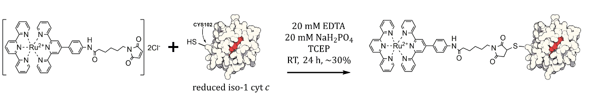
1. Purification of Cytochrome c
Note: This step is not applicable to all proteins. However, it is important to know that a protein obtained from a commercial supplier can contain other, undesired protein isoforms which may need to be removed by further purification13.
- Dissolve 2.4 g of sodium dihydrogen phosphate (Mw = 120 g mol-1) in 1 L of ultrapure water to prepare a buffer solution containing 20 mM NaH2PO4. Adjust the pH with 1 M NaOH to pH 7.
Note: The phosphate buffers used in this protocol should be prepared freshly on a daily basis, and filtered through a 0.2 µm cellulose membrane filter prior to use. - Dissolve 29.22 g of sodium chloride (Mw = 58.44 g mol-1) in 500 ml of the 20 mM NaH2PO4 buffer to make a 20 mM NaH2PO4 and 1 M NaCl buffer. This is the elution buffer for the purification in step 1.6).
- Dissolve 12.0 mg of lyophilized cytochrome c (cyt c) in 6 ml of pH 7 20 mM phosphate buffer.
- Separately dissolve 14.7 mg of dithiothreitol (DTT, Mw = 154.25 g/mol) in 95.3 µl of ultrapure water to prepare a 1 M stock solution.
Note: The DTT stock solution should be prepared freshly, as this reagent is susceptible to oxidative deactivation in aqueous solution. - Pipette 60 µl of the 1 M DTT solution into the protein solution to reduce the cyt c. The color of the solution will change from dark red to light red upon mixing.
- Filter the reduced protein solution through a 0.22 µm low protein binding PVDF syringe filter before injecting onto any chromatographic media.
- Attach a 3.3 ml strong cation exchange column between the injection loop and UV-Vis detector of a Fast Protein Liquid Chromatography (FPLC) instrument.
- Equilibrate the column with 3 column volumes of ultrapure water, followed by 3 column volumes of 20 mM pH 7 phosphate buffer, using a flow rate of 1 ml/min.
- Load 1 ml of reduced crude cyt c into the injection loop, and start a gradient method from 328 - 450 mM NaCl, over 5 column volumes. Monitor the 280 nm and 410 nm channels of the UV-Vis detector, and collect the largest peak.
- Increase the salt concentration to 1 M for 2 column volumes to elute iso-2 cytochrome c, and after the column has been flushed, re-equilibrate the column with 2 column volumes of 20 mM phosphate buffer before injecting the next crude aliquot.
- Repeat steps 1.9-1.10 until all the crude protein has been purified.
- Pool the iso-1 containing fractions and concentrate them using a 3.5 kDa Molecular Weight Cutoff (MWCO) spin filter and centrifuging at 3,000 x g.
- Load the concentrated protein into 3.5 kDa MWCO dialysis cassettes and dialyze against ultrapure water overnight, with 2 changes of water.
- Determine the concentration of the pure, concentrated protein solution by taking 10 µl, diluting it to 100 µl with ultrapure water, and taking an absorbance spectrum. Use a low volume, 100 µl quartz cuvette to obtain protein absorbance spectra. Typically, the undiluted protein concentration is on the order of 50 - 100 µM.
- Use the characteristic 410 nm peak of cyt c to quantify the concentration using the Beer-Lambert law with a molar absorptivity value of 97.6 cm-1 mM-1:
A = ε × c × ι
where A is the absorbance, ε is the molar absorptivity, c is the concentration in mM, and ι is the path length of the cuvette in cm13.
- Use the characteristic 410 nm peak of cyt c to quantify the concentration using the Beer-Lambert law with a molar absorptivity value of 97.6 cm-1 mM-1:
- At this point, divide the protein into 1 ml aliquots and keep frozen at -20 °C until they are needed. Cyt c can be frozen and thawed without loss of structure or function, but repeat cycles will begin to denature the protein.
Note: Proteins tolerate freezing to different degrees. For example, green fluorescent proteins5,9 should not be frozen, instead stored in a refrigerator.
2. Synthesis of Cytochrome c Bioconjugates
- Dissolve 0.9 mg of ruthenium(II) bisterpyridine maleimide (Ru(II) (tpy)2-maleimide, 0.975 μmol, 6 equivalents) in 600 μl of acetonitrile.
Note: If the maleimide used is soluble in water, prepare a stock solution in water instead of acetonitrile. It is important for the maleimide to be soluble in the reaction buffer. Dimethylsulfoxide, N,N-dimethylformamide, and acetonitrile are all commonly used adjuvants to assist in the dissolution of low molecular weight12, often hydrophobic maleimide reactants. - Prepare a buffer solution containing 100 mM phosphate buffer and 100 mM ethylenediaminetetraacetic acid (EDTA, Mw = 292.24 g/mol), which when diluted to 20 mM is at pH 7. Add solid sodium hydroxide until the EDTA is dissolved (several grams typically) and adjust the pH to 7.
- Dissolve 2.87 mg of Tris(2-carboxyethyl)phosphine hydrochloride (TCEP15, Mw = 286.65 g/mol) in 1 ml of ultrapure water to prepare a 10 mM stock solution. This step is important to ensure that the cysteine on the protein is fully reduced prior to maleimide coupling15.
Note: The TCEP stock solution should be prepared freshly for each reaction, as this reagent is susceptible to oxidative deactivation in aqueous solution. - Combine 11.4 ml of ultrapure water with 3 ml of 100 mM phosphate/EDTA stock solution in a 50 ml plastic tube. When diluted further with protein and maleimide stock solutions the buffer concentration will be 20 mM.
Note: If the protein being labelled is a transmembrane protein, ensure that a suitable detergent is added to the buffer to solubilize the protein. If a detergent is not used the membrane protein may slowly precipitate, which will negatively impact reaction yields. Use the detergent in all subsequent purification steps. - Add 0.15 µmol (3 ml of a 50 µM solution, 1 equivalent) of purified cyt c to the phosphate-EDTA buffer.
- Add 7.5 µl of the TCEP stock solution (0.075 µmol, 0.5 equivalents) to the protein solution and leave stirring for 5 min to reduce any protein that may have dimerized due to cysteine oxidation.
Note: Do not add TCEP if the protein of interest does not dimerize readily. Check this by running a protein gel under non-reducing conditions. - Add 600 µl of the acetonitrile solution of Ru(II) (tpy)2-maleimide to the reduced, buffered solution of cyt c, and leave the reaction mixture stirring, in the dark, at RT, for 24 hr.
- Concentrate the reaction mixture using 3.5 kDa MWCO spin filters, centrifuging at 3,000 x g, repeating 2-3 times with fresh 20 mM phosphate buffer until the filtrate runs clear. Remove as much unreacted maleimide from the reaction mixture as possible before the next stage of purification.
Note: At this point, the crude bioconjugation mixture can be stored in the refrigerator, in the dark. It is important to remove unreacted maleimide dye by centrifuge dialysis before storage as the maleimide can slowly and nonspecifically react with lysine residues on the surface of the protein.
3. Purification of Cytochrome c Bioconjugates
- Dissolve 2.4 g of sodium dihydrogen phosphate and 29.22 g of sodium chloride in 1 L of ultrapure water to prepare a buffer solution containing 20 mM NaH2PO4 and 0.5 M NaCl. This is the running buffer for the immobilized metal-affinity chromatography (IMAC) purification.
- Dissolve 17 g of imidazole (Mw = 68.077 g mol-1) in 500 ml of the 20 mM NaH2PO4 and 0.5 M NaCl buffer. This is the elution buffer for the IMAC purification.
- Adjust the pH of both buffers to pH 7 using 1 M NaOH or HCl, and filter them through 0.22 µm regenerated cellulose membranes before use in the FPLC.
- As the IMAC columns purchased are shipped without any metal ions loaded onto the column, prepare the column for a Ni2+-based purification by washing the column with 3 ml of ultrapure water, 3 ml of 100 mM nickel acetate solution, and 6 ml of ultrapure water. If the column is not to be used immediately wash through 3 ml of 20% ethanol and store the column in the refrigerator to prevent bacterial growth.
- Attach a Ni2+-loaded 1 ml IMAC column to the FPLC between the injection loop and UV-Vis detector.
- Equilibrate the column with 3 column volumes of 20 mM phosphate, 0.5 M NaCl buffer using a flow rate of 0.5 ml/min.
- Load 100 µl of crude reaction mixture (filtered through a 0.22 µm syringe filter) on the Ni2+ IMAC column. Wash the column with 3 column volumes of running buffer to elute non-reacted cyt c, followed by an imidazole gradient from 0 to 125 mM over 3 column volumes to elute the ruthenium bisterpyridine-cytochrome c bioconjugate (Ru(II)-cyt c).
- Wash the column with 250 mM imidazole buffer for 5 column volumes, then re-equilibrate the column with 20 mM phosphate, 0.5 M NaCl and repeat step 3.7 until all the crude bioconjugate has been purified.
- Pool the bioconjugate fractions and concentrate them using a 3.5 kDa MWCO spin filter, centrifuging at 3,000 x g.
- Load the concentrated bioconjugate into 3.5 kDa MWCO dialysis cassettes and dialyze against ultrapure water overnight, with 2 changes of water.
- Determine the concentration of the pure, concentrated bioconjugate solution by UV-Vis, using the same molar absorptivity value as iso-1 cytochrome c (97.6 mM-1 cm-1) at 410 nm. Typically, the bioconjugate concentration is on the order of 50 - 100 µM.
- Divide the bioconjugate into 25 µl aliquots and keep frozen at -20 °C until they are needed.
4. Characterization of Cytochrome c Bioconjugates
- Determination of bioconjugate mass by MALDI-TOF MS
- Dissolve 10 mg of caffeic acid in 1 ml of an acetonitrile/water/trifluoroacetic acid solution (80:20:0.1, v/v/v).
- Dilute 5 µl of concentrated protein solution with 5 µl of caffeic acid solution.
- Spot 0.5 µl of caffeic acid solution on the MALDI target plate and allow the solution to dry.
- Spot 0.5 µl the sample/matrix solution on top of this caffeic acid spot, allow the spot to dry. Spot another 0.5 µl of sample matrix on top of this to "sandwich" the sample between layers of matrix, and allow to dry.
- Acquire mass spectra in linear mode using suitable instrument settings for proteins16.
- Investigation of bioconjugates by gel electrophoresis
- Prepare 10 µl of each protein sample to be run by diluting protein samples with premixed lithium dodecyl sulfate (LDS, pH 8.4) buffer (4x) to a final concentration of approximately 20 µg per well. For wells that require reducing conditions, add 1 µl of 500 mM dithiothreitol (DTT) reducing agent (10x).
- Heat samples at 70 °C for 10 min.
- Add 50 ml of premixed 1 M MES, 1 M Tris base, 2% SDS, 20 mM EDTA (pH 7.7) running buffer mixture (20x) from a commercial source to 950 ml of ultrapure water to prepare the gel running buffer.
- Remove the plastic comb from the gel and place the gel in the gel running tank. Fill up the tank with gel running buffer so that the wells are covered with buffer.
- Load samples carefully onto a precast 12% Bis-Tris, 1 mm, 10-well gel using long pipette tips to assist in loading. Load the prestained 10 protein molecular weight marker (3-188 kDa) into a middle well of the gel to aid analysis.
- Run the gel at 200 V constant voltage for 35 min.
- Stain the gel with a commercial Coomassie blue solution for 2 hr, and wash with ultrapure water for 48 hr.
- Determination of bioconjugate purity by UV-Vis spectroscopy
- Prepare 120 µl of 5 µM solutions of Ru(II) (tpy)2-maleimide, iso-1 cyt c, and Ru(II)-cyt c.
- Measure the baseline spectrum of the quartz cuvette containing only ultrapure water, from 250 nm to 650 nm.
- Measure the spectrum of each component, ensuring the cuvette is rinsed and dried between each measurement.
- Plot the absorbance of each component as a function of wavelength, and compare the linear sum of the starting materials with the final product to determine if a 1:1 ratio of Ru(II) (tpy)2-maleimide to cyt c has reacted.
Results
The synthesis of bioconjugates is confirmed by three primary methods: Matrix-Assisted Laser Desorption Ionization Time of Flight Mass Spectrometry (MALDI-TOF MS), polyacrylamide gel electrophoresis, and Ultraviolet-Visible (UV-Vis) spectroscopy, as shown in Figures 2, 3 and 4. A mass increase corresponding to the mass of the appended small molecule, and the lack of an unreacted protein demonstrates the successful covalent linkage of Ru(II) (tpy)2
Discussion
Purification of the starting materials before a bioconjugation is of utmost importance. Proteins obtained from commercial recombinant sources often contain other isoforms of the protein of interest, which can have different surface chemistry and reactivity. For example, in the described bioconjugation, the commercially available cyt c contains a mixture of both iso-1 and iso-2 cyt c12,14,17. Iso-2 and iso-1 forms of cytochrome c are largely homologous, with the main difference being ...
Disclosures
The authors have nothing to disclose.
Acknowledgements
We thank the Australian Research Council (ARC) for ARC Future Fellowship (FT120100101) and ARC Centre of Excellence CE140100036) grants to P.T. and the Mark Wainwright Analytical Centre at UNSW for access to mass spectrometry and NMR facilities.
Materials
| Name | Company | Catalog Number | Comments |
| sodium dihydrogen phosphate | Sigma-Aldrich | 71496 | |
| sodium hydroxide | Sigma-Aldrich | 71691 | |
| sodium chloride | Sigma-Aldrich | 73575 | |
| cytochrome c, from saccaromyces cerevisiae | Sigma-Aldrich | C2436 | |
| dithiothreitol | Sigma-Aldrich | 43819 | |
| TSKgel SP-5PW | Sigma-Aldrich | Tosoh SP-5PW, 07161 | 3.3 ml strong cation exchange column |
| Amicon Ultra-15 | Merck-Millipore | UFC900308 | 3.5 kDa spin filter |
| Slide-A-Lyzer mini dialysis units | Thermo Scientific | 66333 | 3.5 kDa dialysis cassetes |
| Ru(II) bisterpyridine maleimide | Lab made | see ref (14) | |
| acetonitrile | Sigma-Aldrich | A3396 | |
| ethylenediaminetetraacetic acid | Sigma-Aldrich | 03609 | |
| tris(2-carboxyethyl)phosphine hydrochloride | Sigma-Aldrich | 93284 | |
| imidazole | Sigma-Aldrich | 56749 | |
| nickel acetate | Sigma-Aldrich | 244066 | |
| AcroSep IMAC Hypercell column | Pall | via VWR: 569-1008 | 1 ml IMAC column |
| 0.2 micron cellulose membrane filter | Whatman | Z697958 | 47 mm filter for buffers |
| 0.2 micron PVDF membrane filter | Merck-Millipore | SLGV013SL | syringe filters for proteins |
| hydrochloric acid | Sigma-Aldrich | 84426 | Extremely corrosive! Use caution. |
| caffeic acid | Sigma-Aldrich | 60018 | MALDI matrix |
| trifluoroacetic acid | Sigma-Aldrich | 91707 | extremely corrosive! Use caution |
| SimplyBlue SafeStain | Thermo Scientific | LC6060 | Coomassie blue solution |
| NuPAGE Novex 12% Bis-Tris Gel | Thermo Scientific | NP0342BOX | precast protein gels |
| SeeBlue Plus2 Pre-stained Protein Standard | Thermo Scientific | LC5925 | premade protein ladder |
| NuPAGE LDS Sample Buffer (4x) | Thermo Scientific | NP0008 | premade gel sample buffer |
| NuPAGE Sample Reducing Agent (10x) | Thermo Scientific | NP0004 | premade gel reducing agent |
| NuPAGE MES SDS Running Buffer (20x) | Thermo Scientific | NP0002 | premade gel running buffer |
| Voyager DE STR MALDI reflectron TOF MS | Applied Biosystems | ||
| Acta FPLC | GE | Fast Protein Liquid Chromatography | |
| Cary 50 Bio Spectrophotometer | Varian-Agilent | UV-Vis | |
| Milli-Q ultrapure water dispenser | Merck-Millipore | ultrapure water | |
| Low volume UV-Vis Cuvette | Hellma | 105-201-15-40 | 100 microliter cuvette |
References
- Griffin, B. A., Adams, S. R., Tsien, R. Y. Specific covalent labeling of recombinant protein molecules inside live cells. Science. 281, 269-272 (1998).
- Sletten, E. M., Bertozzi, C. R. Bioorthogonal chemistry: Fishing for selectivity in a sea of functionality. Angew. Chem. Int. Ed. 48, 6974-6998 (2009).
- Lyon, R. P., Meyer, D. L., Setter, J. R., Senter, P. D. Conjugation of anticancer drugs through endogenous monoclonal antibody cysteine residues. Meth. Enzymol. 502, 123-138 (2012).
- Natarajan, A., Xiong, C. Y., Albrecht, H., DeNardo, G. L., DeNardo, S. J. Characterization of site-specific ScFv PEGylation for tumor-targeting pharmaceuticals. Bioconjug. Chem. 16, 113-121 (2005).
- Hvasanov, D., et al. One-Pot Synthesis of High Molecular Weight Synthetic Heteroprotein Dimers Driven by Charge Complementarity Electrostatic Interactions. J. Org. Chem. 79, 9594-9602 (2014).
- Thordarson, P., Le Droumaguet, B., Velonia, K. Well-defined protein-polymer conjugates--synthesis and potential applications. Appl. Microbiol. Biotechnol. 73, 243-254 (2006).
- Lutz, J. F., Börner, H. G. Modern trends in polymer bioconjugates design. Prog. Polym. Sci. 33, 1-39 (2008).
- Nicolas, J., Mura, S., Brambilla, D., Mackiewicz, N., Couvreur, P. Design, functionalization strategies and biomedical applications of targeted biodegradable/biocompatible polymer-based nanocarriers for drug delivery. Chem. Soc. Rev. 42, 1147-1235 (2013).
- Wong, C. K., et al. Polymersomes Prepared from Thermoresponsive Fluorescent Protein-Polymer Bioconjugates: Capture of and Report on Drug and Protein Payloads. Angew. Chem. Int. Ed. , 5317-5322 (2015).
- Hermanson, G. T. . Bioconjugate Techniques. , (2013).
- Li, J., Xu, Q., Cortes, D. M., Perozo, E., Laskey, A., Karlin, A. Reactions of cysteines substituted in the amphipathic N-terminal tail of a bacterial potassium channel with hydrophilic and hydrophobic maleimides. Proc. Natl. Acad. Sci. U.S.A. 99 (18), 11605-11610 (2002).
- Peterson, J. R., Smith, T. A., Thordarson, P. Synthesis and room temperature photo-induced electron transfer in biologically active bis(terpyridine)ruthenium(II)-cytochrome c bioconjugates and the effect of solvents on the bioconjugation of cytochrome c. Org. Biomol. Chem. 8, 151-162 (2010).
- Borges, C. R., Sherma, N. D. Techniques for the Analysis of Cysteine Sulfhydryls and Oxidative Protein Folding. Antioxid. Redox Signal. (3), 1-21 (2014).
- Peterson, J. R., Thordarson, P. Optimising the purification of terpyridine-cytochrome c bioconjugates. Chiang Mai J. Sci. 36 (2), 236-246 (2009).
- Hvasanov, D., Mason, A. F., Goldstein, D. C., Bhadbhade, M., Thordarson, P. Optimising the synthesis, polymer membrane encapsulation and photoreduction performance of Ru(II)- and Ir(III)-bis(terpyridine) cytochrome c bioconjugates. Org. Biomol. Chem. 11 (28), 4602-4612 (2013).
- Signor, L., Boeri Erba, E. Matrix-assisted Laser Desorption/Ionization Time of Flight (MALDI-TOF) Mass Spectrometric Analysis of Intact Proteins Larger than 100 kDa. J. Vis. Exp. , e50635 (2013).
- Foucher, M., Verdière, J., Lederer, F., Slonimski, P. P. On the presence of a non-trimethylated iso-1 cytochrome c in a wild-type strain of Saccharomyces cerevisiae). Eur. J. Biochem. 31, 139-143 (1972).
- Müller, M., Azzi, A. Selective labeling of beef heart cytochrome oxidase subunit III with eosin-5-maleimide. FEBS Lett. 184 (1), 110-114 (1985).
- Shen, B. Q., et al. Conjugation site modulates the in vivo stability and therapeutic activity of antibody-drug conjugates. Nat. Biotechnol. 30 (2), 184-189 (2012).
Reprints and Permissions
Request permission to reuse the text or figures of this JoVE article
Request PermissionThis article has been published
Video Coming Soon
Copyright © 2025 MyJoVE Corporation. All rights reserved