
需要订阅 JoVE 才能查看此. 登录或开始免费试用。
Method Article
盖玻片序贯应用评估鼠标透镜的抗压刚度:应变和形态分析
摘要
Age-related increases in eye lens stiffness are linked to presbyopia. This protocol describes a simple, cost-effective method for measuring mouse lens stiffness. Mouse lenses, like human lenses, become stiffer with age. This method is precise and can be adapted for lenses from larger animals.
摘要
眼透镜是透明的器官,折射和光聚焦,以形成在视网膜上的清晰图像。在人类中,睫状肌收缩变形的透镜,从而增加在透镜"光功率集中于附近的物体,被称为住宿的方法。在镜头的刚度与年龄相关的变化都与老花眼,在适应镜头的能力降低,以及由此延伸,需要老花镜。尽管鼠标镜头不适合或开发老花眼,小鼠模型可以为理解镜头病症的宝贵基因工具,并在小鼠中观察到加速老化能够在镜头年龄相关的变化研究。这个协议表明用于确定鼠标透镜刚度,用玻璃盖玻片依次施加压缩负荷增大到透镜的简单,精确和成本效益的方法。有代表性的数据证实,鼠标透镜随着年龄的增长变得更硬,像人类的镜头。这种方法是高度可重复的,并且可以潜在地扩大到从较大的动物机械地测试镜片。
引言
The lens is a transparent and avascular organ in the anterior chamber of the eye that is responsible for fine focusing of light onto the retina. A clear basement membrane, called the lens capsule, surrounds a bulk of elongated fiber cells covered by an anterior monolayer of epithelial cells1,2. Life-long growth of the lens depends on the continuous proliferation and differentiation of epithelial cells at the lens equator into new fiber cells that are added onto previous generations of fiber cells in a concentric manner2. Over time, lens fiber cells undergo compaction, resulting in a rigid center in the middle of the lens called the nucleus1. Accommodation, defined as a dioptric change in the optical power of the eye, occurs in humans when the ciliary muscles contract to deform the lens and allow a true increase in optical power to focus on near objects3-5. In the unaccommodated eye, the lens is held in a relatively flattened state due to tension from zonular fibers. When the ciliary muscles contract, the tension on the lens is released, leading to decreased lens equatorial diameter and increased axial thickness. Age-related changes in the lens cause presbyopia, a progressive loss of lens accommodation, which leads to the need for reading glasses.
Several studies have linked presbyopia to age-related increase in the intrinsic stiffness of the lens6-11. Stiffness is defined as the resistance of an elastic object to deform under applied load. A variety of methods have been used to examine stiffness of human lenses, including spin compression12-14, actuator compression15, probe indentation16, dynamic mechanical analysis 6,10 and bubble-based acoustic radiation force17. While mouse lenses do not accommodate or develop presbyopia, mouse models for lens pathologies are valuable tools because mice are less expensive than larger animals, well characterized genetically and undergo accelerated age-related changes due to rapid aging. A handful of studies have examined mouse lens stiffness with compression methods and demonstrated changes in lens stiffness due to aging or targeted genetic disruptions18-21. Thus, mouse lenses are good models for studying age-related changes in lens stiffness.
This protocol describes a simple and inexpensive, yet precise and reproducible, compression method for determining mouse lens stiffness based on application of glass coverslips onto the lens in conjunction with photographing the lens through a dissection microscope and mirror. This method yields robust strain and morphometric data with an easily fabricated and assembled apparatus. The representative results confirm that mouse lenses increase in stiffness with age.
Access restricted. Please log in or start a trial to view this content.
研究方案
所有动物的程序均按照建议的指南中照顾和实验动物使用由卫生和批准议定书下国家研究院的机构动物护理和使用委员会在斯克里普斯研究所进行。
1.解剖镜
- 根据健康"指南实验动物的护理和使用"的国家机构和认可机构使用动物的协议安乐死的建议只。
- 去核的使用弯钳小鼠眼球。按压围绕与镊子眼组织带来的眼睛从插座中,然后从与镊子插座摘去眼。在解剖盘传送眼睛到新鲜的1X磷酸盐缓冲盐水(PBS)中。
- 切断视神经接近眼球越好。轻轻地,小心地穿过孔插入wher细直镊子进入眼球E中的视神经退出的后路。
- 小心使用剪刀切开成从后角膜边缘的眼球。啮齿动物的透镜占据的眼睛的〜30%。请仔细这些切口,不插入镊子或剪刀太深进入眼内,以免损坏镜头。
- 沿角膜和巩膜在围绕眼球至少一半的方式之间的接合处切割。
- 轻轻地推在角膜上通过在步骤1.4和1.5所取得的开口以除去从眼睛的晶状体。
- 使用精尖镊子直小心删除仍然连在镜头任何大的杂物。目视检查是否有损坏镜头在进行硬度测量之前。
2.测量刚度
- 从使用分析天平同一框体重至少10盖玻片。查找的平均重量的盖玻片。为了保持一致性,使用盖玻片所有实验的同一个盒子。预湿盖玻片和直角镜在1×PBS中在室温下至少2小时开始实验之前。
- 填测量室(参见图1)与65 - 75毫升1×PBS中。测量室由一个内部的机械车间做出来有机玻璃,并在该室草皮被钻床设置为所需的深度与适当的钻头制成。透镜在1×PBS中保持透明,在室温下机械测试的持续时间。

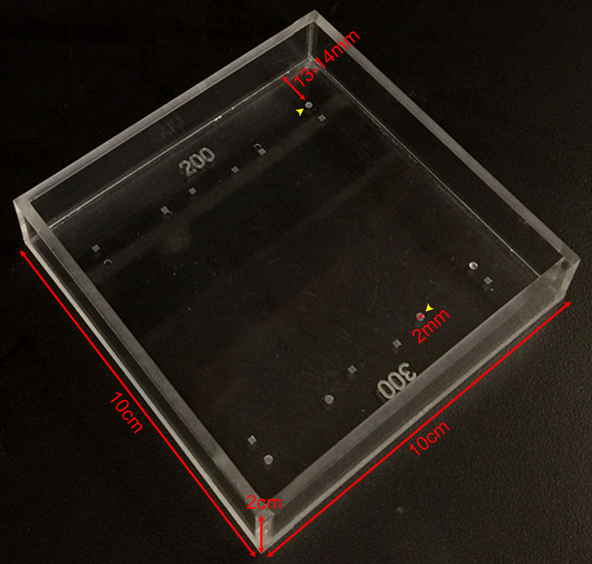
图1:刚度测量室示出了各种不同的深度和形状的草皮层的定做刚度测量室的尺寸的照片。圆形凹坑是200微米或300微米深(黄色箭头)被用于对小鼠透镜的测量结果。草皮是2毫米直径和〜13-从腔边缘14毫米请点击此处查看该图的放大版本。
- 放置直角镜进入室在从将被用于保持透镜的草皮层的恒定距离。确保镜在实验过程中不动。
- 转移解剖镜头测量室仔细抓住钳或弯钳。
- 取卸载透镜的顶视图图片从直接开销。取小鼠透镜照片在30X放大倍数下通过从解剖显微镜(底部),在左,右两侧光纤光源照明。设置光纤电源到最大光强度的80%。根据需要调整基于环境照明,用户偏好和画质电源输出。
- 取卸载透镜的侧视图的图片,这可以通过里可以看到GHT角度的镜子。如果相机未校准,采取在聚焦镜边缘的图片。镜边缘为5mm长,并且该测量可以在以后被用来确定像素/毫米,并作为图像中的比例尺。
- 镜头放置到草皮,并确认镜头在草皮安全,直就位。取装载前透镜的图象。该镜头应在其前或后极部草皮静养。
- 轻轻地放在1盖玻片上的镜头。等待2分钟,以允许蠕变,并采取装载镜头的另一侧查看图片。
- 继续加入盖玻片如在步骤2.8和服用侧视图图片加入每个盖玻片的如在步骤2.8之后,直到总共10盖玻片被应用。
- 删除所有盖玻片。等待2分钟,并除去所有盖玻片后取透镜(内部和外部的草皮的)的侧视图图片。
3.晶状体核测量
- 要determ的INE晶状体核大小,移动镜头到一个干净的培养皿中充满了1X PBS。
- 用细镊子直轻轻解封镜头。
- 脱落的皮质纤维细胞通过滚动手套的手指之间的镜头。剩下的晶状体核会感觉像一个硬的大理石。使用此过程来隔离成人镜头细胞核起1个月的年龄。因为分离的核是一个刚体,不能用此描述的方法进行晶状体核的进一步的机械测试。
- 轻轻漂洗在培养皿在1×PBS中的晶状体核。
- 放置晶状体核放回测量室(未在草皮),并采取晶状体核的图像通过直角反射镜。

图2:恩小鼠镜头由盖玻片压缩 (一)示意图和(B)的照片perimental设置表示在填充用1×PBS测量室200微米深草皮一个2个月大的小鼠透镜。直角反射镜和安装在解剖显微镜的数字照相机中使用由盖玻片压缩过程中收集透镜的图像。 (C)的通过连续增加盖玻片的数字压缩的2个月大的野生型透镜的矢状次照片用于测量轴向和赤道直径和基于盖玻片压缩测试期间计算轴向和赤道菌株提供的原始数据。透镜的反射有时可以在盖玻片(最清晰可见的1盖玻片图像中的)中看到。当进行测量时,忽略该反射并测量到透镜的顶点。 (D)的2个月大的野生型镜头后的压缩和隔离晶状体核矢状美景照片。该压缩后的镜头和孤立的核正坐在草皮之外。比例尺1毫米。这个数字是从Gokh修改在, 等 公共科学图书馆·之一 ,2012 19。 请点击此处查看该图的放大版本。
4.图像分析
- 装货前和使用ImageJ或类似软件的每一装载步骤后测量镜头赤道和轴向的直径。测量每个晶状体核的直径。晶状体核是近球形所以在任何方向的测量就足够了19,21。
- 通过增加使用的草皮的深度修正镜头直径轴。在测量室中,草皮层遮蔽了透镜的轴向厚度为200微米(2个月大的小鼠透镜)或300微米(4月龄和8个月大的小鼠镜头)。
- 使用方程,ε=(D - D 0)计算从透镜直径测量的轴向和赤道菌株/天0,其中ε为应变,d为轴向或电子邮件quatorial直径在给定的负载,而d 0是在零负载的相应的轴向或赤道直径。
- 画出轴向和赤道株作为施加的负荷(毫克)的功能。
- 绘制轴,赤道和核直径。计算并通过由赤道直径除以轴径绘制透镜纵横比。
- 计算和使用等式绘制透镜体积,体积= 4/3×π×R e 2 的 ×R A,其中R e为赤道半径和R a是从在步骤2.6拍摄的照片测量的轴向半径。这个公式假定镜头是一个扁球体(椭球)1.22。
- 计算并用公式,体积= 4/3×π×R N 3,其中R N是透镜核的半径为从在步骤3.5拍摄的照片测量绘制核体积。这个公式假定晶状体核我SA球19,21。
- 计算并绘制细胞核部分作为核体积与透镜体积的比值。
Access restricted. Please log in or start a trial to view this content.
结果
刚度和2-,4-和8个月大的小鼠的镜片的尺寸进行了测量。小鼠从TSRI动物育种设施获得的纯C57BL6菌株背景所有野生型动物,并且每一个透镜装载有1至10个盖玻片。轴向和赤道株计算为通过测量透镜的轴向和赤道直径在加入每个盖玻片的后,再归一直径为相应的卸载每个直径变化的施加载荷的功能。从每个年龄八个透镜进行了测试,结果表示为平均值±标准误差。如图先前...
Access restricted. Please log in or start a trial to view this content.
讨论
使用这种方法来衡量镜片硬度时,有几个关键因素。 ( - 8.5°8)相对于所述腔室(θ)的底部第一,盖玻片在略微倾斜的角度施加到透镜。这将适用于负载的非常小的组分平展,而不是轴向。然而,这种赤道负载可以忽略不计,因为罪θ≈0.1 19。如果此方法适于大透镜,盖玻片到腔室的底部的角将需要被测量以确定赤道负载是否应计入应变计算。其次,它是重要的,以允许透镜到加?...
Access restricted. Please log in or start a trial to view this content.
披露声明
作者什么都没有透露。
致谢
This work was supported by National Eye Institute Grant R01 EY017724 (VMF) and National Institute of Arthritis and Musculoskeletal and Skin Diseases Grant K99 AR066534 (DSG).
Access restricted. Please log in or start a trial to view this content.
材料
| Name | Company | Catalog Number | Comments |
| Fine tip straight forceps | Fine Scientific Tools | 11252-40 | |
| Microdissection scissors, straight edge | Fine Scientific Tools | 15000-00 | |
| Curved forceps | Fine Scientific Tools | 11272-40 | |
| Seizing forceps | Hammacher | HSC 702-93 | Optional |
| Dissection dish | Fisher Scientific | 12565154 | |
| 60 mm Petri dish | Fisher Scientific | 0875713A | |
| 1x phosphate buffered saline (PBS) | Life Technologies | 14190 | |
| 18 x 18 mm glass coverslips | Fisher Scientific | 12-542A | |
| Measurement chamber with divots to hold lenses | Custom-made (see Figure 1) | ||
| Right-angle mirror | Edmund Optics | 45-591 | |
| Light source | Schott/Fostec | 8375 | |
| Illuminated dissecting microscope | Olympus | SZX-ILLD100 | With SZ-PT phototube |
| Digital camera | Nikon | Coolpix 990 |
参考文献
- Lovicu, F. J., Robinson, M. L. Development of the ocular lens. , Cambridge University Press. (2004).
- Piatigorsky, J. Lens differentiation in vertebrates. A review of cellular and molecular features. Differentiation. 19 (3), 134-153 (1981).
- Glasser, A. Restoration of accommodation: surgical options for correction of presbyopia. Clin Exp Optom. 91 (3), 279-295 (2008).
- Keeney, A. H., Hagman, R. E., Fratello, C. J. Dictionary of ophthalmic optics. , Butterworth-Heinemann. (1995).
- Millodot, M. Dictionary of optometry and visual science. 7, Elsevier/Butterworth-Heinemann. (2009).
- Heys, K. R., Cram, S. L., Truscott, R. J. Massive increase in the stiffness of the human lens nucleus with age: the basis for presbyopia. Mol Vis. 10, 956-963 (2004).
- Heys, K. R., Friedrich, M. G., Truscott, R. J. Presbyopia and heat: changes associated with aging of the human lens suggest a functional role for the small heat shock protein, alpha-crystallin, in maintaining lens flexibility. Aging Cell. 6 (6), 807-815 (2007).
- Pierscionek, B. K. Age-related response of human lenses to stretching forces. Exp Eye Res. 60 (3), 325-332 (1995).
- Glasser, A., Biometric Campbell, M. C. optical and physical changes in the isolated human crystalline lens with age in relation to presbyopia. Vision Res. 39 (11), 1991-2015 (1999).
- Weeber, H. A., van der Heijde, R. G. On the relationship between lens stiffness and accommodative amplitude. Exp Eye Res. 85 (5), 602-607 (2007).
- Weeber, H. A., et al. Dynamic mechanical properties of human lenses. Exp Eye Res. 80 (3), 425-434 (2005).
- Fisher, R. F. Elastic properties of the human lens. Exp Eye Res. 11 (1), 143(1971).
- Krueger, R. R., Sun, X. K., Stroh, J., Myers, R. Experimental increase in accommodative potential after neodymium: yttrium-aluminum-garnet laser photodisruption of paired cadaver lenses. Ophthalmology. 108 (11), 2122-2129 (2001).
- Burd, H. J., Wilde, G. S., Judge, S. J. An improved spinning lens test to determine the stiffness of the human lens. Exp Eye Res. 92 (1), 28-39 (2011).
- Glasser, A., Campbell, M. C. Presbyopia and the optical changes in the human crystalline lens with age. Vision Res. 38 (2), 209-229 (1998).
- Pau, H., Kranz, J. The increasing sclerosis of the human lens with age and its relevance to accommodation and presbyopia. Graefes Arch Clin Exp Ophthalmol. 229 (3), 294-296 (1991).
- Hollman, K. W., O'Donnell, M., Erpelding, T. N. Mapping elasticity in human lenses using bubble-based acoustic radiation force. Exp Eye Res. 85 (6), 890-893 (2007).
- Baradia, H., Nikahd, N., Glasser, A. Mouse lens stiffness measurements. Exp Eye Res. 91 (2), 300-307 (2010).
- Gokhin, D. S., et al. Tmod1 and CP49 synergize to control the fiber cell geometry, transparency, and mechanical stiffness of the mouse lens. PLoS One. 7 (11), e48734(2012).
- Sindhu Kumari, S., et al. Role of Aquaporin 0 in lens biomechanics. Biochem Biophys Res Commun. , (2015).
- Fudge, D. S., et al. Intermediate filaments regulate tissue size and stiffness in the murine lens. Invest Ophthalmol Vis Sci. 52 (6), 3860-3867 (2011).
- Kuszak, J. R., Mazurkiewicz, M., Zoltoski, R. Computer modeling of secondary fiber development and growth: I. Nonprimate lenses. Mol Vis. 12, 251-270 (2006).
- Scarcelli, G., Kim, P., Yun, S. H. In vivo measurement of age-related stiffening in the crystalline lens by Brillouin optical microscopy. Biophys J. 101 (6), 1539-1545 (2011).
Access restricted. Please log in or start a trial to view this content.
转载和许可
请求许可使用此 JoVE 文章的文本或图形
请求许可探索更多文章
This article has been published
Video Coming Soon
版权所属 © 2025 MyJoVE 公司版权所有,本公司不涉及任何医疗业务和医疗服务。