
JoVE 비디오를 활용하시려면 도서관을 통한 기관 구독이 필요합니다. 전체 비디오를 보시려면 로그인하거나 무료 트라이얼을 시작하세요.
Method Article
마우스 렌즈의 압축 강성을 평가하는 유리 Coverslips는 순차적으로 응용 프로그램 : 스트레인과 형태 학적 분석
요약
Age-related increases in eye lens stiffness are linked to presbyopia. This protocol describes a simple, cost-effective method for measuring mouse lens stiffness. Mouse lenses, like human lenses, become stiffer with age. This method is precise and can be adapted for lenses from larger animals.
초록
눈 렌즈는 굴절 및 망막에 선명한 화상을 형성하는 광을 집중 투명 기관이다. 인간, 모양체 근육 계약은 가까운 물체에 초점 렌즈 '광 파워, 숙박 시설로 알려진 공정의 증가로 이어지는, 렌즈를 변형합니다. 렌즈 강성 연령 관련 변경 사항이 노안에 연결되었습니다 확장하여 수용 할 수있는 렌즈의 능력 감소하고, 안경을 독서의 필요성. 마우스 렌즈 수용 노안을 개발하지 않더라도, 마우스 모델 이해 렌즈 병리위한 귀중한 유전 적 수단을 제공 할 수 있으며, 마우스에서 관찰 된 가속 열화 렌즈의 연령과 관련된 변화에 대한 연구를 가능하게한다. 이 프로토콜은, 마우스 렌즈 강도를 결정 순차적으로 적용되는 렌즈에 압축 하중을 증가시키는 커버 글라스를 사용하는 단순하고 정확하고 비용 효율적인 방법을 설명한다. 대표적인 데이터는 마우스 렌즈처럼 나이가 엄격한되고 있는지 확인인간의 렌즈. 이 방법은 높은 재현성과 잠재적으로 더 큰 동물 시험 렌즈를 기계적으로까지 확장 할 수 있습니다.
서문
The lens is a transparent and avascular organ in the anterior chamber of the eye that is responsible for fine focusing of light onto the retina. A clear basement membrane, called the lens capsule, surrounds a bulk of elongated fiber cells covered by an anterior monolayer of epithelial cells1,2. Life-long growth of the lens depends on the continuous proliferation and differentiation of epithelial cells at the lens equator into new fiber cells that are added onto previous generations of fiber cells in a concentric manner2. Over time, lens fiber cells undergo compaction, resulting in a rigid center in the middle of the lens called the nucleus1. Accommodation, defined as a dioptric change in the optical power of the eye, occurs in humans when the ciliary muscles contract to deform the lens and allow a true increase in optical power to focus on near objects3-5. In the unaccommodated eye, the lens is held in a relatively flattened state due to tension from zonular fibers. When the ciliary muscles contract, the tension on the lens is released, leading to decreased lens equatorial diameter and increased axial thickness. Age-related changes in the lens cause presbyopia, a progressive loss of lens accommodation, which leads to the need for reading glasses.
Several studies have linked presbyopia to age-related increase in the intrinsic stiffness of the lens6-11. Stiffness is defined as the resistance of an elastic object to deform under applied load. A variety of methods have been used to examine stiffness of human lenses, including spin compression12-14, actuator compression15, probe indentation16, dynamic mechanical analysis 6,10 and bubble-based acoustic radiation force17. While mouse lenses do not accommodate or develop presbyopia, mouse models for lens pathologies are valuable tools because mice are less expensive than larger animals, well characterized genetically and undergo accelerated age-related changes due to rapid aging. A handful of studies have examined mouse lens stiffness with compression methods and demonstrated changes in lens stiffness due to aging or targeted genetic disruptions18-21. Thus, mouse lenses are good models for studying age-related changes in lens stiffness.
This protocol describes a simple and inexpensive, yet precise and reproducible, compression method for determining mouse lens stiffness based on application of glass coverslips onto the lens in conjunction with photographing the lens through a dissection microscope and mirror. This method yields robust strain and morphometric data with an easily fabricated and assembled apparatus. The representative results confirm that mouse lenses increase in stiffness with age.
Access restricted. Please log in or start a trial to view this content.
프로토콜
모든 동물의 절차는 스크립스 연구소의 기관 동물 케어 및 사용위원회의 보건 및 승인 된 프로토콜에 따라 국립 연구소에 의해 실험실 동물의 관리 및 사용을위한 설명서의 권장 사항에 따라 수행 하였다.
1. 렌즈 해부
- 건강 "실험 동물의 관리 및 사용을위한 가이드"의 국립 연구소 및 승인 기관 동물 사용 프로토콜의 권장 사항에 따라 쥐를 안락사.
- 곡선 집게를 사용하여 마우스의 눈을 명백히하다. 소켓에서 눈을 가지고, 다음 집게로 소켓에서 눈을 뽑은 포셉과 눈 주위의 조직을 우울. 해부 접시에 신선한 1X 인산염 완충 식염수 (PBS)에 눈을 전송합니다.
- 가능한 안구에 가까운 시신경을 잘라. 조심스럽게 조심스럽게 구멍 wher을 통해 안구에 잘 직선 핀셋을 삽입전자 시신경은 후방을 종료합니다.
- 조심스럽게 각막의 가장자리에 후방에서 안구에 가위로 절개를합니다. 설치류 렌즈는 눈의 ~ 30 %를 차지한다. 주의 깊게 절개를 확인하고 렌즈 손상을 방지하기 위해 너무 깊은 눈에 핀셋이나 가위를 삽입하지 않습니다.
- 안구 주위에 적어도 절반 방법의 각막과 공막 사이의 접합을 따라 잘라.
- 부드럽게 단계 1.4 및 1.5에서 만든 개구를 통해 안구로부터 렌즈를 제거하기 위해 각막에 누른다.
- 주의 깊게 여전히 렌즈에 부착되어있는 큰 이물질을 제거하기 위해 좋은 팁 직선의 집게를 사용합니다. 시각적으로 강성 측정을 진행하기 전에 손상은 렌즈를 검사합니다.
2. 강성 측정
- 분석 용 저울을 사용하여 동일한 박스에서 적어도 10 커버 슬립을 단다. 커버 슬립의 평균 무게를 찾을 수 있습니다. 일관성을 위해 모든 실험 된 커버와 동일한 상자를 사용합니다. 사전 젖은실험을 시작하기 전에 적어도 2 시간 동안 실온에서 1X PBS에서 커버 슬립 및 직각 미러.
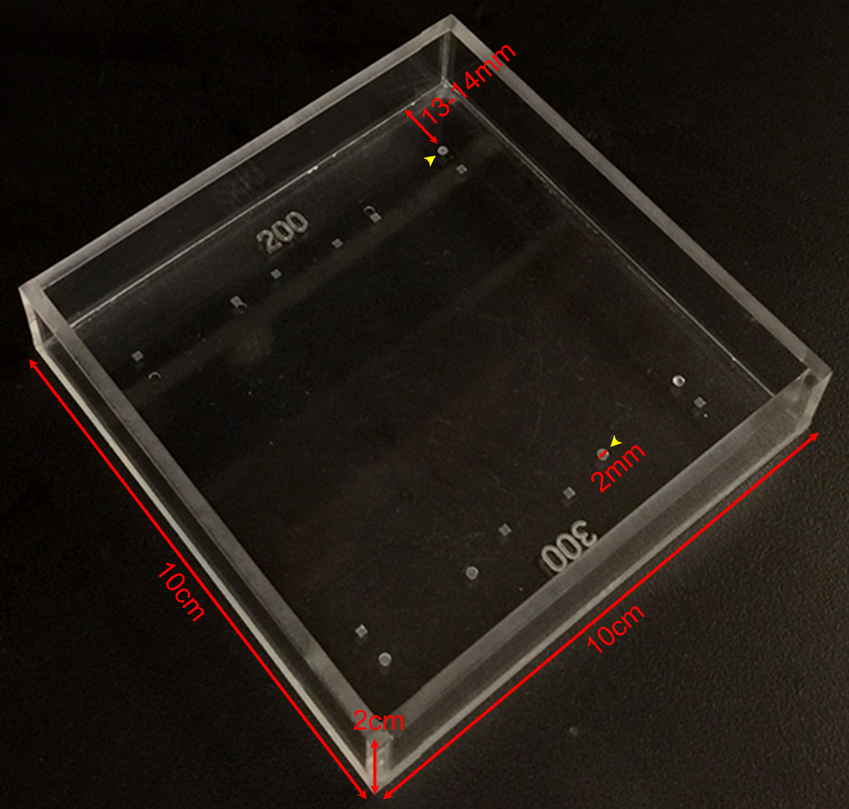
- 1X PBS 75 mL로 - 측정 챔버를 채우기 (65) (도 1 참조). 측정 챔버는 사내 기계 공장으로 플렉시 유리로 만든 후, 챔버 내의 디 보트는 적절한 드릴 비트와 함께 원하는 깊이로 설정 드릴 프레스에 의해 만들어졌다. 렌즈 기계적 시험 기간 동안 실온에서 1X PBS 투명 남아있다.

그림 1 :. 강성 측정 상공 회의소 다른 깊이와 모양의 디 보트의 다양한 사용자 정의 만든 강성 측정 챔버의 크기를 보여주는 사진입니다. 200 μm의 300 μm의 깊이 (노란색 화살촉) 인 라운드 디 보트는 마우스 렌즈의 측정에 사용된다. 디 보트는 2 직경 mm 및 ~ 13입니다-. 챔버의 가장자리에서 14mm 이 그림의 더 큰 버전을 보려면 여기를 클릭하십시오.
- 렌즈를 보유하는 데 사용되는 잔디에서 일정한 거리에있는 실에 직각 거울을 배치합니다. 거울은 실험 기간 동안 이동하지 않습니다 있는지 확인하십시오.
- 신중하게 장악 겸자 또는 곡선 집게로 측정 챔버로 전송 해부 렌즈.
- 직접 오버 헤드 무부하 렌즈의 상위 뷰 사진을 촬영합니다. 현미경 (아래)와 좌우의 광섬유 광원 해부에서 조명으로 30X 배율로 마우스 렌즈 사진을 찍어. 최대 광 강도의 80 %로 광섬유 전원 공급 장치를 설정한다. 필요에 따라 주변 조명, 사용자 선호도 및 화질에 기초하여 상기 전원 공급 장치의 출력을 조정한다.
- 로타리를 통해 알 수있는 무부하 렌즈의 측면도 픽쳐를 취할GHT 각도 거울. 카메라가 보정되지 않은 경우 초점 미러 에지의 사진을 촬영. 거울 가장자리 5 mm 길이, 그리고이 측정 이후의 화소 / mm를 결정하고 이미지의 스케일 바의 역할을하기 위해 사용될 수있다.
- 장소 상기 잔디에 렌즈와 렌즈가 잔디에 안전하고 바로 장착되어 있는지 확인합니다. 로드하기 전에 렌즈의 사진을 촬영합니다. 렌즈는 전방 또는 후방 극의 잔디에서 휴식해야한다.
- 렌즈에 부드럽게 한 커버 슬립을 놓습니다. 크리프를 허용하는 2 분을 기다린로드 렌즈의 또 다른 사이드 뷰 사진을 촬영합니다.
- 단계 2.8로 된 커버를 추가하고 10 커버 슬립의 총이 적용될 때까지 단계 2.8에서와 같이 각 커버 슬립을 첨가 한 후 사이드 뷰 사진 촬영을 계속.
- 모든 커버 슬립을 제거합니다. 2 분을 기다린 모든 된 커버를 제거한 후 렌즈 (내부와 외부 잔디의)의 사이드 뷰 사진을 촬영합니다.
3. 렌즈 핵 측정
- determ하려면렌즈 핵의 크기 이네, 1X PBS로 가득 깨끗한 페트리 접시에 렌즈를 이동합니다.
- 부드럽게 잘 직선 집게를 사용하여 렌즈를 디 캡슐.
- 슬라 오프 장갑을 낀 손가락으로 렌즈를 압연에 의한 대뇌 피질의 섬유 세포. 나머지 렌즈 핵 하드 대리석처럼 느낄 것이다. 세 1 개월부터 성인 렌즈의 핵을 분리하려면이 절차를 사용하십시오. 고립 핵 강체이기 때문에, 렌즈 핵의 상기 기계적 시험이 설명 된 방법을 사용하여 수행 될 수 없다.
- 조심스럽게 페트리 접시에 1X PBS의 렌즈 핵을 씻어.
- 다시 측정 챔버 (안 잔디에서)에 렌즈 핵을 배치하고 오른쪽 각도 거울을 통해 렌즈 핵의 이미지를 촬영.

그림 2 :. 전의 Coverslips는에 의해 압축 된 마우스 렌즈 (A) 도식과 (B) 사진1X PBS로 가득 측정 챔버에서 200 μm의 깊은 잔디에 2 개월 된 마우스 렌즈를 보여주는 perimental 설정. 직각 미러 및 해부 현미경에 장착 된 디지털 카메라에 의해 압축 된 커버하는 동안 렌즈의 이미지를 수집 하였다. (C)를 순차 커버 슬립의 수를 증가시킴으로써 압축 2 개월 야생형 렌즈 시상 뷰 사진 축 적도 직경을 측정하고, 커버 슬립 기반 압축 테스트 중에 축 적도 변형 계산을위한 미가공 데이터를 제공했다. 렌즈의 반사는 때로 커버 슬립 (가장 눈에 잘 한 커버 슬립 이미지)에서 볼 수있다. 측정을 할 때의 반사를 무시하고, 렌즈의 정점까지 측정한다. (D) 상기 2 개월 야생형 렌즈 압축 후의 상기 절연 렌즈 핵의 시상 뷰 사진. 포스트 압축 렌즈와 격리 된 핵은 잔디 외부에 앉아있다. 스케일 바, 1mm. 이 수치는 Gokh에서 수정, 등. PLoS의 하나 2012 년 19. 이 그림의 더 큰 버전을 보려면 여기를 클릭하십시오.
4. 이미지 분석
- 로딩 전에 ImageJ에 또는 유사한 소프트웨어를 이용하여 각각의 적재 단계 후 렌즈 적도 및 축 지름을 측정한다. 각 렌즈 핵의 직경을 측정한다. 어떤 방향에서 측정 19, 21을 충분하므로 렌즈 핵은 거의 구형이다.
- 사용 된 잔디의 깊이를 첨가하여 직경 축 렌즈를 수정한다. 측정 챔버에서 디봇은 렌즈의 축 방향 두께가 200㎛ 인 (2 개월 된 마우스 렌즈) 또는 300 ㎛의 (4 개월, 8 개월 된 마우스 렌즈)을 가려.
- , ε = (D - D 0) 식을 이용하여 렌즈 직경 측정으로부터 축 적도 균주 계산 ε 스트레인 인 / D 0, D는 수직 또는 E이고quatorial 주어진 부하에서 직경 및 D 0 제로 부하에 대응하는 수직 또는 수평 직경입니다.
- (밀리그램)에 부과 된 하중의 함수와 축과 적도 균주를 플롯.
- 축, 적도 핵 직경을 플롯. 계산하고 적도 지름 축 직경 나누어 렌즈 종횡비 플롯.
- 계산 방정식을 사용하여 렌즈 볼륨 플롯 부피 = R의 E는 적도 반경 R A는 단계 2.6에서 찍은 사진으로부터 측정 축 반경이다 4/3 × π × R E × 2 (R) A,. 이 방정식은 렌즈가 편원의 회전 타원체 (타원체) 1,22 가정합니다.
- 계산 단계 3.5에서 찍은 사진에서 측정 된 R N 렌즈 핵의 반경 (R)의 N 3 × 식 체적 = 4/3 × π의 핵을 사용하여 부피를 플롯. 이 방정식은, 렌즈 핵 제가 가정SA 구 19, 21.
- 계산 렌즈 볼륨 핵 부피의 비로 핵 분획을 플롯.
Access restricted. Please log in or start a trial to view this content.
결과
강성 및 2-, 4-, 8 개월 된 마우스 렌즈의 크기를 측정 하였다. 마우스는 TSRI 동물 사육 시설에서 얻은 순수한 C57BL6 변형 배경 모두 야생형 동물이었고, 각각의 렌즈는 1~10 커버 슬립 적재 하였다. 축 적도 균주 각 커버 슬립을 첨가 한 후, 렌즈의 축 적도 직경을 측정 한 다음 해당 언로드 직경 직경 각 변화를 정규화하여 하중의 함수로서 계산 하였다. 각 세부터 여덟 렌즈를 ?...
Access restricted. Please log in or start a trial to view this content.
토론
렌즈의 강도를 측정하는이 방법을 사용할 때 몇 가지 중요한 고려 사항이 있습니다. 챔버 (θ)의 저부에 대하여 - (8.5 ° 8) 우선, 커버 슬립 약간 비스듬한 각도로 렌즈에 적용된다. 이 적도에 축보다는 오히려 부하의 매우 작은 구성 요소를 적용한다. 그러나이 적도 부하가 죄 θ ≈ 0.1 (19) 때문에 무시할 간주됩니다. 이 방법은 큰 렌즈에 적합한 경우, 상기 챔버의 하부에 커버 슬립 각도 적?...
Access restricted. Please log in or start a trial to view this content.
공개
저자는 공개 아무것도 없어.
감사의 말
This work was supported by National Eye Institute Grant R01 EY017724 (VMF) and National Institute of Arthritis and Musculoskeletal and Skin Diseases Grant K99 AR066534 (DSG).
Access restricted. Please log in or start a trial to view this content.
자료
| Name | Company | Catalog Number | Comments |
| Fine tip straight forceps | Fine Scientific Tools | 11252-40 | |
| Microdissection scissors, straight edge | Fine Scientific Tools | 15000-00 | |
| Curved forceps | Fine Scientific Tools | 11272-40 | |
| Seizing forceps | Hammacher | HSC 702-93 | Optional |
| Dissection dish | Fisher Scientific | 12565154 | |
| 60 mm Petri dish | Fisher Scientific | 0875713A | |
| 1x phosphate buffered saline (PBS) | Life Technologies | 14190 | |
| 18 x 18 mm glass coverslips | Fisher Scientific | 12-542A | |
| Measurement chamber with divots to hold lenses | Custom-made (see Figure 1) | ||
| Right-angle mirror | Edmund Optics | 45-591 | |
| Light source | Schott/Fostec | 8375 | |
| Illuminated dissecting microscope | Olympus | SZX-ILLD100 | With SZ-PT phototube |
| Digital camera | Nikon | Coolpix 990 |
참고문헌
- Lovicu, F. J., Robinson, M. L. Development of the ocular lens. , Cambridge University Press. (2004).
- Piatigorsky, J. Lens differentiation in vertebrates. A review of cellular and molecular features. Differentiation. 19 (3), 134-153 (1981).
- Glasser, A. Restoration of accommodation: surgical options for correction of presbyopia. Clin Exp Optom. 91 (3), 279-295 (2008).
- Keeney, A. H., Hagman, R. E., Fratello, C. J. Dictionary of ophthalmic optics. , Butterworth-Heinemann. (1995).
- Millodot, M. Dictionary of optometry and visual science. 7, Elsevier/Butterworth-Heinemann. (2009).
- Heys, K. R., Cram, S. L., Truscott, R. J. Massive increase in the stiffness of the human lens nucleus with age: the basis for presbyopia. Mol Vis. 10, 956-963 (2004).
- Heys, K. R., Friedrich, M. G., Truscott, R. J. Presbyopia and heat: changes associated with aging of the human lens suggest a functional role for the small heat shock protein, alpha-crystallin, in maintaining lens flexibility. Aging Cell. 6 (6), 807-815 (2007).
- Pierscionek, B. K. Age-related response of human lenses to stretching forces. Exp Eye Res. 60 (3), 325-332 (1995).
- Glasser, A., Biometric Campbell, M. C. optical and physical changes in the isolated human crystalline lens with age in relation to presbyopia. Vision Res. 39 (11), 1991-2015 (1999).
- Weeber, H. A., van der Heijde, R. G. On the relationship between lens stiffness and accommodative amplitude. Exp Eye Res. 85 (5), 602-607 (2007).
- Weeber, H. A., et al. Dynamic mechanical properties of human lenses. Exp Eye Res. 80 (3), 425-434 (2005).
- Fisher, R. F. Elastic properties of the human lens. Exp Eye Res. 11 (1), 143(1971).
- Krueger, R. R., Sun, X. K., Stroh, J., Myers, R. Experimental increase in accommodative potential after neodymium: yttrium-aluminum-garnet laser photodisruption of paired cadaver lenses. Ophthalmology. 108 (11), 2122-2129 (2001).
- Burd, H. J., Wilde, G. S., Judge, S. J. An improved spinning lens test to determine the stiffness of the human lens. Exp Eye Res. 92 (1), 28-39 (2011).
- Glasser, A., Campbell, M. C. Presbyopia and the optical changes in the human crystalline lens with age. Vision Res. 38 (2), 209-229 (1998).
- Pau, H., Kranz, J. The increasing sclerosis of the human lens with age and its relevance to accommodation and presbyopia. Graefes Arch Clin Exp Ophthalmol. 229 (3), 294-296 (1991).
- Hollman, K. W., O'Donnell, M., Erpelding, T. N. Mapping elasticity in human lenses using bubble-based acoustic radiation force. Exp Eye Res. 85 (6), 890-893 (2007).
- Baradia, H., Nikahd, N., Glasser, A. Mouse lens stiffness measurements. Exp Eye Res. 91 (2), 300-307 (2010).
- Gokhin, D. S., et al. Tmod1 and CP49 synergize to control the fiber cell geometry, transparency, and mechanical stiffness of the mouse lens. PLoS One. 7 (11), e48734(2012).
- Sindhu Kumari, S., et al. Role of Aquaporin 0 in lens biomechanics. Biochem Biophys Res Commun. , (2015).
- Fudge, D. S., et al. Intermediate filaments regulate tissue size and stiffness in the murine lens. Invest Ophthalmol Vis Sci. 52 (6), 3860-3867 (2011).
- Kuszak, J. R., Mazurkiewicz, M., Zoltoski, R. Computer modeling of secondary fiber development and growth: I. Nonprimate lenses. Mol Vis. 12, 251-270 (2006).
- Scarcelli, G., Kim, P., Yun, S. H. In vivo measurement of age-related stiffening in the crystalline lens by Brillouin optical microscopy. Biophys J. 101 (6), 1539-1545 (2011).
Access restricted. Please log in or start a trial to view this content.
재인쇄 및 허가
JoVE'article의 텍스트 или 그림을 다시 사용하시려면 허가 살펴보기
허가 살펴보기더 많은 기사 탐색
This article has been published
Video Coming Soon
Copyright © 2025 MyJoVE Corporation. 판권 소유