Case Report
Epatectomia laparoscopica S7 con colorazione a fluorescenza positiva
In questo articolo
Riepilogo
Il protocollo dimostra che, sotto guida ecografica laparoscopica, il segmento S7 del fegato può essere colorato con successo perforando i rami viscerale e diaframmatico del tumore, facilitando un'epatectomia anatomica del segmento S7.
Abstract
La resezione epatica laparoscopica per i tumori localizzati nel segmento S7 del fegato adotta tipicamente un approccio chirurgico tradizionale. L'obiettivo principale di questa procedura è sezionare accuratamente il peduncolo epatico del segmento S7. La dissezione del peduncolo epatico del segmento S7 lungo l'ilo epatico richiede un percorso relativamente lungo all'interno del fegato, che aumenta il rischio di perdere l'orientamento e potenzialmente di danneggiare il peduncolo epatico adiacente dei segmenti S5 e S6, compromettendo così il piano di resezione epatica. Un metodo di colorazione positiva è stato utilizzato per perforare direttamente la vena porta corrispondente sotto guida ecografica (comunemente per i segmenti S7 e S8) per colorare in modo specifico il segmento epatico bersaglio, evitando così un'estesa resezione del parenchima epatico e riducendo il danno al tessuto epatico sano circostante. Tuttavia, il metodo di colorazione positiva richiede una base specifica nelle procedure intraoperatorie, che possono essere impegnative per i chirurghi e hanno una certa curva di apprendimento. Attualmente, tecnologie come l'analisi del territorio di ricostruzione tridimensionale, l'ecografia intraoperatoria e l'imaging a fluorescenza con verde indocianina sono popolari e comunemente utilizzate nella resezione epatica laparoscopica. In questo protocollo, sotto guida ecografica laparoscopica, il bacino tumorale è stato perforato attraverso le superfici viscerali e diaframmatiche del fegato per colorare il segmento S7. La resezione laparoscopica del segmento S7 all'interno del fegato anatomico del territorio portale è stata eseguita con successo, confermando ulteriormente la fattibilità e i vantaggi della colorazione a fluorescenza positiva nella resezione epatica laparoscopica in questa fase.
Introduzione
Il carcinoma epatico primario è attualmente il quarto tumore maligno più comune e la seconda causa di morte correlata al cancro in Cina, rappresentando una seria minaccia per la vita e la salute delle persone a livello globale1. Per il carcinoma epatocellulare (HCC), la resezione chirurgica è stata a lungo l'opzione di trattamento primaria. Con i progressi della tecnologia minimamente invasiva, c'è stato un numero crescente di segnalazioni sulla resezione epatica anatomica laparoscopica per il trattamento dell'HCC. Per l'epatectomia laparoscopica localizzata in vari segmenti epatici, compresi i segmenti specializzati (I, IVb, VII e VIII), studi pertinenti hanno dimostrato che questo metodo è sicuro ed efficace2.
Il concetto di resezione epatica anatomica è stato proposto per la prima volta da Makuuchi et al. nel 1985 3,4. La procedura corretta consiste nel segmentare in base alla colorazione del territorio della vena porta ed eseguire la resezione completa della colorazione del territorio della vena porta a cui appartiene il tumore5. Poiché l'HCC si diffonde principalmente lungo la vena porta, in teoria, questo approccio può fornire una migliore efficacia oncologica e ottenere una vera resezione epatica anatomica per tumori in diverse sedi6. Tuttavia, in passato, a causa delle limitazioni della tecnologia e delle attrezzature, questo trattamento era raro. La maggior parte dei centri ha eseguito la resezione epatica anatomica basata sul metodo di segmentazione epatica di Couinaud. Quando il tumore si estende su più segmenti epatici, l'esecuzione di una resezione epatica anatomica può comportare un'eccessiva rimozione di tessuto epatico sano, aumentando così il rischio chirurgico e le complicanze postoperatorie. Inoltre, le potenziali lesioni micrometastatiche possono persistere a causa della resezione incompleta del territorio della vena porta tumorale 7,8.
Con i progressi della tecnologia e delle attrezzature, possiamo definire il territorio della vena porta del tumore sulla base della ricostruzione tridimensionale preoperatoria. Questo aiuta i medici a determinare l'intervallo di resezione, eseguire la puntura ecoguidata e la colorazione dei rami di colorazione del territorio della vena porta a cui appartiene il tumore durante l'intervento chirurgico e determinare il piano della sezione epatica utilizzando l'imaging a fluorescenza verde indocianina per ottenere una resezione epatica anatomica più accurata9. Tuttavia, per la resezione epatica anatomica laparoscopica del segmento epatico S7, l'operazione è più impegnativa perché il peduncolo epatico è profondamente nascosto nel parenchima epatico e localizzato prossimalmente ai lati dorsale e cefalico, determinando così un tempo operatorio più lungo e un trauma maggiore10. Il metodo di colorazione positiva prevede la puntura diretta della vena porta corrispondente sotto guida ecografica (tipicamente utilizzata nei segmenti epatici S7 e S8) in modo che il segmento epatico bersaglio sia colorato direttamente. Questo aiuta il chirurgo a evitare di tagliare una quantità eccessiva di parenchima epatico, massimizzando così la protezione del volume epatico funzionale11. Tuttavia, il metodo di colorazione positiva richiede una specifica base ecografica intraoperatoria, la corretta identificazione dei dotti intraepatici e l'uso appropriato delle tecniche di puntura intraoperatoria della vena porta. Inoltre, pone un'elevata richiesta al chirurgo ed è associato a una curva di apprendimento prerequisito.
Nel paziente qui descritto, il tumore era localizzato nel segmento S7 del fegato. La ricostruzione tridimensionale preoperatoria ha rivelato due rami della vena porta. Poiché il tronco S7 è corto e vicino alla radice della vena porta del segmento S6, è stato perforato lungo le superfici diaframmatiche e viscerali del fegato. Il verde di indocianina è stato iniettato per colorare il segmento epatico bersaglio e il fegato è stato resecato seguendo il segnale fluorescente che ha guidato la procedura e ha assicurato un funzionamento regolare.
Lo scopo del metodo di resezione del segmento S7 del fegato qui dimostrato è quello di promuovere ulteriormente il concetto di resezione epatica anatomica guidata dalla colorazione del territorio portale ed evidenziare i vantaggi della resezione con colorazione positiva del segmento S7. Questa procedura riduce al minimo il volume di tessuto epatico sano rimosso durante la resezione del tumore, massimizzando al contempo l'efficienza della rimozione del tumore.
PRESENTAZIONE DEL CASO:
Un uomo di 30 anni è stato ricoverato all'ospedale Foshan Fosun Chancheng il 2023-02-02. Il paziente è stato trovato con una lesione che occupa spazio nel fegato in un altro ospedale 1 mese prima, senza disagio. Per il resto aveva una storia di buona salute.
Diagnosi, valutazione e pianificazione:
Diagnosi: Carcinoma epatocellulare.
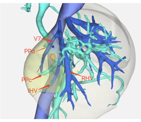
Valutazione: ALT (alanina aminotransferasi): 123 U/L, AST (aspartato aminotransferasi): 34 U/L, emoglobina: 141 g/L, conta piastrinica: 125 x 109 cellule/L, albumina: 40,5 g/L, bilirubina totale: 10,1 μmol/L, creatinina: 67 μmol/L, tempo di protrombina (PT): 14,1 s, antigene di superficie dell'epatite B positivo, HBV DNA (DNA del virus dell'epatite B): 3,51 x 10⁶ UI/L, protrombina anormale (PIVKA-II): 21 mAU/mL, AFP (alfa-fetoproteina): 56,29 μg/L, CA199 (antigene dei carboidrati199): <0,8 U/mL, CEA (antigene carcinoembrionale): 4,65 U/mL, colinesterasi: 7128 U/L, grado A di Child-Pugh. TC (tomografia computerizzata) e risonanza magnetica (risonanza magnetica) potenziata (gadoxetato disodico) dell'addome superiore: 1 cm di massa nel segmento S7 del fegato, ricostruzione tridimensionale, analisi del territorio (vedi Figura 1). Il volume epatico rimanente era del 78,8%.

Figura 1: Analisi della ricostruzione tridimensionale. La posizione del tumore, la ricostruzione tridimensionale del portale e delle vene epatiche correlate al tumore e i vasi sanguigni importanti vicino al tumore. Abbreviazioni: v7 = segmenti di 7 rami di una vena epatica; PPC = vena porta posteriore C; PPD = vena porta posteriore D; IHV = vena epatica interterritoriale; RHV = vena epatica destra. Clicca qui per visualizzare una versione più grande di questa figura.
Piano: è stata pianificata l'epatectomia laparoscopica S7 con colorazione positiva alla fluorescenza. Fase 1: La TC e la ricostruzione tridimensionale sono state utilizzate per l'analisi del territorio. Il tumore era localizzato nei territori della vena porta posteriore C (PPc) e della vena porta posteriore D (PPd) del segmento S7 del fegato (riferendosi alla classificazione della vena porta posteriore destra degli studiosi giapponesi4; fare riferimento alla Figura 2). Fase 2: L'ecografia intraoperatoria ha mostrato che la vena porta a cui apparteneva il tumore aveva due rami vascolari. Fase 3: I peduncoli epatici bersaglio PPc e PPd sono stati recisi e le vene tra le regioni epatiche S6 e S7 e la vena epatica destra sono state completamente esposte sotto guida fluorescente. Passaggio 4: resezione del tumore guidata dalla colorazione fluorescente.

Figura 2. TAC preoperatoria. (A) Sezione TC della vena porta associata al tumore di PPd (freccia rossa). (B) Sezione TC della vena porta associata al tumore di PPc. Clicca qui per visualizzare una versione più ampia di questa figura.
Protocollo
Questo protocollo ha seguito le linee guida del Comitato Etico per la Ricerca Umana dell'Ospedale Foshan Fosun Chancheng. Il consenso informato scritto è stato ottenuto dal paziente per la partecipazione a questo studio.
1. Preparazione preoperatoria
- Preparazione del paziente: Il paziente è stato posto in posizione supina, con la testa sollevata e i piedi abbassati e inclinati a sinistra di circa 30°. È stata somministrata l'anestesia generale, compresa l'intubazione tracheale. Sono stati eseguiti la disinfezione addominale e il drappeggio dell'area chirurgica.
- Disposizione del trocar: un trocar di 1,2 cm (foro di osservazione) è stato inserito dopo aver tagliato la pelle orizzontalmente 1 cm a destra dell'ombelico con un coltello chirurgico. Quindi, un trocar di 1,0 cm è stato inserito all'intersezione della linea medioclavicolare, 5 cm sotto il margine costale destro, un trocar di 0,5 cm è stato inserito sotto il margine costale destro e l'ascella, un trocar di 1,2 cm è stato inserito sotto il processo xifoideo e un trocar di 0,5 cm 3 cm a sinistra del punto medio della linea che collega l'ombelico e il processo xifoideo. Il chirurgo sta alla destra e l'assistente alla sinistra del paziente. La telecamera è stata posizionata nel foro di osservazione.
- Esplorazione addominale: sono state eseguite ecografie intraoperatorie lungo le vene portale ed epatiche per determinare la relazione tra il tumore e il dotto, confermata dalla ricostruzione tridimensionale. L'esplorazione laparoscopica del fegato e della cavità addominale non ha rivelato altre lesioni o metastasi. La localizzazione ecografica del portale anteriore (AP) e della PP ha rivelato che la PP era di tipo B.
NOTA: L'ecografia intraoperatoria valuta le dimensioni del tumore, la posizione, le metastasi intraepatiche e la sua relazione con i vasi sanguigni circostanti.
2. Procedura chirurgica
- Separazione del legamento periepatico: un coltello ad ultrasuoni è stato utilizzato per tagliare i legamenti rotondi e falciformi del fegato. Il secondo portale epatico è stato sezionato, esponendo la radice della vena epatica destra. Il legamento coronarico destro e il legamento triangolare destro sono stati tagliati. Un punto è stato utilizzato per legare le tre vene epatiche corte dorsalmente al lato destro della vena cava inferiore per liberare completamente il fegato destro.
- Banda di occlusione: un coltello ad ultrasuoni è stato utilizzato per liberare le aderenze intorno alla cistifellea per esporre il forame dei Venturi. Le pinze gastriche gastriche sono state utilizzate lungo il forame di Venturi e una fascia di occlusione è stata posizionata nel primo portale epatico.
- Puntura e resezione
- La PPc era visibile attraverso la porta operativa principale destra sulla superficie diaframmatica e la PPd era visibile sulla superficie viscerale. La PPc e la PPd sono state perforate utilizzando la guida dell'ecografia intraoperatoria con una sonda e un foro di puntura.
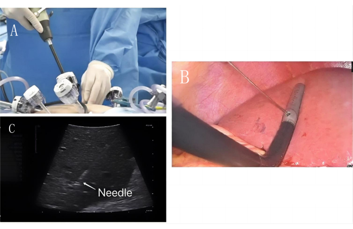
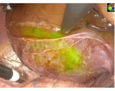
- Puntura della superficie diaframmatica (Figura 3 e Figura 4): la sonda è stata inserita nel foro operativo principale sotto il margine costale destro. La PPC era visibile sulla superficie diaframmatica e il lungo diametro della PPC era esposto. È stato selezionato il punto di puntura dalla radice del PPC. Per pungere il dotto biliare è stato utilizzato un ago per colangia transepatica percutanea (PTC) da 21G. È stato utilizzato il metodo con una faccia, tre punte e quattro dita orizzontali.
- Da un lato, il punto medio delle aste di regolazione sinistra e destra è stato utilizzato per gli ultrasuoni intraoperatori come punto di mira e l'asta della sonda è stata utilizzata come piano verticale per la puntura in piano. I tre punti selezionati sono stati il punto di puntura della pelle, il foro di puntura della sonda intraoperatoria e il punto di puntura del peduncolo epatico bersaglio. La lunghezza delle quattro dita orizzontali è stata utilizzata per misurare il punto di puntura della pelle approssimativamente all'intersezione del piano verticale dell'asta della sonda e della pelle davanti al trocar.
- L'ago PTC è stato tenuto in modo che lo smusso sia rivolto verso il lato distale ventrale. Il nucleo dell'ago è stato rimosso e 3 mL di verde indocianina 0,025 mg/mL sono stati iniettati lentamente. La superficie diaframmatica è stata visualizzata utilizzando l'imaging a fluorescenza.
- Puntura della superficie viscerale (Figura 5 e Figura 6): la posizione del foro di puntura è stata selezionata nell'ambito del processo xifoideo per l'inserimento della sonda. La PPd è stata selezionata come punto di puntura e 3 mL di diluizione di verde indocianina 0,025 mg/mL sono stati iniettati lentamente.
- A questo punto, la superficie viscerale del segmento S7 del fegato è stata visualizzata in fluorescenza e utilizzata per determinare il margine di resezione. Una corda di trazione elastica è stata utilizzata per tirare il segmento S7 del fegato dal bordo inferiore del segmento S6 del fegato all'addome inferiore sinistro.
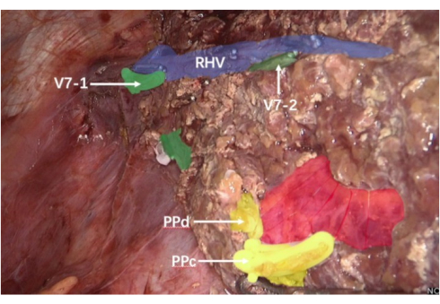
- Il tessuto epatico è stato tagliato dal lato caudale a quello cefalico e lungo il confine tra i segmenti fluorescente e non fluorescente, lungo la vena epatica interterritoriale (IVH) tra i segmenti epatici S6 e S7 e la vena epatica destra (Figura 7).
- Lungo il bordo destro della vena epatica destra, la vena da reflusso S7 del fegato è stata legata utilizzando clip di legatura e scollegata dai peduncoli epatici a due rami della sezione epatica S7. Un bisturi a ultrasuoni e l'elettrocoagulazione bipolare sono stati utilizzati per tagliare il tessuto epatico utilizzando i bordi dell'imaging fluorescente sotto bassa pressione venosa centrale assistita da anestesia.
- Emostasi del fegato residuo: il fegato residuo è stato attentamente controllato e i punti di sanguinamento sono stati chiusi uno per uno utilizzando l'elettrocoagulazione bipolare. Per chiudere l'incisione è stata utilizzata una sutura antibatterica Vicryl rivestita.
- Per il dolore postoperatorio, sono stati somministrati analgesici per via endovenosa. Durante l'osservazione postoperatoria, sono stati misurati i cambiamenti nella funzionalità epatica e nei livelli di bilirubina.

Figura 3: Puntura della superficie diaframmatica. (A) L'ecografia intraoperatoria entra lungo il foro operatorio principale. (B) L'ago per puntura PTC laparoscopico viene utilizzato per perforare lungo il foro di fissazione ecografica intraoperatoria. (C) Ago per puntura PTC (freccia bianca) Puntura PPc Immagine della vena porta in PPc sotto guida ecografica. Clicca qui per visualizzare una versione più grande di questa figura.

Figura 4: Colorazione positiva della puntura della superficie diaframmatica. Immagine di colorazione positiva della vena porta in PPc e contrassegnare i bordi macchiati con un coltello ad ultrasuoni. L'area verde mostra il territorio del portale a cui appartiene PPc. Clicca qui per visualizzare una versione più grande di questa figura.

Figura 5: Puntura della superficie viscerale. (A) L'ecografia intraoperatoria entra lungo il foro dell'assistente operativo. (B) L'ago per puntura PTC laparoscopico viene utilizzato per perforare lungo il foro di fissazione ecografica intraoperatoria. (C) Ago da puntura PTC (freccia bianca) Immagine della vena porta PPd sotto guida ecografica. Clicca qui per visualizzare una versione più grande di questa figura.

Figura 6: Colorazione positiva per puntura della superficie viscerale. Immagine di colorazione positiva della vena porta PPd e contrassegnare i bordi macchiati con un coltello ad ultrasuoni. L'area verde mostra il territorio del portale a cui PPd appartiene. Clicca qui per visualizzare una versione più grande di questa figura.

Figura 7: IHV e vena epatica destra. L'area viola mostra la vena epatica destra; l'area verde mostra segmenti di 7 rami della vena epatica; l'area gialla è una sezione trasversale della PPC e della PPD; L'area rossa è il peduncolo epatico posteriore destro. Abbreviazioni: v7-1= Il primo dei rami del segmento S7 della vena epatica; v7-2= Il secondo dei rami del segmento S7 della vena epatica; RHV = vena epatica destra; PPc: ramo C della vena porta nel lobo posteriore destro del fegato; PPd: ramo D della vena porta nel lobo posteriore destro del fegato. Clicca qui per visualizzare una versione più grande di questa figura.
Risultati
In questo caso clinico, è stata eseguita con successo la resezione epatica anatomica guidata dalla colorazione laparoscopica del territorio portale del segmento S7, con colorazione positiva lungo le superfici diaframmatiche e viscerali (Figura 6 e Figura 7). È stata eseguita la resezione di un singolo tumore di 1 cm. Il tempo operatorio è stato di 210 minuti e la perdita di sangue intraoperatoria è stata di 100 ml. La durata del ricovero postoperatorio è stata di 7 giorni e il paziente è ancora in follow-up continuo. Il follow-up viene eseguito ogni 2 mesi per 2 anni, inclusi test di funzionalità epatica, test dei marcatori tumorali ed esami basati sull'imaging (ecografia, TC o risonanza magnetica). La risonanza magnetica post-operatoria è mostrata nella Figura 8.
Esame del campione: I risultati dell'esame patologico postoperatorio hanno rivelato HCC di grado III (moderatamente scarsamente differenziato) con un margine chirurgico negativo. Immunoistochimica: AFP (+), CK19 (+), glipicano-3 (+), epatocita (+), CD10 (-), CD34 (+, trasformazione capillare), CK7 (-) e Ki-67 (+, circa il 30% nell'area dell'hotspot).

Figura 8: Risonanza magnetica postoperatoria. La risonanza magnetica del paziente dopo l'intervento. Clicca qui per visualizzare una versione più grande di questa figura.
Discussione
Attualmente, la chirurgia epatica sta entrando in un'era di chirurgia minimamente invasiva. Ci sono state segnalazioni di resezione anatomica di vari segmenti epatici in pazienti con HCC, e la sicurezza di questa operazione è stata verificata2. La sua efficacia in oncologia ha attirato una crescente attenzione e l'approccio dell'epatectomia anatomica proposto da Shindoh et al. prevede una resezione completa basata sulla colorazione del territorio correlato al tumore6. Limitata dalle tecniche e dalle attrezzature precedenti, solo la segmentazione epatica di Couinaud può essere utilizzata per ottenere un concetto simile a quello della resezione anatomica.
È ben noto che l'HCC metastatizza principalmente lungo la vena porta, con la maggior parte dei pazienti che sviluppano trombi tumorali della vena porta. Il 32° Incontro Annuale della Società Giapponese di Chirurgia Epatobiliare e Pancreatica e l'Expert Consensus Meeting on Precise Anatomy for Minimally Invasive Hepatobiliary and Pancreatic Surgery hanno chiarito e unificato la definizione di epatectomia anatomica, ovvero la rimozione completa del parenchima epatico PT8. La segmentectomia epatica anatomica è chiaramente definita come la completa rimozione del territorio che colora i segmenti epatici dominati dal peduncolo epatico di terzo livello. Con lo sviluppo dell'analisi della colorazione del territorio di ricostruzione tridimensionale e dell'imaging a fluorescenza con verde indociania, è stata raggiunta l'epatectomia anatomica basata sulla colorazione del territorio della vena porta portante tumore9. Le persone hanno iniziato a scoprire che l'ambito di resezione della resezione epatica anatomica nel bacino della vena porta differisce significativamente da quello della resezione epatica anatomica classica. Questa deviazione potrebbe portare a micrometastasi residue nel bacino della vena porta e a recidiva locale. Quando il tumore si estende su più segmenti epatici, l'ambito di resezione dell'epatectomia anatomica classica è significativamente più ampio di quello dell'epatectomia anatomica nel bacino della vena porta, rendendo difficile ottenere una resezione precisa e minimamente invasiva per soddisfare le esigenze cliniche.
La resezione epatica anatomica guidata dalla colorazione laparoscopica del territorio portale utilizza la ricostruzione tridimensionale preoperatoria per delineare il bacino della vena porta personalizzato e portatore di tumore. Il metodo è stato combinato con un sistema di imaging fluorescente verde indocianina durante l'intervento chirurgico per garantire una resezione precisa.
In precedenza, per l'epatectomia anatomica classica, Ferrero et al. hanno sezionato una piccola quantità di tessuto epatico dal lato dorsale per controllare accuratamente il peduncolo epatico del segmento S7, hanno cercato il tronco principale della vena epatica destra utilizzando l'approccio della vena epatica cefalica e hanno seguito il tronco principale per recidere il fegato e completare la resezione anatomica S7 del fegato2. Morise et al. hanno eseguito la resezione epatica anatomica laparoscopica S7 utilizzando un approccio toracico9, mentre GoroHonda et al. hanno resecato il segmento S7 del fegato utilizzando l'approccio caudale dorsale12. Chen et al. hanno sostenuto l'epatectomia ortotopica laparoscopica utilizzando S713; tuttavia, per la resezione epatica anatomica del segmento S7 guidata dalla colorazione laparoscopica del territorio portale, se il peduncolo epatico bersaglio viene ottenuto attraverso l'approccio chirurgico classico, è necessario scindere una grande quantità di parenchima epatico. Se la colorazione viene eseguita su questa base, alcuni segmenti epatici che appartengono allo spartiacque tumorale potrebbero non essere colorati a causa del tessuto epatico già tagliato. Ciò influenzerà l'efficacia della colorazione e della rimozione del tumore.
Il passaggio chiave nella resezione epatica anatomica del segmento S7 guidata dalla colorazione laparoscopica del territorio portale è la puntura precisa del peduncolo epatico bersaglio sotto guida ecografica intraoperatoria. Solo fissando il territorio correlato al tumore e i rami di colorazione è possibile eseguire la colorazione a fluorescenza per guidare l'intervento chirurgico. Il metodo di colorazione laparoscopica con puntura della vena porta ha una curva di apprendimento ripida, che richiede non solo competenza nella comprensione dell'anatomia del fegato, ma anche una solida base per l'interpretazione dei risultati ecografici durante la laparoscopia. Questo metodo rappresenta quindi una sfida tecnica riconosciuta a livello internazionale.
Sebbene i principianti possano apprendere rapidamente questa tecnologia utilizzando i fori guida per la puntura su alcune apparecchiature a ultrasuoni perché l'angolo tra il tunnel di puntura e la sonda è fissato a 60°, richiede una selezione precisa del punto di puntura attraverso la parete addominale; In caso contrario, potrebbe verificarsi un errore di foratura14. Per risolvere questo problema, proponiamo un metodo a un lato, a tre punti e a quattro dita orizzontali: da un lato, il punto medio delle aste di regolazione sinistra e destra è stato utilizzato come punto di mira e l'asta della sonda è stata utilizzata come piano verticale per la puntura in piano. Sui tre punti, il punto di puntura della pelle, il foro di puntura della sonda intraoperatoria e il punto di puntura del peduncolo epatico bersaglio e, sulle quattro dita orizzontali, la pelle. Il punto di puntura era localizzato approssimativamente all'intersezione delle quattro dita orizzontali, dal piano verticale dell'asta della sonda e dalla pelle davanti al trocar. La vena porta è stata perforata dalle superfici diaframmatiche e viscerali del fegato per ottenere una resezione laparoscopica guidata dalla colorazione del territorio portale del segmento anatomico S7 del fegato e verificare la fattibilità e la sicurezza dell'operazione.
Durante la procedura di puntura, abbiamo evidenziato diversi dettagli critici che richiedono attenzione, tra cui il tipo di ago da puntura, la posizione del chirurgo, l'angolo e la direzione della punta dell'ago e la velocità con cui l'assistente somministra il farmaco. La dimensione dell'ago per puntura dovrebbe idealmente variare da 18 G a 21 G (0,8 mm-1,2 mm) e la portata dell'iniezione deve essere attentamente controllata. La posizione del chirurgo segue generalmente il principio controlaterale del tumore: se il tumore si trova sul lato destro del fegato, il chirurgo si trova sul lato sinistro e viceversa. La punta dell'ago è stata inserita nel fegato con un angolo verso l'alto e diretta verso l'estremità distale del peduncolo epatico bersaglio per prevenire il reflusso del farmaco e la contaminazione degli altri rami durante l'iniezione. La direzione della punta dell'ago era la seguente: una punta, tre macchie e quattro dita orizzontali, con l'ago che entrava nel fegato rivolto verso l'alto. Una volta che la punta dell'ago è entrata nel fegato, le regolazioni sono state ridotte al minimo per evitare danni al fegato e la direzione della punta dell'ago potrebbe andare persa a causa dello spostamento della sonda durante la procedura. La sonda deve quindi essere ruotata delicatamente per individuare la punta dell'ago piuttosto che regolare la direzione dell'ago per allinearsi con il piano degli ultrasuoni. Se la direzione della punta dell'ago non è coerente con la direzione del peduncolo epatico bersaglio, la punta può essere ritirata e combinata con l'ecografia e l'ago può essere inserito di nuovo. Per quanto riguarda la velocità di somministrazione del farmaco, è fondamentale mantenere il controllo sulla velocità di iniezione sotto la visualizzazione diretta dell'ecografia per garantire che il farmaco segua la direzione del flusso sanguigno della vena porta.
Il fallimento della colorazione laparoscopica con puntura della vena porta si divide principalmente in due aspetti: troppi rami del tumore che non possono essere completamente colorati e fallimento della colorazione causato dal reflusso del farmaco. Per quanto riguarda queste due limitazioni, se si riscontra che più rami della vena porta forniscono sangue al tumore durante l'intervento chirurgico, ma la ricostruzione 3D preoperatoria non riflette questo, la colorazione parziale dello spartiacque e i confini della fluorescenza possono essere utilizzati per scindere il parenchima epatico, localizzare e bloccare il peduncolo epatico e quindi staccare il parenchima epatico rimanente, secondo la linea ischemica o la colorazione inversa. Se i dettagli di cui sopra non vengono presi in considerazione e si verifica il reflusso del farmaco, il dispositivo a fluorescenza può essere regolato in modalità bianco e nero per migliorare il contrasto.
Tuttavia, rispetto agli attuali segmenti epatici anatomici laparoscopici S7 e S815, il metodo di colorazione positiva per aspirazione della vena porta presenta i seguenti vantaggi. In teoria, il segmento epatico colorato positivamente dello spartiacque della vena porta è la presentazione più vicina allo spartiacque anatomico reale, che può evitare efficacemente il volume epatico residuo non funzionale e ridurre il rischio di micrometastasi tumorali e recidive16. Il posizionamento accurato del peduncolo epatico bersaglio senza sezionare il portale epatico e danneggiare il parenchima epatico può evitare efficacemente complicanze postoperatorie come la perdita di bile. Con la premessa di una colorazione riuscita, la completa separazione del parenchima epatico lungo l'interfaccia fluorescente previene efficacemente i danni alla vena epatica e riduce il rischio di sanguinamento.
Ulteriori ricerche su studi randomizzati controllati di resezione anatomica epatica laparoscopica del territorio portale per il trattamento dell'HCC dovrebbero essere condotte per indagare la superiorità di questo approccio per l'oncologia. Inoltre, l'importanza degli ultrasuoni nella chirurgia epatobiliare sta diventando sempre più evidente.
Divulgazioni
Gli autori non hanno nulla da rivelare.
Riconoscimenti
Gli autori non hanno nulla da rivelare.
Materiali
| Name | Company | Catalog Number | Comments |
| Coated Vicryl Plus Antibacterial Suture | Ethicon, Inc. | 3650118 | The product is suitable for the placement and/or ligation of soft tissues |
| Color Doppler ultrasound diagnostic scanner | BK Medical | 20153251933 | intraoperative ultrasound |
| Disposable laparoscopic puncture device and puncture sheath | Jiangsu Fenghe Medical Equipment Co., Ltd | 20182021588 | Used for laparoscopic examination and surgical procedures, to puncture the abdominal wall tissue of the human body and establish a working channel for abdominal surgery |
| Four way curved electron convex array laparoscopic intraoperative probe | BK Medical | 20153251933 | Used for intraoperative examination and interventional treatment in various laparoscopic surgeries |
| HAKKO SONOGUIDE PTC NEEDLE | Baguang Trading (Shanghai) Co., Ltd | 20172146872 | Percutaneous liver bile duct puncture needle |
| Indocyanine Green for Injection | DANDONG YICHUANG PHARMACEUTICAL | ICP-09018669-1 | Assessment of liver reserve function and liver imaging |
| WECK Hem-o-lok | Teleflex Medical | 20143466018 | Ligation of blood vessels or tissues |
Riferimenti
- Han, B. F., et al. Cancer incidence and mortality in China, 2022. J Natl Cancer Cent. 4 (1), 47-53 (2024).
- Ishizawa, T., et al. Laparoscopic segmentectomy of the liver: from segment I to VIII. Ann Surg. 256 (6), 959-964 (2012).
- Makuuchi, M., et al. Ultrasonically guided subsegmentectomy. Surg Gynecol Obstet. 161 (4), 346-350 (1985).
- Takamoto, T., Makuuchi, M. Precision surgery for primary liver cancer. Cancer Biol Med. 16 (3), 475-485 (2019).
- Cho, A., et al. Relation between hepatic and portal veins in the right paramedian sector: proposal for anatomical reclassification of the liver. World J Surg. 28, 8-12 (2004).
- Shindoh, J., et al. Complete removal of the tumor-bearing portal territory decreases local tumor recurrence and improves disease-specific survival of patients with hepatocellular carcinoma. J Hepatol. 64 (3), 594-600 (2016).
- Shindoh, J., et al. The intersegmental plane of the liver is not always flat - tricks for anatomical liver resection. Ann Surg. 251 (5), 917-922 (2010).
- Ciria, R., et al. A snapshot of the 2020 conception of anatomic liver resections and their applicability on minimally invasive liver surgery. A preparatory survey for the expert consensus meeting on precision anatomy for minimally invasive HBP surgery. J Hepatobiliary Pancreat Sci. 29 (1), 41-50 (2022).
- Zheng, J., et al. Laparoscopic anatomical portal territory hepatectomy with cirrhosis by takasaki's approach and indocyanine green fluorescence navigation (with Video). Ann Surg Oncol. 27 (13), 5179-5180 (2020).
- Kawaguchi, Y., et al. Difficulty of laparoscopic liver resection: proposal for a new classification. Ann Surg. 267 (1), 13-17 (2018).
- Liang, X., et al. Laparoscopic anatomical portal territory hepatectomy using Glissonean pedicle approach (Takasaki approach) with indocyanine green fluorescence negative staining: how I do it. HPB. 23 (9), 1392-1399 (2021).
- Ferrero, A., et al. Laparoscopic right posterior anatomic liver resections with Glissonean pedicle -first and venous craniocaudal approach. Surg Endosc. 35 (1), 449-455 (2021).
- Morise, Z. Laparoscopic liver resection for posterosuperior tumors using caudal approach and postural changes: a new technical approach. World J Gastroenterol. 2016 (47), 10267-10274 (2016).
- Okuda, Y., et al. Intrahepatic Glissonean pedicle approach to segment 7 from the dorsal side during laparoscopic anatomic hepatectomy of the cranial part of the right liver. J Am Coll Surg. 226 (2), e1-e6 (2018).
- Cao, J., et al. Totally laparoscopic anatomic S7 segmentectomy using in situ split along the right intersectoral and intersegmental planes. Surg Endosc. 35 (1), 174-181 (2021).
- Wang, X., Tong, H., Li, J., Wang, H. I. Indocyanine green fluorescence-guided laparoscopic anatomical segmentectomy of liver segment 6: Surgical strategy and technical details. Ann Surg Oncol. 31 (10), 6546-6550 (2024).
Ristampe e Autorizzazioni
Richiedi autorizzazione per utilizzare il testo o le figure di questo articolo JoVE
Richiedi AutorizzazioneEsplora altri articoli
This article has been published
Video Coming Soon