
Aby wyświetlić tę treść, wymagana jest subskrypcja JoVE. Zaloguj się lub rozpocznij bezpłatny okres próbny.
Method Article
An Intestinal Gut Organ Culture System for Analyzing Host-Microbiota Interactions
* Wspomniani autorzy wnieśli do projektu równy wkład.
W tym Artykule
Podsumowanie
This article presents a unique method for analyzing host-microbiome interactions using a novel gut organ culture system for ex vivo experiments.
Streszczenie
The structure of the gut tissue facilitates close and mutualistic interactions between the host and the gut microbiota. These cross-talks are crucial for maintaining local and systemic homeostasis; changes to gut microbiota composition (dysbiosis) associate with a wide array of human diseases. Methods for dissecting host-microbiota interactions encompass an inherent tradeoff among preservation of physiological tissue structure (when using in vivo animal models) and the level of control over the experiment factors (as in simple in vitro cell culture systems). To address this tradeoff, Yissachar et al. recently developed an intestinal organ culture system. The system preserves a naive colon tissue construction and cellular mechanisms and it also permits tight experimental control, facilitating experimentations that cannot be readily performed in vivo. It is optimal for dissecting short-term responses of various gut components (such as epithelial, immunological and neuronal elements) to luminal perturbations (including anaerobic or aerobic microbes, whole microbiota samples from mice or humans, drugs and metabolites). Here, we present a detailed description of an optimized protocol for organ culture of multiple gut fragments using a custom-made gut culture device. Host responses to luminal perturbations can be visualized by immunofluorescence staining of tissue sections or whole-mount tissue fragments, fluorescence in-situ hybridization (FISH), or time-lapse imaging. This system supports a wide array of readouts, including next-generation sequencing, flow cytometry, and various cellular and biochemical assays. Overall, this three-dimensional organ culture system supports the culture of large, intact intestinal tissues and has broad applications for high-resolution analysis and visualization of host-microbiota interactions in the local gut environment.
Wprowadzenie
The intestine is a highly complex organ containing a wide range of cell types (epithelial cells, immune system cells, neurons, and more) organized in a particular structure that allows cells to interact and communicate with one another and with the luminal content (microbiota, food, etc.)1. Currently, the research toolbox available for analyzing host-microbiota interactions includes in vitro cell cultures and in vivo animal models2. In vivo animal models provide a physiological tissue construction3 but with poor experimental control and limited ability to manipulate the experiment conditions. In vitro culture systems, on the other hand, use primary cells or cell lines that can be supplemented with microbes4, offering tight control over the experiment parameters but lack the cellular complexity and the tissue architecture. Modern in vitro assays allow the advanced use of healthy and pathological human tissue samples, like epithelial organoids derived from mouse or human sources5,6, and samples that mimic the mucosal microenvironment7. Another example is the 'gut on a chip' assay, which includes the human colonic epithelial cell line (Caco2), extracellular matrix and microfluidic channels to mimic the physiological condition of the gut invariant8. However, as advanced and innovative as in vitro samples may be, they do not maintain a normal tissue architecture or naïve cellular composition.
To address that, Yissachar et al. recently developed an ex vivo organ culture system9 (Figure 1) that maintains intact gut fragments ex vivo, benefiting from the advantages of both in vivo and in vitro models. This ex vivo gut organ culture system is based on a custom-made culture device that supports a multiplexed culture of six colon tissues, allowing examining experimental inputs under comparable conditions while controlling the system's inputs and outputs. Recent works have demonstrated that this system is valuable for analyzing intestinal responses to individual gut bacteria9, whole human microbiota samples10 and microbial metabolites11. This system allows, for the first time, the study of these early host-microbiota interactions with a high level of control over the host, microbial and environmental components. Furthermore, it allows monitoring and manipulating the system throughout the experiment, in real-time.

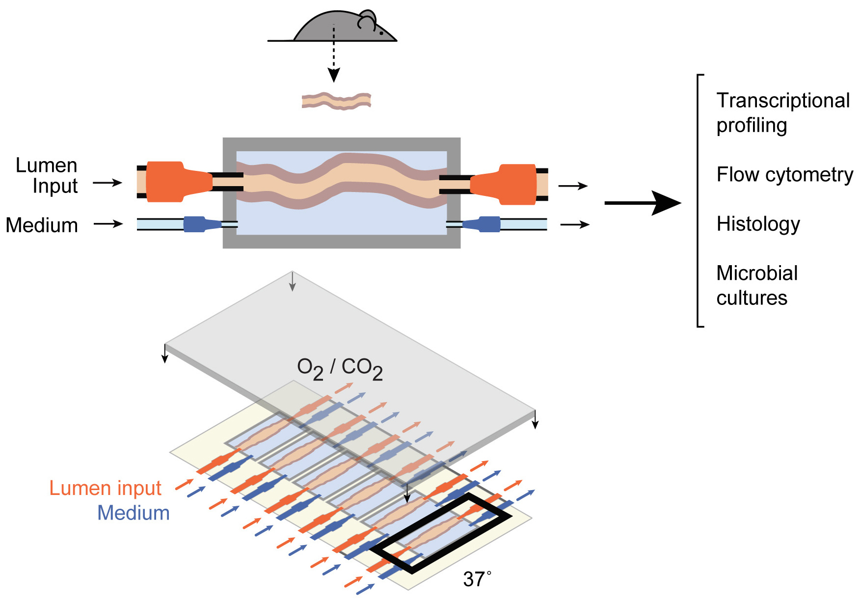
Figure 1: Schematics of the gut culture device. A whole intestinal tissue fragment is attached to the output and input ports of the chamber (top), with pumps regulating the medium flow inside the lumen and in the external medium chamber. The entire device (bottom) contains 6 such chambers. This figure has been modified from Yissachar et al. 2017. Please click here to view a larger version of this figure.
Protokół
This protocol follows the animal care guidelines approved by the ethics committee for animal welfare.
1. Experiment preparation
- Fabrication of the gut organ culture device (3 days)
- Using a 3D printer, print the reusable plastic molds for the organ culture device (the device has 6 wells, with 24 small and large holes, and for the device cover lid) (3D files attached).
NOTE: These plastic molds may be used for fabrication of numerous devices. - Insert the blunt-end needles (22 G & 18 G) to the appropriate position within the device mold and cast approximately 20 g of polydimethylsiloxane (PDMS) mix (1:10 weight ratio, base to catalyst) for one set of the device and lid.
- Place the molds in a vacuum chamber for 30 min, to remove air bubbles from the PDMS mix.
- Incubate the molds at 55 °C overnight, to complete PDMS polymerization.
- When the PDMS is set, pull out the needles from the mold and carefully release the culture device and lid from the plastic molds.
- Remove PDMS residues from the well outline using a surgical blade. Attach the PDMS device and the device cover onto a cover glass (75 mm x 50 mm micro slides) using non-toxic silicon adhesive and leave the parts to set overnight (apply the glue to the smooth side of the device).
- Insert twelve 22 G needles for the lumen and twelve 18 G needles for the well. Fix all the needles in place using silicone and let it set overnight. Insert two 18 G needles into the cover lid for proper air flow in and out of the gut organ culture device.
- Check all the needles for leaks using a water-filled syringe. Check that there is no leaking from the wells by filling the wells with water.
- Place two surgical knots on each 22 G needle that will be connected to the colon. Place the device and the lid in an autoclave paper bag and sterilize in an autoclave.
- Using a 3D printer, print the reusable plastic molds for the organ culture device (the device has 6 wells, with 24 small and large holes, and for the device cover lid) (3D files attached).
- Culture medium (0.5 h)
- In a biological hood, mix the following (in 50 mL tube): 37 mL of Iscove's Modified Dulbecco's Medium (IMDM), 10 mL of KSR serum replacement, 1 mL of B27 supplement, 0.5 mL of N2 supplement, 0.5 mL of 1 M HEPES buffer, and 0.5 mL of non-essential amino acids.
- Filter, and store the complete medium at 4 °C.
- Tubing and surgical tool preparation
- Cut the appropriate length of tubes for the input lumen, input well, output lumen, and output well (12 short tubes and 12 long tubes). Connect an appropriate adaptor to each side of the tube.
- Prepare the surgical tools: straight scissors, 4x thin forceps, and 2x sharp forceps.
- Place the tubes and the surgical tools in an autoclave paper bag and sterilize them using an autoclave.
- Prepare the luminal input (desired stimulation - bacteria, stool, drugs, etc.).
- Before the experiment, determine the bacterial load of the bacterial culture, by serial dilutions12, and culture under aerobic and/or anaerobic conditions.
- After calculating the bacterial load, dilute the bacterial cultures in sterile tissue culture medium to obtain the required bacterial concentration. For fecal samples, filter using a 100 µm strainer.
NOTE: For nonbacterial stimulation (drugs, metabolites, etc.), dilute the substance to the required concentration using the gut culture medium.
2. Experiment setup preparation
- Inside the organ culture incubator, turn on the heater unit, and set it to 37 °C.
- Set up the pumps as well as the input and output syringes.
- Tissue culture medium input
- In a biological or laminar flow hood, fill the input well syringes with complete culture medium. The final volume depends on the experiment's duration and the flow rate; usually 1 mL/h plus additional medium for purge.
- Connect the tubes to the syringes using the Luer-lock adaptor. Place the filled syringes in the syringe pumps.
- Purge the input syringes. Make sure that the well medium flows out from all tubes into a waste glass.
- Luminal input
- Fill the luminal input syringes with stimulation treatment (bacteria, drugs, etc.). The volume depends on the duration of the experiment and the flow rate (usually 30 µL/h plus additional medium for purge).
- Connect the tubes to the syringes using the Luer-lock adaptor. Place the filled syringes in the syringe pumps.
- Purge input syringes. Make sure that the stimulation flow out of all the tubes into a waste glass. Be careful not to contaminate the different stimulations.
- Outputs
- Place the empty syringes in the output syringe pumps (set pump mode to 'withdrawal').
- Device setup
- Set the regulator that controls the gas mixture (95% O2 + 5% CO2) flow rate for a gentle, minimal flow.
- Examine the device in a laminar hood.
- Flush the needles of the device with sterile IMDM to wash the needles.
- Add 500 µL of sterile culture medium into each well of the device.
- Preparation for tissue dissection
- Put the sterile surgical tools inside the laminar hood.
- Fill a 10 mL syringe with sterile IMDM and connect a sterile (autoclaved) 22 G blunt-end needle for flushing of the colon.
3. Organ cultures
- Mice sacrifice and tissue dissection
- In a laminar hood, sacrifice 12-14 day old mice by decapitation. Spray the mice with 70% ethanol and place the mice on a plastic plate.
- Using sharp scissors and forceps, dissect the mouse and take out the digestive tract from the stomach to the anus by cutting all the fat and connective tissues. Cut the colon and place it on a new plate.
NOTE: Minimize the contact with the colon tissue. Do not touch the middle part of the colon tissue. Hold the tissue gently and only at the edges of the tissue.
- Colon flush and wash
- Under a dissection microscope, gently flush the colon content with sterile IMDM (into the proximal side) with the prepared 10 mL syringe (step 2.7.2). After removing the feces from the intestinal tissue, place the colon in a new 6 well plate filled with 0.5 mL of sterile IMDM.
- Connecting the colon to the device
- Take the tissue and carefully connect it onto the 22 G needle and make a tight tie with the two threads. At this point, it is imperative to maintain the correct orientation of the colon to the lumen flow (proximal = input, distal = output). Repeat steps 3.2-3.3 for all 6 tissues.
- Verify that the input needle Luer locks are empty from the medium. If not, empty them. Add stimulations to the input needle Luer lock (to avoid entry of air bubbles into the lumen). Repeat this step for each colon with the appropriate stimulation.
- Check that all the tissues are connected and place the cover lid on top of the device.
- Connecting the organ culture device to the pumps
- Place the device into the pre-heated temperature-controlled chamber (37 °C).
- Connect gas flow
- Connect the gas adaptor to the cover lid using the appropriate input needle.
- Connect the input and output tubes to the device.
NOTE: Connect the proximal colon side to the input tubs.
- Purge the lumen with the stimulation inputs.
- Gently flow luminal stimulation through the gut and verify medium flow in the output tubes.
- Wash external medium.
- Wash the external medium 3 times (set the pumps at a rate of 600 µL/min for both input and output well). Each wash takes 1 min (starting by emptying the well).
- Start the experiment.
- Start the pumps at the following rates:
Flow rate: lumen: input- 30 µL/h, output- 35 µL/h
External medium: input- 1000 µL/h, output- 950 µL/h
NOTE: Experiment time can vary between 30 min to 24 h.
- Start the pumps at the following rates:
- End of experiment (up to 24 h for colon organ cultures)
- Disconnect all the tubes from the device.
- Disconnect the tissues from the needles and continue to the desired readouts.
Wyniki
The gut organ culture system maintains tissue viability ex vivo. The evaluation of the tissue viability was done throughout the culture period. Colon tissue fragments were incubated in the gut organ culture system and fixed following 2/12/24 h culture. The intestinal epithelial cell (IEC) layer integrity was validated by immunofluorescence staining using E-cadherin and cytokeratin-18 antibodies. Likewise, mucus-filled goblet cells in the colonic epithelium and mucus secretion within the lumen were detected as we...
Dyskusje
This article describe an optimized protocol for ex vivo gut organ cultures that Yissachar et al. have recently developed (published9 and unpublished data). The gut organ culture system supports multiplexed culture of intact intestinal fragments while maintaining luminal flow. It provides full control over the intra- and extra-luminal environment (including stimulation dose, exposure time and flow rate) and preserves the naïve intestinal tissue structure and its cellular complexity
Ujawnienia
The authors have nothing to disclose.
Podziękowania
We thank past and present members of the Yissachar lab for their valuable contributions in optimizing the gut organ culture system protocol. We thank Yael Laure for critical editing of the manuscript. This work was supported by the Israel Science Foundation (grant No. 3114831), the Israel Science Foundation - Broad Institute Joint Program (grant No. 8165162), and the Gassner Fund for Medical Research, Israel.
Materiały
| Name | Company | Catalog Number | Comments |
| Device | |||
| 18 Gauge Blunt Needle | Mcmaster | 75165a754 | |
| 22 Gauge Blunt Needle | Mcmaster | 75165a758 | |
| All Purpose Adhesive Selant 100% Silicone | DAP | 688 | |
| Cubic Vacuum Desiccator VDC-21+ 2 Shelves | AAAD4021 | ||
| Glass Slide 1 mm Thick | Corning | 2947-75X50 | |
| Mini Incubator im-10 | AAH24315K | ||
| MPC 301E Vacuum PUMP | VI-412711 | ||
| Plastic Quick Turn Tube Coupling Plugs | Mcmaster | 51525k121 | |
| plastic Quick Turn Tube Coupling Sockets | Mcmaster | 52525k211 | |
| Sylgard 184 Silicone Elastomer | Dow | Polydimethylsiloxane, PDMS | |
| Tubing | Mcmaster | 6516t11 | |
| Zortrax M200 | Zortrax | Zortrax Z-SUITE, Autodesk Fusion 360 | |
| Zortrax M200 Materials: z-ultrat | Zortrax | ||
| Medium | |||
| B27 Supplement (50x), Serum Free | Thermo Fisher Scientific | 17504044 | |
| HEPES Buffer (1M) | Thermo Fisher Scientific | 15630056 | |
| Iscove's Modified Dulbecco's Medium with Phenol Red (1x) | Thermo Fisher Scientific | 12440061 | |
| Knock-Out Serum | Thermo Fisher Scientific | 10828028 | |
| N2 Supplement (100x) | Thermo Fisher Scientific | A1370701 | |
| Non Essential Amino Acid (100x) | Thermo Fisher Scientific | 11140035 | |
| Surgical Tools | |||
| Large Scissors | Aseltech | 11-00-10 | |
| Sharp Forceps | F.S.T | 11297-10 | |
| Silk Braided Surgical Thread | SMI | 8010G | |
| Straight Scissors | F.S.T | 14091-09 | |
| Thin Forceps | F.S.T | 11051-10 | |
| Organ System | |||
| 0.1 µm Filter | Life Gene | ||
| 0.22 µm Filter | Life Gene | ||
| 5 mL Luer Lock Syringe | B-D | 309649 | |
| Greenough Stereo Microscope | ZEISS | Stemi 305 | |
| Recirculating Precision Air Heater "CUBE" | CUBE-2-LIS | ||
| Syringe Pump | new era pump systems inc | nep-ne-1600-em |
Odniesienia
- Mowat, A. M., Agace, W. W. Regional specialization within the intestinal immune system. Nature Reviews Immunology. 14 (10), 667-685 (2014).
- Pearce, S. C., et al. Intestinal in vitro and ex vivo Models to Study Host-Microbiome Interactions and Acute Stressors. Frontiers in Physiology. 9 (1584), (2018).
- Hooper, L. V., et al. Molecular analysis of commensal host-microbial relationships in the intestine. Science. 291 (5505), 881-884 (2001).
- Haller, D., et al. Non-pathogenic bacteria elicit a differential cytokine response by intestinal epithelial cell/leucocyte co-cultures. Gut. 47 (1), 79-87 (2000).
- Sato, T., et al. Single Lgr5 stem cells build crypt-villus structures in vitro without a mesenchymal niche. Nature. 459 (7244), 262-265 (2009).
- Sato, T., et al. Long-term expansion of epithelial organoids from human colon, adenoma, adenocarcinoma, and Barrett's epithelium. Gastroenterology. 141 (5), 1762-1772 (2011).
- Tsilingiri, K., et al. Probiotic and postbiotic activity in health and disease: comparison on a novel polarised ex-vivo organ culture model. Gut. 61 (7), 1007-1015 (2012).
- Gazzaniga, F. S., et al. Harnessing Colon Chip Technology to Identify Commensal Bacteria That Promote Host Tolerance to Infection. Frontiers in Cellular and Infection Microbiology. 11, 638014 (2021).
- Yissachar, N., et al. An Intestinal Organ Culture System Uncovers a Role for the Nervous System in Microbe-Immune Crosstalk. Cell. 168 (6), 1135-1148 (2017).
- Duscha, A., et al. Propionic Acid Shapes the Multiple Sclerosis Disease Course by an Immunomodulatory Mechanism. Cell. 180 (6), 1067-1080 (2020).
- Grosheva, I., et al. High-Throughput Screen Identifies Host and Microbiota Regulators of Intestinal Barrier Function. Gastroenterology. 159 (5), 1807-1823 (2020).
- Blaize, J. F., Corbo, C. P. Serial Dilutions and Plating: Microbial Enumeration. Journal of Visualized Experiments. , (2021).
- Ivanov, I. I., et al. Induction of intestinal Th17 cells by segmented filamentous bacteria. Cell. 139 (3), 485-498 (2009).
- Schnupf, P., et al. Growth and host interaction of mouse segmented filamentous bacteria in vitro. Nature. 520 (7545), 99-103 (2015).
- Chung, H., et al. Gut immune maturation depends on colonization with a host-specific microbiota. Cell. 149 (7), 1578-1593 (2012).
- Atarashi, K., et al. Th17 Cell Induction by Adhesion of Microbes to Intestinal Epithelial Cells. Cell. 163 (2), 367-380 (2015).
Przedruki i uprawnienia
Zapytaj o uprawnienia na użycie tekstu lub obrazów z tego artykułu JoVE
Zapytaj o uprawnieniaThis article has been published
Video Coming Soon
Copyright © 2025 MyJoVE Corporation. Wszelkie prawa zastrzeżone