Method Article
A Novel Cell Injection Method with Minimum Invasion
In This Article
Summary
This method eliminates any major invasion during cell injections caused by the cell suspension solution.
Abstract
Directly injecting cells into tissues is a necessary process in cell administration and/or replacement therapy. The cell injection requires a sufficient amount of suspension solution to allow the cells to enter the tissue. The volume of the suspension solution affects the tissue, and this can cause major invasive injury as a result of the cell injection. This paper reports on a novel cell injection method, called slow injection, that aims to avoid this injury. However, pushing out the cells from the needle tip requires a sufficiently high injection speed according to Newton's law of shear force. To solve the above contradiction, a non-Newtonian fluid, such as gelatin solution, was used as the cell suspension solution in this work. Gelatins solution have temperature sensitivity, as their form changes from gel to sol at approximately 20 °C. Therefore, to maintain the cell suspension solution in the gel form, the syringe was kept cooled in this protocol; however, once the solution was injected into the body, the body temperature converted it to a sol. The interstitial tissue fluid flow can absorb excess solution. In this work, the slow injection technique allowed cardiomyocyte balls to enter the host myocardium and engraft without surrounding fibrosis. This study employed a slow injection method to inject purified and ball-formed neonatal rat cardiomyocytes into a remote area of myocardial infarction in the adult rat heart. At 2 months following the injection, the hearts of the transplanted groups showed significantly improved contractile function. Furthermore, histological analyses of the slow-injected hearts revealed seamless connections between the host and graft cardiomyocytes via intercalated disks containing gap junction connections. This method could contribute to next-generation cell therapies, particularly in cardiac regenerative medicine.
Introduction
Cell administration and replacement are promising novel therapeutic strategies for heavily damaged organs. Among these novel therapeutic strategies, cardiac regenerative medicine has attracted considerable attention. However, the inflammation caused by injuries mediates scar formation in several organs1,2,3,4. The human heart consists of approximately 1010 cardiomyocytes; therefore, theoretically5,6, it must be treated with more than 109 cardiomyocytes. Administering a large number of cardiomyocytes via traditional injection methods may lead to significant tissue injuries7. This method provides a novel cell injection method with minimal tissue invasion.
Cell administration into the organ parenchyma requires injection(s). However, a discrepancy exists in that the injection itself may lead to tissue injury. Tissue injury causes local inflammation and incurable scarring in organs and tissues, as well as impaired regenerative ability8,9,10. The mammalian heart has an extremely high propensity to develop scars instead of regenerating because it requires immediate injury repair in order to endure the high blood pressure caused by its continuous pumping function11. Ablation therapy utilizes this high propensity toward scar formation and blocks the circuit likely to undergo scar formation using arrhythmia12. In a previous study, it was observed that the scar tissue isolated the injected cardiomyocytes in the host myocardium. Thus, this represents the next target issue that needs to be overcome to obtain improved therapeutic efficacy in cardiac regenerative medicine.
Tissue interstitial fluid flow plays a vital role in conveying oxygen and nutrients to cells and removing the excreted waste from cells. The physiological speed of interstitial fluid flow in each tissue/organ is different (the range is 0.01-10 µm/s)13. To the best of the author's knowledge, there are no data regarding the capacity of individual tissues/organs to support extra amounts of fluid without pathological edema; however, this experiment attempts to use a slow injection speed to possibly reduce tissue injury, and the results can be used to determine the practicality of this concept.
Protocol
The animal experiments were conducted according to the Kansai-Medical University ethical guidelines for animal experiments and were approved by the ethical committees (approval number: 23-104). All the animals were raised under a constant light-dark cycle in a specific pathogen-free environment. All the sterilized surgical tools, such as scissors, forceps, and retractors, were autoclaved and dried thoroughly.
1. Preparation of the neonatal rat cardiomyocyte balls
- Neonatal rat heart collection
- For the heart collection, follow a procedure similar to that described in a previous report7.
- Immerse neonatal (0-2 days after birth) Sprague-Dawley (SD) rats in povidone-iodine and 70% ethanol solutions sequentially, and then transfer them into an air-tight case filled with vaporized isoflurane (the concentration should be over 10% v/v) for deep anesthesia.
- After the confirmation of unconsciousness by a loss of locomotor activity, decapitate the ratwhile holding the body from the back with the hand, and then cut 2-4 mm of tissue from the frontal center of the ribcage to the caudal and then in the rostral direction using sharp scissors.
- Grip the back skin to pull the cut open and push out the heart from the ribcage. Cut the ventricles using scissors and immerse them in phosphate-buffered saline (PBS) without calcium or magnesium (PBS(−)).
- Dispersion of the neonatal rat cardiomyocytes
- Mince the collected ventricles dispersed in a minimal amount of Ads buffer (Ads buffer: 116 mM NaCl, 20 mM HEPES, 12.5 mM NaH2PO4, 5.6 mM glucose, 5.4 mM KCl, 0.8 mM MgSO4, pH 7.35) in an autoclaved concave glassware into small pieces (1 mm x 1 mm) using curved scissors.
- Transfer the minced tissues and a micro-magnetic stirrer into a 50 mL centrifugation tube, and disperse the tissues into single cells with 0.1% collagenase, 0.1% trypsin, 20 µg/mL DNase I, and 50 nM tetramethylrhodamine methyl ester in Ads buffer at 37 °C by stirring for 30 min.
- Separate the aggregates and dispersed cells through natural sedimentation, collect only the dispersed cells into a tube, and digest the residual cell aggregates again with the same digestion medium.
- Continue this procedure until all the cells are completely dissociated. To confirm the complete dispersion of the cells, observe the tubes under a microscope (4x objective lens).
- Collect the dispersed cells using centrifugation at 150 x g for 5 min and dissociate them in 1-2 mL of Ads buffer.
- Fluorescence-activated cell sorting of cardiomyocytes
- Analyze the cells using fluorescence-activated cell sorting (FACS) using 556-601 nm bandpass filters to detect red fluorescence signals.
- Carefully perform pre-gating to eliminate doublet fractions14. The gate settings for doublet elimination should be as per the manufacturer's instructions.
- Set the forward scatter on the x-axis and the red fluorescence signal on the y-axis. Three populations were observed: a lowermost population containing erythrocytes and dead cells, a middle population containing non-cardiomyocytes, including fibroblasts and endothelial cells, and a top population containing pure ventricular cardiomyocytes7.
- Preparation of red fluorescence-labeled cardiomyocyte balls
- Selectively sort the cardiomyocytes, and centrifuge for 5 min at 150 x g. Completely dissolve the cell pellets in 1 mL of alpha-modified minimal essential medium (alpha-MEM) containing 10% heat-inactivated fetal bovine serum (FBS).
- Measure the cell concentration using a hemocytometer and dilute the cell suspension solution to 3,000 cells/mL with alpha-MEM 10% FBS.
- Distribute them into cell non-adhesive 96-well plates (100 µL per well), centrifuge for 5 min at 100 x g, and culture for 2-3 d in a cell culture incubator with 5% CO2 at 37 °C.
- Before the injection experiments, harvest a cardiomyocyte ball from each well via aspiration with the culture medium using a 1,000 µL pipette, and collect them in a 15 mL tube.
- Stain with PKH26 following the manufacturer's instructions for tracking after engraftment.
2. Preparations for the slow injection method
- Preparation of the gelatin stock solution
- Weigh the gelatin, dissolve it in Ads buffer to produce a 10% w/v solution, and autoclave it.
- Production of a cell injection device
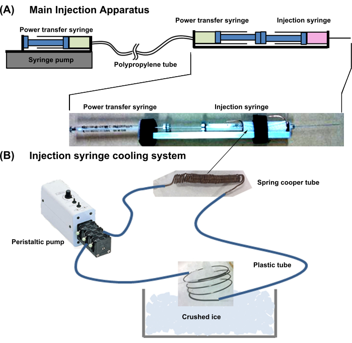
- Overall device design: Prepare the apparatus shown in Figure 1. This system is combined with a syringe cooling apparatus and main injection apparatus.
- Prepare the main injection apparatus described below:
- For a neonatal cardiomyocyte ball injection, use a 29 G, 50 mm long needle equipped with a syringe.
- Connect a power transfer syringe (18G, 1 mL) back-to-back using a cell injection syringe (Figure 1). Connect the needle of the power transfer syringe to a thin polypropylene tube with low lumen expandability.
- Then, connect the other side of the power transfer tube to the needle of the same syringe (18G, 1 mL), and set it in a syringe pump. Fill the two power transfer syringes and tubes with water without any air bubbles.
NOTE: When one plunger of the power transfer syringe is pushed in, the pressure is directly transferred to the other syringe, and the plunger protrudes.
- Set up the injection syringe cooling system as described below.
- Wind a copper tube (external diameter = 1 mm; internal diameter = 0.3 mm; thickness = 0.35 mm) tightly around the cell-containing part of the cell injection syringe, leaving 10 mm of excess pipe at both ends.
- Connect the copper tube to flexible plastic tubing. Further, connect the other ends of the plastic tubing to an external pump, fill the line with cooling water, and cool the water by immersing the excess copper tube into crushed ice (Figure 1).
NOTE: This cooling system maintains the cell suspension solution in the cylinder at approximately 2 °C.
3. Development of a rat myocardial infarction model by obtuse coronary artery occlusion
- Anesthetize male immune-deficient nude rats (F344/Njcl-rnu/rnu) with air containing 3% isoflurane. Insert a cannula into the trachea and connect it to the respirator.
- Connect a cannula to an isoflurane vaporizer with a controller to maintain an 3% isoflurane concentration and, thus, sustain sufficient anesthesia. Confirm no response to pain stimuli. Apply veterinarian ointment on the eyes to prevent dryness.
- Fix the limbs with cut surgical tapes on a 40 °C warmed surgical plate. Twist the body of the rat to the right of the body axis and use the left armpit as the surgical field.
- Remove the hair in the surgical field using a depilatory cream and wipe the skin with povidone-iodine. Using sharp scissors, cut a 1.5 cm incision in the skin and pectoralis major muscle.
- Confirm the third intercostal space and rip the intercostal muscles and costal pleura using micro forceps with blunt tips. Keep the chest open by using a retractor. Gently remove the thin pericardium with forceps.
- To construct an infarction model of the lateral wall of the heart, find the position 1 mm caudal to the tip of the left atrium to identify the obtuse coronary artery, pass a 7-0 silk suture, scoop up tissue that is 2.5 mm wide and 2.5 mm deep from dorsal to ventral, and ligate the tissue tightly.
- Confirm successful ligation by the weak contraction distal from the ligature. After gently removing the retractor, place a 5-0 silk suture between the second and fourth intercostal spaces, and close the thoracotomy.
- Decrease the isoflurane concentration to 1%. Gently suture the muscle and skin with 5-0 silk. Decrease the isoflurane concentration to 0% and wait for approximately 5 min until spontaneous breathing starts.
- Topically apply 2 mg/mL lidocaine in saline to the incision. Administrate 1 mL of saline via a subcutaneous injection. Apply veterinarian ointment topically to prevent infections.
- Remove the rat from the intubation tube, and return to the animal cage; then, raise the rats in individual cages for 1 week.
- Analyze the changes in the cardiac systolic pump function using echocardiography.
NOTE: The cardiac function will be reduced owing to lateral myocardial infarction.
4. Echo-guided percutaneous cell transplantation using the slow injection method
- Pre-warm 10% gelatin stock at 37 °C until it becomes liquid.
- Dilute 10% gelatin stock with pre-warmed Ads buffer to obtain the final injectable 5% w/v gelatin solution (100 µL is required per animal).
- Suspend 96 cardiomyocyte balls (total: 28,800 cardiomyocytes/animal) in 100 µL of pre-warmed injection solution.
- Load the suspension prepared in step 4.3 into the cell injection syringe, avoiding the aspiration of excess air.
- To eliminate bubbles in the syringe, hold it vertically with the needle upward, tap the syringe, and collect any bubbles at the upper ridge of the syringe.
NOTE: During this step, observe the cardiomyocyte balls gradually settle onto the rubber seal of the plunger. - Maintain the vertical position of the syringe, push up the plunger slowly, and discard the bubbles and excess cell suspension solution until 20 µL of the cell suspension remains in the syringe. Push the plunger carefully at a constant speed so that the cardiomyocyte ball remains resting on the rubber seal of the plunger.
- Immerse the capped syringe directly in an ice bath for 5 min.
- Place a cooled syringe in the injection apparatus. Tightly fix the injection syringe apparatus settled on a fine movement device on the animal stage using an X-Y-Z position-adjustable handmade clamp. The fine movement device can move the needle position using the x-direction 20 mm slide, y-direction swing, and z-direction bowing movements.
- Anesthetize the rats in a sealed box filled with air containing 3% isoflurane. Confirm anesthesia by no response to pain stimuli. Apply veterinarian ointment on the eyes to prevent dryness.
- Fix the limbs with cut surgical tapes on a 40 °C warmed echo plate. To sustain sufficient anesthesia, ensure the inhaled air contains an approximately 3% concentration of isoflurane.
- Remove the hair at the injection field (2 cm in diameter) and chest with depilatory cream and wipe the skin with povidone-iodine.
- Apply echo-gel on the chest and echo probe. Place the echo probe close to the chest along the cranial and caudal axes and start to obtain B-mode echocardiography following the manufacturer's manual.
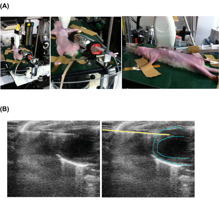
- Advance the tip of the injection needle into the myocardium at the frontal view (Figure 2 and Supplementary Video 1).
- To activate the main syringe pump, push the Start button, and rotate the dial to adjust to a predetermined number for an injection speed of approximately 0.02 µL/s. Apply veterinarian ointment topically around the needling position to prevent infections.
NOTE: It is necessary to determine the appropriate dial number for the intended injection speed using an injection solution. After the injection, remove the injection needle using an exemplary movement system. - Decrease the isoflurane concentration to 0% and wait for approximately 5 min until the animal regains sufficient consciousness to maintain sternal recumbency.
5. Evaluation of cardiac function
- Anesthetize the rats in a sealed box filled with air containing 3% isoflurane. Confirm anesthesia by no response to pain stimuli. Apply veterinarian ointment on the eyes to prevent dryness.
- Apply electrifying cream for ECG acquisition on the limb tips. Fix the limbs with cut surgical tapes on a 40 °C warmed echo plate. Use a physiological monitoring system to detect real-time ECG and heart rate.
- According to the manufacturer's instructions, first determine the long-axis angle using the B-mode echo image showing the left ventricular apex to the outflow tract, and then rotate the echo-probe 90° to change to the short-axis view.
- Using only the caudal to rostral axis fine movement system of the animal stage, adjust the short-axis view to the papillary muscle level. Then, change the image mode to M-mode by pressing the M-mode button, and record a video for 5 s by pressing the Cine-loop button.
- To analyze and calculate the fraction shortening using the software, press the Measure button and a vertical line tool to define the end-systolic and end-diastolic left ventricular internal dimensions. The software automatically calculates the percent fraction shortening (FS).
- Decrease the isoflurane concentration to 0% and wait for approximately 5 min until the animal regains sufficient consciousness to maintain sternal recumbency.
6. Immunohistochemistry
- Fix the euthanized body (perform euthanasia as in step 1.1.3) on the table using the cut surgical tapes, excise the hearts, wash with PBS, and immerse the heart in 4% paraformaldehyde/PBS.
- Dissect the hearts into three sections, immerse them in 40% sucrose for cryoprotection, embed in an optimal cutting temperature (OCT) compound, and freeze them at −80 °C.
- Attach the cryosectioned tissues (8 µm thickness) to aminosilane-coated glass slides. After sufficient drying using non-warmed wind generated by a general hair dryer, immerse the glass slides in tris-buffered saline containing 0.2% Tween-20 (TBS-T).
- Immerse them in a blocking solution for 30 min at 25 °C. Pour 100 µL of the primary antibody-containing blocking solution onto the slide, and incubate overnight at 4 °C with paraffin sealing to keep the antibody solution spread on the tissue.
NOTE: The antibody concentration is shown in the Table of Materials. - Lay the slides horizontally in a handmade high-humidity box utilizing the natural vapor from wet paper to prevent evaporation. Wash the slides three times with TBS-T.
- Treat with the secondary antibody-containing blocking agent for 1 h at 25 °C in the same manner as the primary antibody. After three washes, observe the fluorescence signals using a fluorescence microscope and confocal laser microscope.
NOTE: The antibody concentration is shown in the Table of Materials. - Statistical analysis: For the cell volume comparison, as shown in Figure 3A, perform a non-paired t-test; to assess the cardiac functional recovery following cardiomyocyte administration using the slow injection method, as shown in Figure 4A, use a paired t-test. In this work, differences were considered statistically significant at P < .01. Error bars represent the standard deviation.
Results
Effects of the slow injection on cell survival and collagen deposition
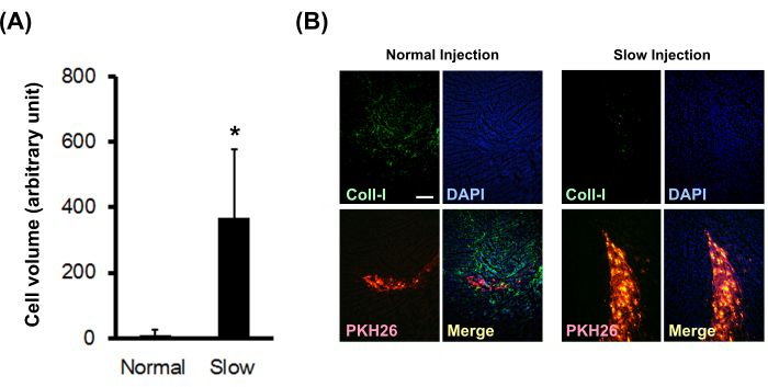
Neonatal rat cardiomyocyte balls labeled with PKH26 were injected into normal nude rat myocardium using a normal or slow injection method. The results showed that the slow injection method significantly increased the engrafted cell volume (Figure 3A) and significantly decreased on-site type I collagen deposition (Figure 3B).
Effects of slow injection on treatment efficacy in a rat infarction model
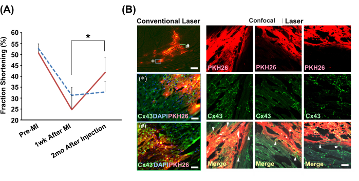
The echocardiograph-guided slow injection method was used to inject neonatal rat cardiomyocyte balls or PBS(−) into the infarcted hearts of model rats. The cell-injected group alone exhibited significant improvement in heart contraction function after 2 months (Figure 4A). Immunohistochemical analyses revealed a seamless connection between the engrafted cells and host myocytes via intercalated disks containing gap junctions (Figure 4B).

Figure 1: Schematic of the whole injection system. (A) Main injection apparatus. (B) Injection syringe cooling system. Please click here to view a larger version of this figure.

Figure 2: Echocardiography-guided percutaneous slow injection. (A) Setting of the animal, echo probe, and injection apparatus. (B) Echocardiographic view of the injecting syringe and heart. Note that the left and the right pictures are the same, but a yellow line has been added to indicate the needle position. Please click here to view a larger version of this figure.

Figure 3: Effect of the slow injection method on the engrafted cell volume and collagen deposition. (A) The engrafted cell volumes (N = 3) were calculated from serial sections. The error bars indicate standard deviations. *P < 0.01 in a non-paired t-test. (B) Immunohistochemical staining for type I collagen. The scale bar indicates 200 µm. Please click here to view a larger version of this figure.

Figure 4: Improvements in heart function and histological integration with cardiomyocyte balls engrafted by the slow injection method. (A) Representative echocardiograph M-mode views. The graph shows the transition of fraction shortenings in the cardiomyocyte ball-transplanted group (solid red line; N = 4) and vehicle (cell suspension solution for the slow injection method) group (dotted blue line; N = 3). Abbreviations: MI = myocardial infarction; Cx43 = connexin 43; DAPI = 4',6-diamidino-2-phenylindole; PKH26 = red fluorescent cell membrane label. The error bars indicate the standard deviations. *P < 0.01 in a paired t-test. (B) Immunohistochemical analyses of the relationship between the engrafted cardiomyocyte balls and host cardiomyocytes. The conventional laser microscopic observations using a 2x objective lens are shown in the left column. Zoomed-in versions (using a 20x objective lens) of two areas shown in the box, labeled with * and #, are presented below. Scale bars: top image = 300 µm; * and # = 30 µm. Confocal laser microscopic images using a 20x objective lens are shown along for comaparison. Three positions are shown. In the merged images, the arrowheads indicate the existence of gap junctions (Cx43) directly connecting the graft and host cardiomyocytes. The scale bar indicates 30 µm. Please click here to view a larger version of this figure.
Supplementary Video 1: Echo-guided slow injection method. The B-mode echocardiogram in the frontal view shows the injection needle tip advanced into the myocardium. Please click here to download this File.
Discussion
One of the critical points in the successful performance of the slow injection method is the preparation of an effective injection system using a powerful syringe pump and a strong pressure transfer tube. A high-pressure system is required to push gel out from the tip of a fine needle. The second critical point is the stabilization of the heart. The beating of the heart against an injection needle advanced into the myocardium can injure the tissue. In this study, an echo-guided injection was conducted to avoid the animals undergoing a second open chest injury and to administer the cell injection in a stabilized heart with the lungs inflated. Moreover, in some applications for larger animals or humans, some injection devices attached onto the heart should be considered as part of the strategic design of the application. For open chest injections into the hearts of small animals, the use of a long, flexible needle is recommended given their higher heart rates.
In this work, the slow injection method significantly increased the surviving cardiomyocyte volume compared to the normal injection method. The normal injection causes cell damage via shear stress15. In contrast, the slow injection method does not cause such stress theoretically because it uses a non-Newtonian solution in addition to the slow injection.
In terms of local fibrosis, the interstitial space around the normally injected surviving cardiomyocytes showed strong and widespread type I collagen deposition. In contrast, the type I collagen signals around the engrafted cardiomyocytes grafted using the slow injection method were much weaker and more limited. This suggests that the slow injection method caused significantly less damage. The slow injection of neonatal cardiomyocytes into the adult myocardium significantly improved the contractile function of the infarcted heart. The histological analyses suggested that grafting the cardiomyocytes using the slow injection method resulted in direct connections and functional coupling with the host cardiomyocytes. This phenomenon explains the mechanism of the functional recovery of the host myocardium. To the best of our knowledge, this is the first report of engrafted neonatal cardiomyocytes with large-scale seamless connections to the host adult cardiomyocytes. The functional connections with the host myocardium via electrical and mechanical coupling may make the engrafted cardiomyocytes mature and allow them to act as functional myocytes that contribute to the host heart function. Long-term physical force interactions between the host and graft cardiomyocytes are crucial for full maturation. Therefore, 2 months might be required after the injection for the functional recovery of the infarcted heart. The time-dependent recovery of the patient's heart function may be an expected phenomenon in therapeutic applications, and this can be a hallmark of the successful establishment of de novo functional coupling and integration between the host and grafted cardiomyocytes.
The slow injection method can be performed during open chest surgery. In addition, this method can be applied to mice. For future applications in human therapy, we still need to resolve several issues. The injection speed should be optimized by considering the buffer capacity of the interstitial fluid flow in each human target organ. Xeno-free materials, such as human gelatin or biodegradable synthetic materials, should be applied. Clinical GMP-grade slow injection apparatus, such as compact organ-specific disposable tools or a reusable wide-organ applicable apparatus, should be developed.
Disclosures
The author has nothing to disclose.
Acknowledgements
This study was supported by a grant from JSPS KAKENHI (Grant No. 23390072 and 19K07335) and AMED (Grant No. A-149).
Materials
| Name | Company | Catalog Number | Comments |
| 18-gauge needle & tuberculin, 1 mL | Terumo | NN1838R, SS-01T | |
| 29-gauge 50 mm-long needle | Ito Corporation, Tokyo, Japan | 14903 Type-A | |
| A copper tube | General Suppliers | outer diameter, 1 mm; inner diameter, 0.3 mm; thickness, 0.35 mm | |
| Ads Buffer | Each ingredient was purchased from Fuji Film Wako Chemical Inc., Miyazaki, Japan | Hand made, Composition: 116 mM NaCl, 20 mM HEPES, 12.5 mM NaH2PO4, 5.6 mM glucose, 5.4 mM KCl, 0.8 mM MgSO4, pH 7.35 | |
| alpha-MEM | Fuji Film Wako Chemical Inc., Miyazaki, Japan | 051-07615 | |
| Anti-collagen type I rabbit polyclonal antibody (H+L) | Proteintech | 14695-1-AP | using dilution 1:100 |
| Anti-Connexin-43 rabbit polyclonal antibody (H+L) | Sigma Aldrich | C6219 | using dilution 1:100 |
| Anti-rabbit IgG (H+L) donley polyclonal antibody-AlexaFluo488 | Thermo Scientific | A21206 | using dilution 1:300 |
| blocking solution (Blocking One) | Nacalai Tesque, Kyoto, Japan | 03953-95 | |
| collagenase | Fuji Film Wako Chemical Inc., Miyazaki, Japan | 034-22363 | |
| confocal laser microscope | Carl Zeiss Inc., Oberkochen, Germany | LSM510 META | |
| DNase I | Sigma-Aldrich | DN25 | |
| FACS Aria III | Becton Dickinson, Franklin Lakes, NJ, USA | ||
| fetal bovine serum | BioWest, FL, USA | S1820-500 | |
| fine movement device (Micromanipulator) | Narishige Co., Ltd., Tokyo, Japan | M-44 | |
| fluorescence microscope | Nikon Instruments, Tokyo, Japan | Eclipse Ti2 | |
| gelatin from bovine skin | Sigma-Aldrich | G9382 | dissolving in PBS (-) to 10%, and autoclaving it |
| Neonatal Sprague-Dawley (SD) rats | Japan SLC Inc., Shizuoka, Japan | 0–2 d after birth | |
| non-adhesive 96-well plates (spheloid plate) | Sumitomo Bakelite, Tokyo, Japan | MS-0096S | |
| Optimal Cutting Temperature (OCT) Compound | Sakura Finetek USA, Inc., CA, USA | Tissue-Tek OCT compound | |
| peristaltic pump (for cooling system) | As One Co., Osaka, Japan | SMP-23AS | |
| PKH26 | Sigma-Aldrich | PKH26GL | |
| Stir Bar, Micro, Magnetic, PTFE, Length x Dia. in mm: 5 x 2 | Chemglass life sciences LLC, NJ, USA | CG-2003-120 | |
| syringe | Ito Corporation, Tokyo, Japan | MS-N25 | |
| syringe pump with remote controller | As One Co., Osaka, Japan | MR-1, CT-10 | |
| tetramethylrhodamine methyl ester | Thermo Fisher Scientific, Waltham, MA, USA | T668 | |
| trypsin | DIFCO, Becton Dickinson, Franklin Lakes, NJ, USA | 215240 | |
| Tween-20 | Fuji Film Wako Chemical Inc., Miyazaki, Japan | 167-11515 | |
| veterinarian ointment | Fujita Pharmaceutical Co., Ltd. | Hibikusu ointment #WAK-95832 | |
| Vevo 2100 Imaging System | Fujifilm VisualSonics, Inc., Toronto, Canada | Vevo 2100 | |
| Vevo 2100 Imaging System software version 1.0.0 | Fujifilm VisualSonics, Inc., Toronto, Canada | Vevo 2100 | |
| Weakly curved needle with ophthalmic thread | Natsume Seisakusho Co., Ltd., Tokyo, Japan | C7-70 |
References
- Chavkin, N. W., et al. The cell surface receptors Ror1/2 control cardiac myofibroblast differentiation. Journal of the American Heart Association. 10 (13), e019904(2021).
- Li, H., et al. The cell membrane repair protein MG53 modulates transcription factor NF-κB signaling to control kidney fibrosis. Kidney International. 101 (1), 119-130 (2022).
- Liu, X., Liu, Y., Khodeiry, M. M., Lee, R. K. The role of monocytes in optic nerve injury. Neural Regeneration Research. 18 (8), 1666-1671 (2023).
- Weber, F., Treeck, O., Mester, P., Buechler, C. Expression and function of BMP and activin membrane-bound inhibitor (BAMBI) in chronic liver diseases and hepatocellular carcinoma. International Journal of Molecular Sciences. 24 (4), 3473(2023).
- Tohyama, S., et al. Distinct metabolic flow enables large-scale purification of mouse and human pluripotent stem cell-derived cardiomyocytes. Cell Stem Cell. 12 (1), 127-137 (2013).
- Hattori, F., Fukuda, K. Strategies for replacing myocytes with induced pluripotent stem in clinical protocols. Transplantation Reviews. 26 (3), 223-232 (2012).
- Hattori, F., et al. Nongenetic method for purifying stem cell-derived cardiomyocytes. Nature Methods. 7 (1), 61-66 (2010).
- Fernandes, S., et al. Human embryonic stem cell-derived cardiomyocytes engraft but do not alter cardiac remodeling after chronic infarction in rats. Journal of Molecular and Cellular Cardiology. 49 (6), 941-949 (2010).
- Shiba, Y., et al. Allogeneic transplantation of iPS cell-derived cardiomyocytes regenerates primate hearts. Nature. 538 (7625), 388-391 (2016).
- Wendel, J. S., et al. Functional effects of a tissue-engineered cardiac patch from human induced pluripotent stem cell-derived cardiomyocytes in a rat infarct model. Stem Cells Translational Medicine. 4 (11), 1324-1332 (2015).
- Hattori, F. Technology Platforms for Heart Regenerative Therapy Using Pluripotent Stem Cells. Stem Cells and Cancer Stem Cells, Volume 7: Therapeutic Applications in Disease and Injury. , 33-45 (2012).
- Tao, S., et al. Ablation lesion characterization in scarred substrate assessed using cardiac magnetic resonance. JACC: Clinical Electrophysiology. 5 (1), 91-100 (2019).
- Rutkowski, J. M., Swartz, M. A. A driving force for change: interstitial flow as a morphoregulator. Trends in Cell Biology. 17 (1), 44-50 (2007).
- Hattori, F. How to purify cardiomyocytes for research and therapeutic purposes. Cardiac Regeneration using Stem Cells. Fukuda, K., Yuasa, S. , CRC Press. Boca Raton, FL. (2013).
- Li, M., Tian, X., Zhu, N., Schreyer, D. J., Chen, X. Modeling process-induced cell damage in the biodispensing process. Tissue Engineering. Part C, Methods. 16 (3), 533-542 (2010).
Reprints and Permissions
Request permission to reuse the text or figures of this JoVE article
Request PermissionExplore More Articles
This article has been published
Video Coming Soon
Copyright © 2025 MyJoVE Corporation. All rights reserved